. 1999 Dec;73(12):10061-9.
doi: 10.1128/JVI.73.12.10061-10069.1999.

Putative RNA capping activities encoded by brome mosaic virus: methylation and covalent binding of guanylate by replicase protein 1a

Affiliations

Putative RNA capping activities encoded by brome mosaic virus: methylation and covalent binding of guanylate by replicase protein 1a

T Ahola et al. J Virol. 1999 Dec.

Abstract

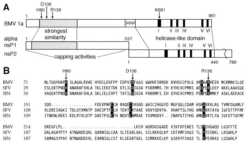
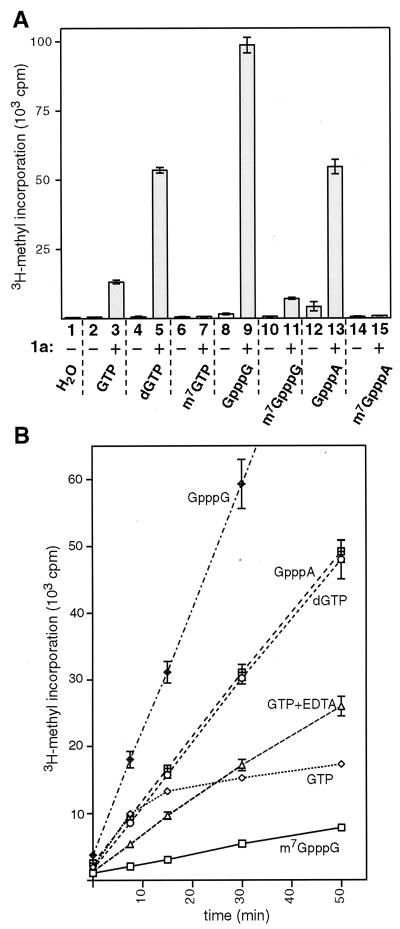
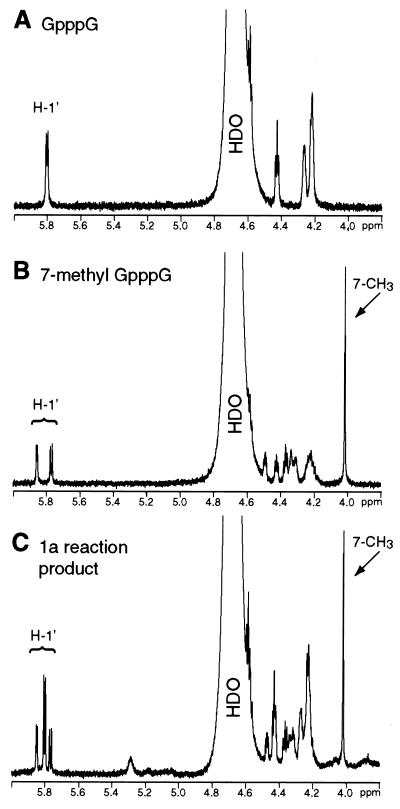
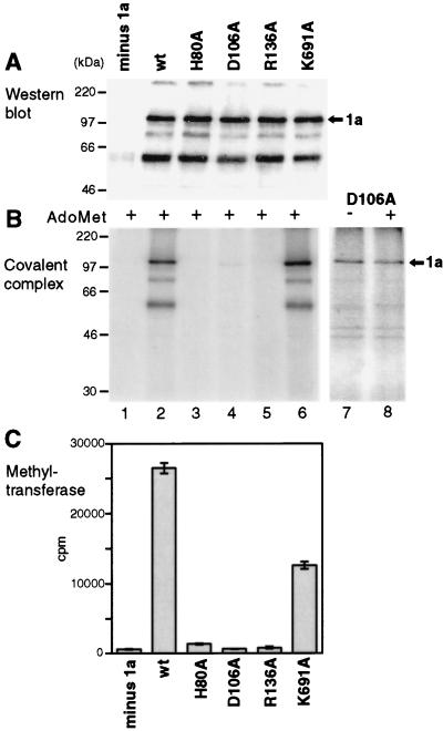
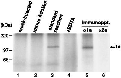
Brome mosaic virus (BMV) RNA replication is directed by two virus-encoded proteins, 1a and 2a. The amino-terminal half of 1a is a distant homolog of alphavirus nonstructural protein nsP1, which has been implicated in capping viral RNAs. In this study, we examined the enzymatic activities of BMV 1a expressed in yeast, where the protein is fully functional in RNA replication. 1a methylated GTP, dGTP, and the cap analogs GpppG and GpppA, using S-adenosylmethionine (AdoMet) as the methyl donor. Product analysis by nuclear magnetic resonance spectroscopy showed that 1a methylation was specific for guanine position 7. Additionally, 1a interacted with GTP to form a covalent 1a-m(7)GMP complex. This reaction was specific for GTP, required AdoMet, and was accompanied by transfer of (3)H-methyl from AdoMet to the covalent 1a-guanylate complex. The covalent complex could be immunoprecipitated by 1a antibodies. The 1a-m(7)GMP complex was inhibited in catalyzing further methyltransferase reactions. Mutation of conserved amino acids in the N-terminal half of 1a reduced both methyltransferase and covalent complex formation activities to very low or undetectable levels. Covalent 1a-guanylate complex formation took place in similar, AdoMet-dependent fashion in extracts of BMV-infected barley protoplasts. These results show that BMV 1a has activities similar to those of alphavirus nsP1, demonstrating conservation of these putative capping functions across a wide span of sequence divergence within the alphavirus-like superfamily. Conservation of this unusual combination of functions also supports the inference that the superfamily caps viral RNAs by an unusual pathway proceeding via a m(7)GMP intermediate.


Figures

FIG. 1
FIG. 1
Comparison of BMV 1a with alphavirus replicase proteins nsP1 and nsP2. (A) Schematic showing BMV 1a and alphavirus nsP1 and nsP2. The region of most significant similarity between alphavirus nsP1s and the N-terminal half of BMV 1a is shaded (2, 4, 35). The C-proximal BMV 1a and alphavirus nsP2 helicase-like domains have six highly conserved motifs, numbered I to VI. In 1a, the nsP1 and nsP2-related domains are separated by a proline-rich linker region, marked PPP. The BMV 1a residues mutated in this study are marked at the top with arrows. (B) Sequence alignment of BMV 1a and the nsP1 proteins of the alphaviruses Semliki Forest virus (SFV) and Sindbis virus (SIN) in the region of their strongest similarity (shown shaded in Fig. 1A). The most strongly conserved residues in the alphavirus-like superfamily are highlighted, and residues mutated in this study are marked with arrows.
FIG. 2
FIG. 2
Isolation of 1a-containing membranes. The total lysate from yeast cells expressing BMV 1a (lane 1) was subjected to flotation in a discontinuous sucrose gradient, consisting of 1 ml of 10% sucrose, 5 ml of 50% sucrose, and 6 ml of 60% sucrose (originally containing the lysate). After centrifugation, a floated membrane fraction (top 3 ml; lane 2), an intermediate fraction (next 3 ml; lane 3), the sample loading layer (bottom 6 ml; lane 4), and a resuspended pellet fraction (lane 5) were collected. The floated fraction was diluted with buffer and subjected to a second centrifugation to concentrate the membranes. This yielded supernatant (lane 6) and pellet (lane 7) fractions. All fractions were analyzed by SDS-PAGE and Western blotting and probed with antiserum against the N-terminal amino acids 1 to 502 of 1a (33). Equal percentages of each fraction were loaded on the gel, so that the recovery of 1a protein can be estimated from direct comparisons of the various lanes. Positions of molecular weight markers are shown at the left, and the position of full-length 1a is shown at the right.
FIG. 3
FIG. 3
Analysis of covalent complex formation between 1a and 7-methylated guanylate by SDS-PAGE and autoradiography. Portions of 1a-enriched membrane fractions prepared as in Fig. 2, lane 7, were incubated under standard conditions (see Materials and Methods) with [α-32P]GTP and unlabeled AdoMet (A) or Ado[methyl-3H]Met and unlabeled GTP (B) (lane 3). In control experiments, unlabeled AdoMet (A) or GTP (B) was omitted (lane 2), 5 mM EDTA was added to the standard reaction (lane 4), or membranes from cells not expressing 1a were incubated under standard conditions (lane 1). In each panel, the standard reaction mixture (lane 3) was also subjected to immunoprecipitation with anti-1a or anti-2a antiserum (lanes 5 and 6). (C) Vaccinia virus capping enzyme (lanes marked V) or 1a was incubated with [α-32P]GTP or [8-3H]GTP, as indicated, under the same conditions, in the presence of unlabeled AdoMet. Arrows mark the position of full-length 1a, arrowheads indicate the vaccinia virus capping enzyme, and positions of coelectrophoresed molecular weight markers are shown at the left.
FIG. 4
FIG. 4
Substrate specificity of covalent complex formation. 1a was incubated under standard conditions (see Materials and Methods) with the indicated, 32P-labeled nucleotide substrates in the presence of AdoMet. Greek letters indicate the labeled phosphate position in each case; 1 mM pyrophosphate (PPi; lane 9) or phosphate (Pi; lane 10) was included as indicated. All samples were analyzed by SDS-PAGE and autoradiography. The arrow marks the position of 1a, and positions of molecular weight markers are shown at the left.
FIG. 5
FIG. 5
Methyltransferase activity of 1a. (A) The substrates indicated below the bars were assayed as methyl group acceptors in standard reactions with membrane fractions isolated from yeast lacking 1a (−) and yeast expressing 1a (+). Two independent membrane preparations of each type were assayed, each in duplicate, with each of the substrates shown. Radioactivity incorporated to the substrate was measured by scintillation counting, and the average counts per minute for each condition is displayed by the histogram bars. Standard deviations are indicated by error bars. (B) Kinetics of the methyltransferase reaction catalyzed by 1a were studied by withdrawing aliquots from larger-scale reactions at 0, 7.5, 15, 30, and 50 min. The incorporated radioactivity was measured as described above, and the average counts per minute for each time point is displayed. The error bars indicate standard deviation and are included for all points, but in some cases they are obscured by the symbols used to plot average values. Two independent 1a-containing membrane preparations were assayed, each in duplicate, with each of the substrates shown. Standard reaction conditions including 2 mM MgCl2 were used except for the curve labeled GTP+EDTA, for which case the divalent cations were replaced with 5 mM EDTA. The curve for GpppG linearly continues to the 50-min time point (not shown).
FIG. 6
FIG. 6
Identification of the 1a methyltransferase reaction product. Expansions (6.0 to 3.8 ppm) of 500-MHz 1H NMR spectra of 0.5 mM solutions of GpppG (A) and 7-methyl-GpppG (B) in D2O are shown. For panel C, a 1a-containing membrane fraction was incubated with GpppG and AdoMet, and the partially purified mixture of unreacted GpppG and the methylated reaction product was isolated as described in Materials and Methods. Similar expansion of the NMR spectrum of a D2O solution of this 1a reaction product-substrate mixture is shown. Peaks due to H-1′ and 7-CH3 (10) and the large peak due to residual 1H-containing water (HDO) are labeled.
FIG. 7
FIG. 7
Effects of point mutations on enzymatic activities of 1a. Membrane fractions from yeast lacking 1a (lane 1), expressing wt 1a (lane 2), or expressing the indicated point mutant derivatives of 1a (lanes 3 to 6) were analyzed by Western blotting with 1a antiserum (A). The same fractions were assayed for covalent m7GMP complex formation in the presence of AdoMet (B) and for guanine-7-methyltransferase activity (C). The histogram bars of panel C show averages and standard deviations from assays of two complete sets of such fractions, each in duplicate. A longer exposure of the covalent guanylate binding activity of mutant D106A is shown in panel B, lanes 7 and 8, assayed in the absence or presence of AdoMet, as indicated. Arrows mark the position of 1a, and positions of molecular weight markers are shown at the left.
FIG. 8
FIG. 8
Covalent guanylate complex formation by 1a derived from BMV-infected plant cells. Portions of BMV-infected barley protoplast membrane fractions were incubated under standard conditions (see Materials and Methods) with [α-32P]GTP and unlabeled AdoMet (lane 3). In control experiments, unlabeled AdoMet was omitted (lane 2), 5 mM EDTA was added to the standard reaction (lane 4), or membranes from mock-infected barley protoplasts were incubated under standard conditions (lane 1). Four volumes of the standard reaction mixture (lane 3) was also subjected to immunoprecipitation with anti-1a or anti-2a antiserum (lanes 5 and 6). All samples were analyzed by SDS-PAGE and autoradiography.

Similar articles

Cited by

References

    1. Ahlquist P. Bromovirus RNA replication and transcription. Curr Opin Genet Dev. 1992;2:71–76. - PubMed
    1. Ahlquist P, Strauss E G, Rice C M, Strauss J H, Haseloff J, Zimmern D. Sindbis virus proteins nsP1 and nsP2 contain homology to nonstructural proteins from several RNA plant viruses. J Virol. 1985;53:536–542. - PMC - PubMed
    1. Ahola T, Kääriäinen L. Reaction in alphavirus mRNA capping: formation of a covalent complex of nonstructural protein nsP1 with 7-methyl-GMP. Proc Natl Acad Sci USA. 1995;92:507–511. - PMC - PubMed
    1. Ahola T, Laakkonen P, Vihinen H, Kääriäinen L. Critical residues of Semliki Forest virus RNA capping enzyme involved in methyltransferase and guanylyltransferase-like activities. J Virol. 1997;71:392–397. - PMC - PubMed
    1. Allison R, Thompson C, Ahlquist P. Regeneration of a functional RNA virus genome by recombination between deletion mutants and requirement for cowpea chlorotic mottle virus 3a and coat genes for systemic infection. Proc Natl Acad Sci USA. 1990;87:1820–1824. - PMC - PubMed

Publication types

MeSH terms

LinkOut - more resources