. 1999 Dec;73(12):10070-8.
doi: 10.1128/JVI.73.12.10070-10078.1999.

In vitro infection of ovine cell lines by Jaagsiekte sheep retrovirus

Affiliations

In vitro infection of ovine cell lines by Jaagsiekte sheep retrovirus

M Palmarini et al. J Virol. 1999 Dec.

Abstract

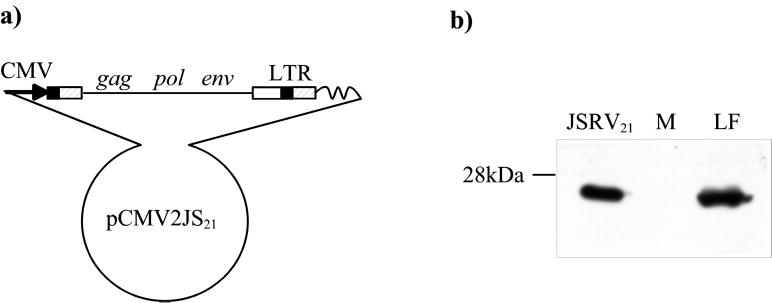
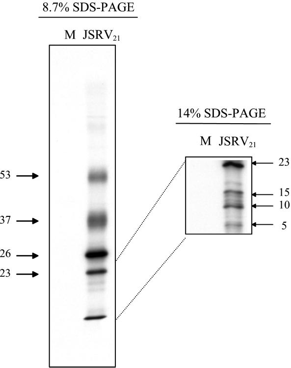
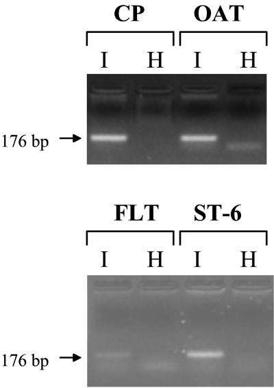
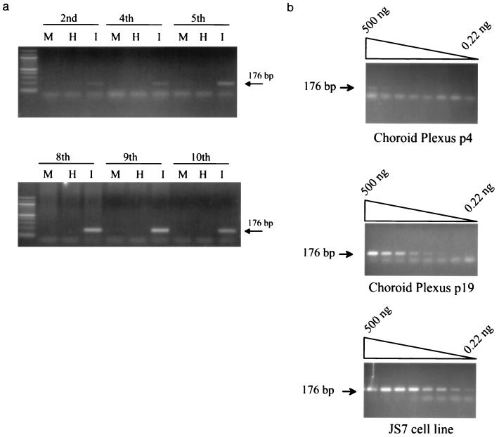
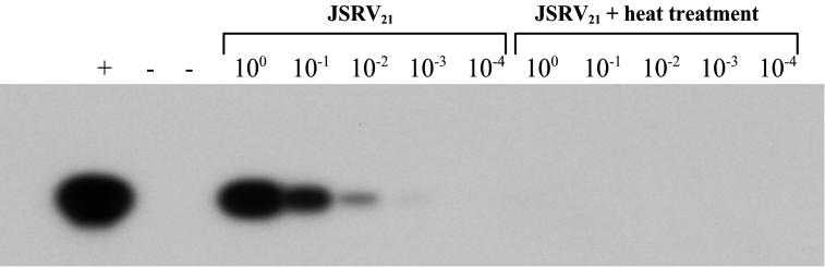
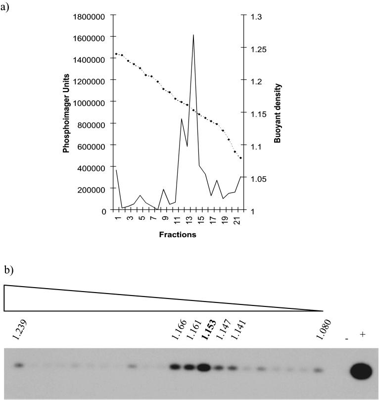
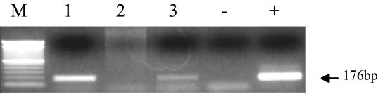
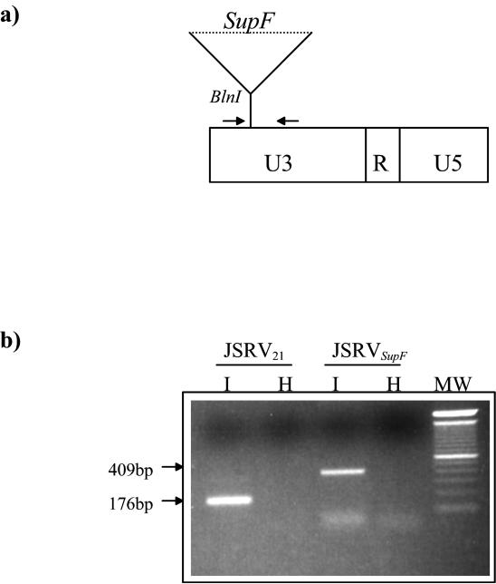
Sheep pulmonary adenomatosis (SPA), also known as jaagsiekte or ovine pulmonary carcinoma, is a contagious lung cancer of sheep, originating from type II pneumocytes and Clara cells. Previous studies have implicated a type D retrovirus (jaagsiekte sheep retrovirus [JSRV]) as the causative agent of SPA. We recently isolated a proviral clone of JSRV from an animal with a spontaneous case of SPA (JSRV(21)) and showed that it harbors an infectious and oncogenic virus. This demonstrated that JSRV is necessary and sufficient to induce SPA. A major impediment in research on JSRV has been the lack of an in vitro tissue culture system for the virus. The experiments reported here show the first successful in vitro infection with this virus, using the JSRV(21) clone. JSRV(21) virus was obtained by transiently transfecting human 293T cells with a plasmid containing the JSRV(21) provirus driven by the human cytomegalovirus immediate-early promoter. Virus produced in this manner exhibited reverse transcriptase (RT) activity that banded at 1.15 g/ml in sucrose density gradients. Infection of concentrated JSRV(21) into ovine choroid plexus (CP), testes (OAT-T3), turbinate (FLT), and intestinal carcinoma (ST6) cell lines resulted in establishment of infection as measured by PCR amplification. Evidence that this reflected genuine infection included the fact that heat inactivation of the virus eliminated it, the levels of viral DNA increased with passage of the infected cells, and the infected cells released active RT as measured by the sensitive product enhancement RT assay. The RT activity released from the infected cells banded at 1.15 g/ml, and JSRV(21) provirus was transmitted from infected cells to uninfected ones by cocultivation. However, the amount of virus released from infected cells was low. These results suggest that the JSRV receptor is present on many ovine cell types and that the observed restriction of JSRV expression in vivo to tumor cells might be controlled by factors other than the viral receptor. Finally we tagged the U3 of pJSRV(21) with the bacterial supF gene, an amber suppressor tRNA gene. The resulting clone, termed pJSRV(supF), is infectious in vitro. It may be a useful tool for future studies on viral DNA integration, since the normal sheep genome contains 15 to 20 copies of highly JSRV-related endogenous sequences that cross-react with many JSRV hybridization probes.


Figures

FIG. 1
FIG. 1
Organization of the pCMV2JS21 construct and in vitro synthesis of JSRV21 particles. (a) pCMV2JS21 is a plasmid containing a modified version of the integrated JSRV21 provirus in which the U3 region of the upstream LTR was replaced by the human cytomegalovirus immediate-early promoter. (b) SDS-PAGE and Western blotting of 300-fold-concentrated supernatant from 293T cells transiently transfected with pCMV2JS21. The filters were probed with a rabbit polyclonal antiserum toward the major capsid protein (CA) of JSRV. Lung fluid collected from an SPA-affected animal and concentrated in the same way as the 293T supernatant was used as a positive control (LF). Concentrated supernatant from mock-transfected 293T cells was used as a negative control (M). The 26-kDa JSRV-CA protein is indicated.
FIG. 2
FIG. 2
Proteins in JSRV21 virions. Labeled JSRV21 was prepared from transfected 293T cells that had been labeled with [35S]methionine 24 h prior to supernatant harvest. Viral particles were purified by isopynic centrifugation, and the fractions corresponding to 1.14 to 1.16 g/ml were pooled and analyzed by SDS-PAGE on 8.7 and 15% polyacrylamide gels followed by autoradiography (JSRV21). 293T cells that were mock-transfected were labeled and processed in parallel (M). No radioactivity banded at 1.14 to 1.16 g/ml from tissue culture supernatant from the mock-transfected culture. JSRV21 virions contained major bands of 53, 37, 26, and 23 kDa visible in the 8.7% polyacrylamide gel (left). An additional band migrated at the bottom of the 8.7% polyacrylamide gel that resolved into three bands of 15, 10, and 5 kDa in the 14% polyacrylamide gel. The sizes of the radioactive proteins were calculated from the mobilities of protein size markers.
FIG. 3
FIG. 3
In vitro infection of ovine cell lines. Four different ovine cell lines were infected with JSRV21 (I) or in parallel with heat-inactivated virus (H), and the cultures were serially passaged. PCR for exogenous JSRV with primers from the U3 region of the LTR was carried out on 500 ng of infected-cell DNA. The diagnostic 176-bp product is indicated by an arrow. PCR assays were carried out on CP cells at passage 7 after infection and on the other three cell lines at passage 5.
FIG. 4
FIG. 4
In vitro infection of CP cells. (a) PCR for JSRV DNA was carried out on 500 ng of DNA from infected CP cells at different passages. Analysis of cells infected with heat-treated JSRV21 (I) or mock-infected cells (M) of equivalent passage numbers was also carried out. The passages postinfection are indicated at the top of each gel. The intensity of the amplified 176-bp product intensified in the later passages (passages 8 through 10). (b) PCR for JSRV DNA on threefold dilutions of infected CP cell DNA at passage 4 postinfection are compared with the same cells at passage 19 postinfection. The specific amplimer of 176 bp is indicated by an arrow. The faster-migrating bands are primer-dimers. The passage 19 cells had approximately 34 times as much JSRV21 DNA as the passage 4 cells did. PCR on serial dilutions of JS7 cell line (9) DNA is also shown. JS7 cells contain one copy of JSRV provirus per cell (2a). The results indicate that passage 19-infected CP cells have approximately 32 less DNA per cell than do JS7 cells.
FIG. 5
FIG. 5
RT activity released from infected CP cells. RT activity was tested by PERT assay on 10-fold dilutions of 33-fold-concentrated supernatant from infected CP cells (passage 12) or on equivalently concentrated supernatant from CP cells infected with heat-treated JSRV21 at the same passage. Southern blot hybridization with an MS2-specific probe was used for PCR product detection. Note the presence of the specific MS2 band only in the JSRV21-infected cells. Controls: +, a PERT reaction with purified Moloney murine leukemia virus RT (10−4 U); −, a PERT reaction with buffer A alone.
FIG. 6
FIG. 6
Buoyant density of virus produced by infected CP cells. Supernatant from JSRV21-infected CP cells (passage 19) was concentrated and fractionated by isopycnic centrifugation in a 20 to 55% continuous sucrose gradient. Gradient fractions were assayed for RT activity by the PERT assay. (a) RT activity (solid line) expressed in PhosphorImager units taken from samples visualized in panel b; the broken line shows density (grams per milliliter). The peak of RT activity is in the fraction with a density of 1.15 g/ml. (b) PERT reactions from each gradient fraction visualized by Southern blotting for MS-2 DNA; +, Moloney murine leukemia virus RT (10−4 U); −, buffer A control.
FIG. 7
FIG. 7
Transfer of JSRV infection from infected CP cells. U3 PCR for exogenous JSRV DNA was carried out on 500 ng of genomic DNA. The parental infected and uninfected CP cells before cocultivation are shown in lanes 1 and 2, respectively. In lane 3 are shown CP cells 8 weeks after cocultivation. +, JSRV21 plasmid DNA; −, distilled water. M, 100-bp ladder (Gibco).
FIG. 8
FIG. 8
JSRVsupF is infectious in vitro. (a) Schematic diagram of the JSRVsupF LTR. The supF bacterial tRNA suppressor gene was inserted into the U3 region of pCMV2JS21. The primers used in this study for the PCR detection of JSRV provirus (JSRV U3-PCR) are downstream and upstream of the supF gene (bold arrows); the resulting PCR product obtained from the amplification of JSRVsupF will be 409 bp, versus the 176 bp for the wild-type JSRV LTR. (b) CP cells were infected with JSRVsupF or wild-type JSRV21 obtained from transfected 293T cells and assayed for infection by PCR analysis. The results, from passage 2 after infection, show successful infection by JSRVsupF. Cells were infected in parallel with untreated virus (I) or virions heat inactivated at 65°C for 15 min (H). Note the different sizes of the PCR products obtained from choroid plexus cells infected with JSRV21 versus JSRVsupF.

References

    1. Bai J, Zhu R Y, Stedman K, Cousens C, Carlson J, Sharp J M, DeMartini J C. Unique long terminal repeat U3 sequences distinguish exogenous jaagsiekte sheep retroviruses associated with ovine pulmonary carcinoma from endogenous loci in the sheep genome. J Virol. 1996;70:3159–3168. - PMC - PubMed
    1. Bradac J, Hunter E. Polypeptides of Mason-Pfizer monkey virus. I. Synthesis and processing of the gag-gene products. Virology. 1984;138:260–275. - PubMed
    1. DeMartini, J. C., and J. M. Sharp. Unpublished data.
    1. DiFronzo N L, Holland C A. A direct demonstration of recombination between an injected virus and endogenous viral sequences, resulting in the generation of mink cell focus-inducing viruses in AKR mice. J Virol. 1993;67:3763–3770. - PMC - PubMed
    1. Dion A S, Williams C J, Pomenti A A. The major structural proteins of murine mammary tumor virus: techniques for isolation. Anal Biochem. 1977;82:18–28. - PubMed

Publication types

LinkOut - more resources