. 1999 Dec;73(12):10146-57.
doi: 10.1128/JVI.73.12.10146-10157.1999.

Physical and functional interaction between the Y-box binding protein YB-1 and human polyomavirus JC virus large T antigen

Affiliations

Physical and functional interaction between the Y-box binding protein YB-1 and human polyomavirus JC virus large T antigen

M Safak et al. J Virol. 1999 Dec.

Abstract

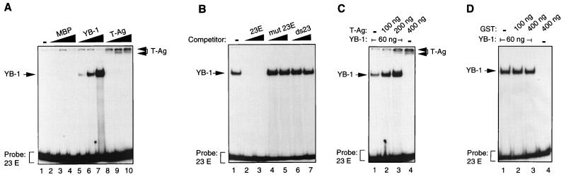
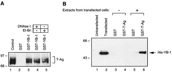
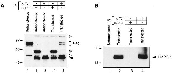
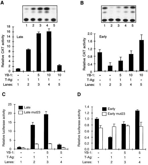
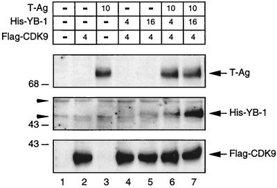
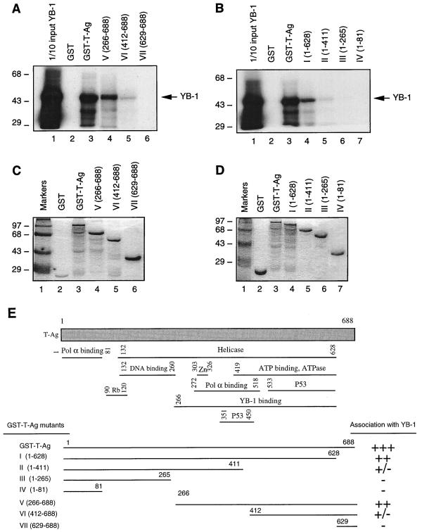
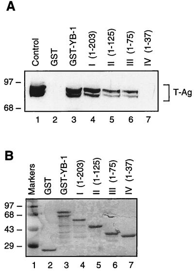
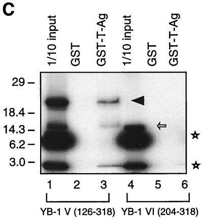
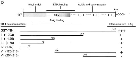
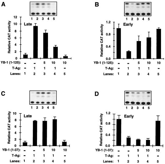
Y-box binding protein YB-1 is a member of a family of DNA and RNA binding proteins which have been shown to affect gene expression at both the transcriptional and translational levels. We have previously shown that YB-1 modulates transcription from the promoters of the ubiquitous human polyomavirus JC virus (JCV). Here we investigate the physical and functional interplay between YB-1 and the viral regulatory protein large T antigen (T-antigen), using JCV as a model system. Results of mobility band shift assays demonstrated that the efficiency of binding of YB-1 to a 23-bp single-stranded viral target sequence was significantly increased when T-antigen was included in the binding reaction mixture. Affinity chromatography and coimmunoprecipitation assays demonstrated that YB-1 and T-antigen physically interact with each other. Additionally, results of transcription studies demonstrated that these two proteins interact functionally on the JCV early and late gene promoters. Whereas ectopic expression of YB-1 and T-antigen results in synergistic transactivation of the viral late promoter, YB-1 alleviates T-antigen-mediated transcriptional suppression of the viral early promoter activity. Furthermore, we have localized, through the use of a series of deletion mutants, the sequences of these proteins which are important for their interaction. The T-antigen-interacting region of YB-1 is located in the cold shock domain of YB-1 and its immediate flanking sequences, and the YB-1-interacting domain of T-antigen maps to the carboxy-terminal half of T-antigen. Results of transient transfection assays with various YB-1 mutants and T-antigen expression constructs confirm the specificity of the functional interaction between YB-1 and T-antigen. Taken together, these data demonstrate that the cellular factor YB-1 and the viral regulatory protein T-antigen interact both physically and functionally and that this interaction modulates transcription from the JCV promoters.


Figures

FIG. 1
FIG. 1
T-antigen induces binding of YB-1 to the 23-bp early strand (23E) in electrophoretic mobility shift assays. (A) Interaction of bacterially expressed MBP, MBP–YB-1 fusion protein, and baculovirus-expressed T-antigen with the 23-bp early single-stranded DNA probe. The 23E probe was incubated with 30, 60, or 240 ng of MBP alone (lanes 2 to 4, respectively) or MBP–YB-1 fusion protein (lanes 5 to 7, respectively) or with 100, 200, or 400 ng of T-antigen (T-Ag) (lanes 8 to 10, respectively). DNA-protein complexes were resolved on a 6% polyacrylamide gel under native conditions. Positions of MBP–YB-1 and T-antigen complexes with the 23E probe are indicated by arrows. (B) Competitive band shift assay. The 23E probe was incubated with MBP–YB-1 protein in the absence (lane 1) or presence of a 50- or 250-fold molar excess of unlabeled competitor DNA as indicated. 23E represents the 23-bp wild-type early single-stranded oligonucleotide (5′-CAGTTTTAGCCAGCTCCTCCCTA-3′) (lanes 2 and 3), mut 23E represents a mutant 23E single-stranded oligonucleotide (5′-AGTATTACACAGATATTTATTAC-3′) (lanes 4 and 5), and ds23 represents the wild-type double-stranded 23-bpse (lanes 6 and 7). (C) T-antigen induces binding of YB-1 to the 23-bp early single strand. The 23E probe was incubated with recombinant YB-1 (MBP–YB-1), either alone (lane 1) or with baculovirus-expressed T-antigen (lanes 2 and 3), or with T-antigen alone (lane 4). Formed DNA-protein complexes were resolved on a 6% polyacrylamide gel under native conditions. Respective protein concentrations used for the binding reactions are shown above the panel. The protein concentration used for all binding reactions was kept constant by addition of MBP. MBP–YB-1:23E and T-Ag:23E complexes are indicated by arrows. (D) Bacterially expressed, purified GST protein does not induce binding of YB-1 to the 23-bp early single strand. The 23-bp probe was incubated with recombinant YB-1, either alone (lane 1) or in combination with GST (lanes 2 and 3), or with GST alone (lane 4). Formed DNA-protein complexes were resolved as described for panel C. Respective protein concentrations used for the binding reactions are shown above the panel. MBP–YB-1:23E complex is indicated by an arrow. In all panels, brackets indicate free probe.
FIG. 2
FIG. 2
In vitro interaction between YB-1 and T-antigen. (A) Bacterially expressed GST (lane 2) or GST–YB-1 (lane 3) was immobilized on GST-Sepharose beads and incubated with whole-cell extract prepared from hamster glial cells constitutively expressing T-antigen (HJC-15b). Additionally, whole-cell extracts from HJC-15b cells, treated with DNase I (0.2 U/μg of protein) or ethidium bromide (100 ng/ml) (Et-Br), were also incubated with GST–YB-1 (lanes 4 and 5, respectively). HJC-15b whole-cell extract was loaded as a migration control (lane 1). Nonbinding proteins were removed from the column by extensive washing, and proteins interacting with GST or GST–YB-1 were resolved by SDS–10% PAGE and analyzed by Western blotting with an anti-T-antigen antibody (Ab-2 416). The bracket indicates T-antigen (T-Ag). (B) Whole-cell extracts prepared from untransfected HJC-15b cells (lanes 1, 3, and 4) and from HJC-15b cells transfected with a histidine-tagged YB-1 expression plasmid (pEBV-YB-1) (lanes 2, 5, and 6) were incubated with either GST alone (lanes 3 and 5) or GST–T-antigen (GST-T-Ag) (lanes 4 and 6) as indicated. After being washed, proteins interacting with GST or GST–T-antigen were resolved by SDS–10% PAGE and analyzed by Western blot analysis with anti-T7 antibody for detection of His-tagged YB-1. Whole-cell extracts from HJC-15b cells either untransfected (lane 1) or transfected with pEBV-His-YB-1 expression plasmid (lane 2) were loaded as negative and positive migration controls, respectively. The arrow indicates histidine-tagged YB-1. The positions of molecular mass markers (in kilodaltons) are shown on the left of each panel.
FIG. 3
FIG. 3
T-antigen coimmunoprecipitates with YB-1. (A) Coimmunoprecipitation of T-antigen with His–YB-1. Coimmunoprecipitation experiments were performed as described in Materials and Methods. Antibodies used for the respective lanes are shown at the top. HJC-15b whole-cell extract was loaded as a migration control (lane 1). The bracket indicates T-antigen (T-Ag). An arrowhead indicates the position of immunoglobulin heavy chain detected by the secondary antibody. Open arrows indicate nonspecific bands. IP, immunoprecipitation; α-T7, anti-T7 antibody; α-pre, preimmune serum. (B) Direct immunoprecipitation of His-tagged YB-1 with anti-T7 antibody, demonstrating that this antibody works in immunoprecipitation assays. Whole-cell extract (40 μg) from hamster glial cells (HJC-15b) transfected with the pEBV-YB-1 expression plasmid was immunoprecipitated with preimmune serum or with anti-T7 as indicated. The immunocomplexes were analyzed by Western blotting for His-tagged YB-1, using anti-T7. Whole-cell extracts from HJC-15b cells either untransfected or transfected with His-tagged YB-1 were loaded as negative and positive controls (lanes 1 and 2, respectively). The arrow indicates His-tagged YB-1. The positions of molecular mass markers (in kilodaltons) are shown on the left of each panel.
FIG. 4
FIG. 4
Functional interaction between T-antigen and YB-1. (A) T-antigen and YB-1 synergistically transactivate the JCV late gene promoter in U-87MG cells. A reporter plasmid (7.5 μg) containing the JCV late gene promoter was transfected into U-87MG cells alone or together with YB-1 and T-antigen expression plasmids (T-Ag). Expression plasmid DNA concentrations used in the transfections are indicated at the bottom of the panel (in micrograms per plate). The data are represented as CAT activity relative to basal-level expression of the promoter. The results of a representative CAT assay are shown at the top. (B) YB-1 alleviates T-antigen-mediated transcriptional suppression from the JCV early promoter. Experiments similar to those detailed for panel A were also performed with a reporter plasmid containing the JCV early gene promoter. The results are expressed as CAT activity relative to basal-level expression of the promoter. (C and D) Experiments similar to those detailed for panel A were also performed with luciferase plasmids containing early and late wild-type promoters as well as early and late promoters containing point mutations within the 23-bp region (Late mut23 and Early mut23). Reporter activity of each plasmid is expressed as luciferase activity relative to basal-level expression of the wild-type promoter. Results shown in each panel represent the means of data from three independent experiments. Bars indicate standard deviations.
FIG. 5
FIG. 5
Stability of YB-1 protein is increased in the presence of T-antigen. U-87MG cells were either transfected with CMV–T-antigen alone (lane 3) or cotransfected with pEBV-His-YB-1 and CMV–T-antigen expression plasmids (lanes 6 and 7). Lanes 4 and 5 received only pEBV-His-YB-1. Expression plasmid concentrations used in the transfections are indicated above the panels. The DNA concentrations for the transfection mixtures were normalized by addition of the respective empty expression vector DNA. A flag-tagged CDK9 expression plasmid was also included in the transfection mixture as an internal control for transfection efficiency. Thirty micrograms of whole-cell lysate was resolved by SDS–10% PAGE followed by immunoblotting. The different strips were cut out from the same blot and analyzed for T-antigen (T-Ag), His-tagged YB-1, and flag-tagged CDK9. Whole-cell lysate from untransfected cells (lane 1) was loaded as a negative control. The arrowheads indicate nonspecific bands. The arrows indicate the positions of the respective proteins. The positions of molecular mass markers (in kilodaltons) are shown to the left of the panels.
FIG. 6
FIG. 6
Localization of the domain of T-antigen that interacts with YB-1. (A and B) GST–T-antigen (GST-T-Ag) and N-terminal (A) and C-terminal (B) T-antigen deletion mutants immobilized on glutathione-Sepharose beads were incubated with in vitro-translated [35S]methionine-labeled YB-1. The Sepharose beads were washed extensively, and bound proteins were resolved by SDS–10% PAGE and analyzed by autoradiography. One-tenth of the input YB-1 used in each reaction was loaded as a migration control (lane 1 in each panel). The arrows indicate the position of in vitro-translated [35S]methionine-labeled YB-1. (C and D) SDS–10% PAGE analysis of GST, GST–T-antigen, and T-antigen N-terminal (C) and C-terminal (D) deletion mutants. (E) Summary of the results obtained from in vitro mapping assays. A schematic representation of T-antigen is shown at the top (not to scale). The abilities of T-antigen and its deletion mutants to interact with YB-1 are shown on the right (+++, specific interaction; ++, reduced interaction; +/−, minimal interaction; and −, no interaction). Pol α binding, polymerase α binding domain. The positions of molecular mass markers (in kilodaltons) are shown to the left of panels A to D.
FIG. 7
FIG. 7
Localization of the domain of YB-1 that interacts with T-antigen. (A) Whole-cell extract from hamster glial cells constitutively expressing T-antigen (HJC-15b) was incubated with either GST alone (lane 2) or C-terminal deletion mutants of GST–YB-1 fusion proteins immobilized on glutathione-Sepharose beads. Bound complexes were washed extensively, resolved by SDS–8% PAGE, and analyzed for T-antigen by Western blotting with an anti-T-antigen antibody (Ab-2 416). HJC-15b whole-cell extract was loaded as a migration control (lane 1). The bracket indicates T-antigen. (B) SDS–10% PAGE analysis of GST and of GST–YB-1 and its C-terminal deletion mutants. (C) Two different 35S-labeled, in vitro-translated N-terminal deletion mutants of YB-1 [YB-1 (126–318) and YB-1 (204–318)] were incubated with GST (lanes 2 and 5) or GST–T-antigen (T-Ag) (lanes 3 and 6). Bound proteins were resolved by SDS-PAGE and analyzed by autoradiography. Lanes 1 and 4 contain 1/10 of the amount used in the pull-down experiments with YB-1 (126–318) and YB-1 (204–318), respectively. An arrowhead and an open arrow designate the positions of the in vitro-translated N-terminal deletion mutants YB-1 (126–318) and YB-1 (204–318), respectively. The asterisks denote nonspecific products of the in vitro transcription-translation reactions. (D) A schematic representation of full-length YB-1 is shown at the top. The abilities of YB-1 and its deletion mutants to interact with T-antigen are depicted on the right (+++, specific interaction; ++ or +, reduced interaction; and −, no interaction). The positions of molecular mass markers (in kilodaltons) are shown to the left of panels A to C.
FIG. 7
FIG. 7
Localization of the domain of YB-1 that interacts with T-antigen. (A) Whole-cell extract from hamster glial cells constitutively expressing T-antigen (HJC-15b) was incubated with either GST alone (lane 2) or C-terminal deletion mutants of GST–YB-1 fusion proteins immobilized on glutathione-Sepharose beads. Bound complexes were washed extensively, resolved by SDS–8% PAGE, and analyzed for T-antigen by Western blotting with an anti-T-antigen antibody (Ab-2 416). HJC-15b whole-cell extract was loaded as a migration control (lane 1). The bracket indicates T-antigen. (B) SDS–10% PAGE analysis of GST and of GST–YB-1 and its C-terminal deletion mutants. (C) Two different 35S-labeled, in vitro-translated N-terminal deletion mutants of YB-1 [YB-1 (126–318) and YB-1 (204–318)] were incubated with GST (lanes 2 and 5) or GST–T-antigen (T-Ag) (lanes 3 and 6). Bound proteins were resolved by SDS-PAGE and analyzed by autoradiography. Lanes 1 and 4 contain 1/10 of the amount used in the pull-down experiments with YB-1 (126–318) and YB-1 (204–318), respectively. An arrowhead and an open arrow designate the positions of the in vitro-translated N-terminal deletion mutants YB-1 (126–318) and YB-1 (204–318), respectively. The asterisks denote nonspecific products of the in vitro transcription-translation reactions. (D) A schematic representation of full-length YB-1 is shown at the top. The abilities of YB-1 and its deletion mutants to interact with T-antigen are depicted on the right (+++, specific interaction; ++ or +, reduced interaction; and −, no interaction). The positions of molecular mass markers (in kilodaltons) are shown to the left of panels A to C.
FIG. 8
FIG. 8
Functional interaction of T-antigen with YB-1 deletion mutants. (A and B) Transcriptional activity of deletion mutant YB-1 (1–125), in the presence or absence of T-antigen (T-Ag), on CAT reporter plasmids (7.5 μg) containing the viral late (A) and early (B) promoters. The concentrations of expression plasmids are indicated at the bottom of the respective panels (in micrograms per plate). Representative CAT data are shown above the graphs, and quantitative analysis of results is expressed as CAT activity relative to the basal-level expression of the promoter. (C and D) Transcriptional activity of deletion mutant YB-1 (1–37), in the presence or absence of T-antigen, on CAT reporter plasmids (7.5 μg) containing late (C) and early (D) promoters. The experimental design was virtually identical to that described for panels A and B. The results shown in each panel represent the means of data from three independent experiments. Bars indicate standard deviations.

References

    1. Ansari S A, Safak M, Gallia G L, Sawaya B E, Amini S, Khalili K. Interaction of YB-1 with human immunodeficiency virus type and TAR RNA modulates viral promoter activity. J Gen Virol. 1999;80:229–236. - PubMed
    1. Asakuno K, Kohno K, Uchiumi T, Kubo T, Sato S, Isono M, Kuwano M. Involvement of a DNA binding protein, MDR-NF1/YB-1, in human MDR1 gene expression by actinomycin D. Biochem Biophys Res Commun. 1994;199:1428–1435. - PubMed
    1. Bargou R C, Jurchott K, Wagener C, Bergmann S, Metzner S, Bommert K, Mapara M Y, Winzer K J, Dietel M, Dorken B, Royer H D. Nuclear localization and increased levels of transcription factor YB-1 in primary human breast cancers are associated with intrinsic MDR1 gene expression. Nat Med. 1997;3:447–450. - PubMed
    1. Bayarsaihan D, Enkhmandakh B, Lukens L N. Y-box proteins interact with the S1 nuclease-sensitive site in the chicken alpha 2(1) collagen gene promoter. Biochem J. 1996;319:203–207. - PMC - PubMed
    1. Berger J R, Concha M. Progressive multifocal leukoencephalopathy: the evolution of a disease once considered rare. J Neurovirol. 1995;1:5–18. - PubMed

Publication types

MeSH terms

LinkOut - more resources