. 1999 Dec;73(12):10164-72.
doi: 10.1128/JVI.73.12.10164-10172.1999.

A hydrophobic patch in ecotropic murine leukemia virus envelope protein is the putative binding site for a critical tyrosine residue on the cellular receptor

Affiliations

A hydrophobic patch in ecotropic murine leukemia virus envelope protein is the putative binding site for a critical tyrosine residue on the cellular receptor

T Zavorotinskaya et al. J Virol. 1999 Dec.

Abstract

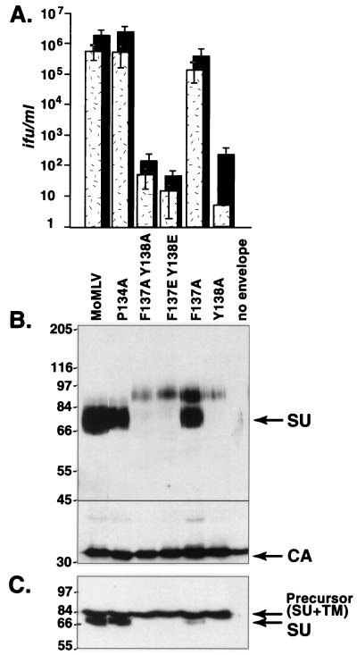
In the receptor for ecotropic murine leukemia viruses, tyrosine 235 contributes a critical hydrophobic side chain to the virus-receptor interaction (14). Here we report that tryptophan 142 in ecotropic Moloney murine leukemia virus envelope protein is essential to virus binding and infection. Replacement of tryptophan 142 by alanine or serine resulted in misfolding. However, replacement by methionine (W142M) allowed correct folding of the majority of glycoprotein molecules. W142M virus showed a marked reduction in virus binding and was almost noninfectious, suggesting that tryptophan 142 is involved in receptor binding. In contrast, W142Y virus containing a replacement of tryptophan 142 with an aromatic residue (tyrosine) was as efficient as wild-type virus in infection and binding of cells expressing the wild-type receptor. However, W142Y virus was 100-fold less efficient than wild-type virus in infection of cells expressing a mutant receptor containing tryptophan instead of the critical tyrosine. These results strongly support tryptophan 142 being an essential residue on the virus envelope protein that interacts directly with the critical hydrophobic residue at position 235 of the ecotropic receptor. Tryptophan 142 forms one side of a shallow hydrophobic pocket on the surface of the envelope protein, suggesting that it might comprise the complete putative binding site for tyrosine 235. We discuss the implications of our findings with respect to two models of the envelope protein trimer. Interestingly, both models place tryptophan 142 at the interface between adjacent subunits of the trimer.


Figures

FIG. 1
FIG. 1
Substitution of phenylalanine 137 did not affect infection, whereas substitution of tyrosine 138 markedly reduced infection. (A) Infectious titers on mouse NIH 3T3 cells bearing the endogenous ecotropic MLV receptor (stippled bars) and human 293 cells expressing the exogenous receptor (black bars). Titers were calculated from the endpoint dilutions (n = 4) after exposure to virions pseudotyped with envelope proteins containing the indicated substitutions. Each value is the average of results from five independent experiments. MoMLV, wild-type ecotropic Mo MLV; no envelope, supernatant or lysate of cells transfected with a virus genome that lacks an env gene; ifu, infectious units. (B) Western blot analysis of virions containing mutant envelope proteins. Samples were denatured by boiling them in the presence of 100 mM dithiothreitol, chilled on ice, and separated on sodium dodecyl sulfate (SDS)–8% polyacrylamide gels. Then proteins were transferred onto nitrocellulose membranes (Protran; Schleicher & Schuell). The membrane was cut into two parts at the position indicated by the black line, roughly that of the 45-kDa molecular mass standard. Envelope proteins (SU and precursor) were detected on the top portion by incubation with goat anti-Rauscher gp70 antiserum and structural CA protein was detected on the bottom portion by incubation with goat anti-Rauscher p30 antiserum. Subsequent incubation with mouse anti-goat or mouse anti-rabbit antiserum conjugated to horseradish peroxidase (Sigma) was performed, and immunoblots were developed with Renaissance chemiluminescence substrate (NEN). (C) Western blot analysis of virus producer cell lysates. Proteins were separated on SDS–8% polyacrylamide gels and then blotted to anti-SU antiserum. Numbers to the left of panels B and C show molecular masses in kilodaltons.
FIG. 2
FIG. 2
Replacement of tryptophan 142 with alanine, serine, or methionine, but not with tyrosine, profoundly reduced infection. Substitution of tyrosine for tryptophan 142 did not affect envelope protein processing or virus infectivity, but replacement of that residue with an alanine, serine, or methionine residue greatly reduced virus entry and interfered with envelope protein processing. (A) Virus titers on NIH 3T3 cells (stippled bars) and human 293 cells expressing the exogenous wild-type receptor (black bars). ifu, infectious units. (B) Western blot analysis of virions containing mutant env genes. (C) Western blot analysis of virus producer cell lysates. Molecular masses (in kilodaltons) are noted at the left of the blots.
FIG. 3
FIG. 3
The 85-kDa envelope protein species represents a hyperglycosylated form of SU in F137A virions but represents uncleaved precursor protein in W142M virions. (A) Digestion with glycosidase F to remove N-linked carbohydrates. Sucrose gradient-purified wild-type and mutant virions were incubated overnight in the absence (−) or presence (+) of excess glycosidase F, and then proteins were separated on SDS–8% polyacrylamide gels and immunoblotted with anti-SU (top portion) or anti-CA (bottom portion) antiserum. To determine if glycosidase digestion had been completed, lysates of wild-type Mo MLV producer cells were also analyzed. (B) Immunoblot analysis of virions with anti-TM antiserum. Virion proteins separated on SDS–6 to 15% polyacrylamide gradient gels were immunoblotted with rabbit anti-p15E antiserum (1:250; gift of Alan Rein) with Immobilon membranes to determine if the 85-kDa species contained TM sequences. After detection of the anti-TM antiserum-reactive proteins (left blot), membranes were washed and incubated with anti-SU antiserum (right blot). Molecular mass markers (in kilodaltons) are noted at the left.
FIG. 4
FIG. 4
Virus binding. Binding assays were performed essentially as described previously (4, 11) with modifications as previously reported (20). No virus, nonspecific binding of antiserum in the absence of virus to 293 cells stably expressing receptor; No receptor, binding of wild-type Mo MLV to parental 293 cells lacking ecotropic receptor. The mean fluorescence intensities of the no-receptor and no-virus peaks were 0.2 and 0.3, respectively. Infectivity was summarized from the data in Fig. 1 and 2. +, titer within 10-fold of that of wild-type Mo MLV; −, titer 10,000-fold less than that of wild-type Mo MLV.
FIG. 5
FIG. 5
A marked loss of infection resulting from a tryptophan 142-to-tyrosine change in the envelope protein correlates with a change from tyrosine to tryptophan in the critical position corresponding to tyrosine 235 in the receptor. Gray bars, wild-type Mo MLV; black bars, W142Y virus stock; white bars, W142M virus stock. Human cells expressing the altered receptor and human cDNAs have been previously described (14). (A) TCID50s for human cells expressing the wild-type or mutant receptor cDNAs. Receptor mutants contained the following changes: a single E237V change (M-65) (Ygv), Y235W and E237V changes (M-38W) (Wgv), Y237H and E237V changes (M-38H) (Hgv), and Y235M and E237V changes (M-38M) (Mgv). (B) TCID50s for human cells expressing cDNAs encoding mutant proteins of the homologous human protein. In two independent titrations, wild-type Mo MLV showed a TCID50 of 2 × 10³ infectious units (ifu) per ml on cells expressing the human protein containing proline 242-to-tryptophan and valine 244-to-glutamic acid changes (Wge), whereas W142Y virus showed a TCID50 of 20 ifu/ml on parallel cultures of the WGE cells. Human homologs contained the following changes in positions 242 and 244, corresponding to Y235 and E237 of the receptor, respectively: P242Y and V237E changes (H-40) (Yge), P242F and V237E changes (H-40F) (Fge), and P242W and V237E changes (H-40W) (Wge). Values are the average TCID50s from two experiments using virus generated from two independent transfections in endpoint dilution titration (n = 4), except that values for the FGE cells represent a single titration. No infections by W142M virus were observed on HGV, MGV, FGE, or WGE cells. Amino acids are indicated by the single-letter amino acid code.
FIG. 6
FIG. 6
A model for the putative receptor binding site. All residues are represented as space-filled amino acids and are numbered according to the Mo MLV sequence starting with the N-terminal alanine on mature SU. Shaded residues represent those previously shown to be critical for receptor binding (D84, R83, and R95) and the residues in the hydrophobic patch (W142, W152, Y151, and Y210). (A) The left image shows the side view of SU. The middle image shows an enlargement of the hydrophobic patch from the same view as that shown in the left image. The arrow indicates the proposed binding site of tyrosine 235 in the receptor. The right image shows the top surface of the view seen in the left image. (B) The left image shows a diagram of the SU trimer model proposed by Fass and coworkers (6, 7); the right image shows a diagram of an alternative trimer model generated as described in Materials and Methods. (C) Alignment of the residues comprising the putative binding patch. Numbers to the right and left of the sequence indicate the amino acid positions beginning with the N-terminal residues of mature SU. Numbers above the sequence indicate the positions in Mo MLV. Boxes indicate the structural features in ecotropic MLV SU. Mo MLV, ecotropic Moloney MLV; Fr MLV, ecotropic Friend MLV; CasBrE, ecotropic MLV CasBrE; 4070A, amphotropic MLV; MCF, Mink cell focus-forming virus strain 1233; Xeno, xenotropic MLV strain NZB; GALV, gibbon ape leukemia virus strain SEATO.

Similar articles

Cited by

References

    1. Albritton L M, Kim J W, Tseng L, Cunningham J M. Envelope-binding domain in the cationic amino acid transporter determines the host range of ecotropic murine retroviruses. J Virol. 1993;67:2091–2096. - PMC - PubMed
    1. Bae Y, Kingsman S M, Kingsman A J. Functional dissection of the Moloney murine leukemia virus envelope protein gp70. J Virol. 1997;71:2092–2099. - PMC - PubMed
    1. Battini J L, Heard J M, Danos O. Receptor choice determinants in the envelope glycoproteins of amphotropic, xenotropic, and polytropic murine leukemia viruses. J Virol. 1992;66:1468–1475. - PMC - PubMed
    1. Cosset F L, Morling F J, Takeuchi Y, Weiss R A, Collins M K L, Russell S J. Retroviral retargeting by envelopes expressing an N-terminal binding domain. J Virol. 1995;69:6314–6322. - PMC - PubMed
    1. Davey R A, Zuo Y, Cunningham J M. Identification of a receptor-binding pocket on the envelope protein of Friend murine leukemia virus. J Virol. 1999;73:3758–3763. - PMC - PubMed

Publication types

MeSH terms

LinkOut - more resources