. 1999 Dec;73(12):10214-23.
doi: 10.1128/JVI.73.12.10214-10223.1999.

Altering the cellular location of an antigen expressed by a DNA-based vaccine modulates the immune response

Affiliations

Altering the cellular location of an antigen expressed by a DNA-based vaccine modulates the immune response

P J Lewis et al. J Virol. 1999 Dec.

Abstract

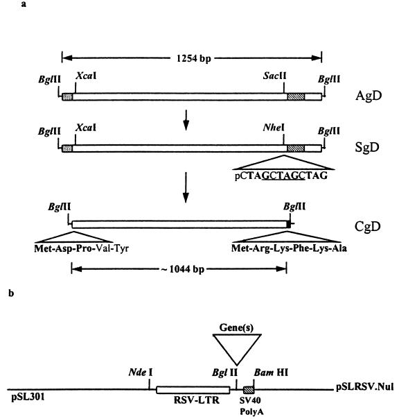
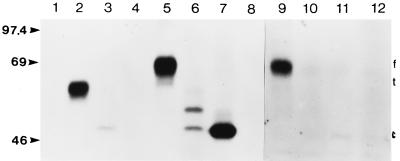
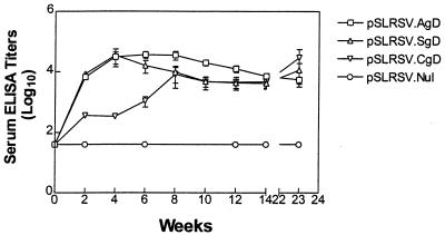
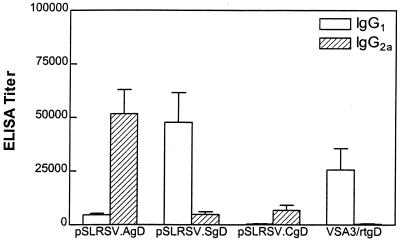
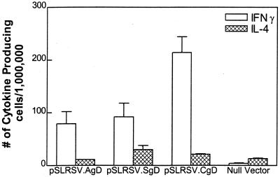
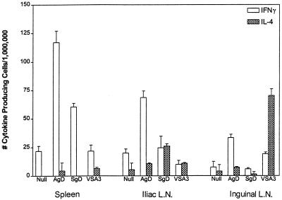
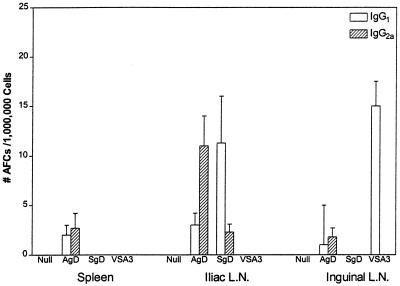
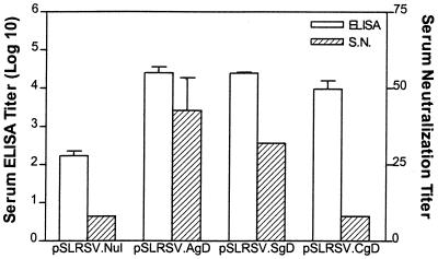
The potential for DNA vaccines encoding mutated versions of the same antigen to modulate immune responses in C3H/HeN mice was investigated. We created expression plasmids that encoded several versions of glycoprotein D (gD) from bovine herpesvirus 1, including authentic membrane-anchored glycoprotein (pSLRSV.AgD), a secreted glycoprotein (pSLRSV.SgD), and an intracellular protein (pSLRSV.CgD). Immunization of an inbred strain of mice with these plasmids resulted in highly efficacious and long-lasting humoral and cell-mediated immunity. We also demonstrated that the cell compartment in which plasmid-encoded gD was expressed caused a deviation in the serum immunoglobulin (Ig) isotype profile as well as the predominant cytokines secreted from the draining lymph node. Immunization of C3H/HeN mice with DNA vaccines encoding cell-associated forms of gD resulted in a predominance of serum IgG2a and gamma interferon-secreting cells within the spleens and draining lymph nodes. In contrast, mice immunized with a secreted form of this same antigen displayed immune responses characterized by greater levels of interleukin 4 in the draining lymph node and IgG1 as the predominant serum isotype. We also showed evidence of compartmentalization of distinct immune responses within different lymphoid organs.


Figures

FIG. 1
FIG. 1
Diagrammatic depiction of expression cassettes. Panel a depicts membrane-anchored (AgD), secreted (SgD), and cytosolic (CgD) forms of BHV-1 gD. The expression cassettes all utilized the high-copy-number pSL301 plasmid backbone. The evolution of secreted and cytosolic versions of BHV-1 gD is shown as are details regarding relevant changes in coding sequences, start codons (CgD), and termination codons. The NheI restriction sequence (underlined) and triple stop codons (boldface) are shown for gene SgD. Novel amino acids translated at the amino and carboxy termini of CgD are in boldface. Gene and construct designations are indicated to the immediate right of each diagram. All genes encoding full-length or truncated versions of BHV-1 gD were inserted at the BglII site (▿) shown in panel b. Panel b depicts the null vector (pSLRSV.Nul) with the RSV-LTR enhancer or promoter and the short polyadenylation sequence derived from SV40.
FIG. 2
FIG. 2
Autoradiograph of immunoprecipitated forms of membrane-anchored, secreted, and cytosolic BHV-1 gD. Preconfluent COS-7 cells transiently transfected with pSLRSV.AgD, pSLRSV.SgD, pSLRSV.CgD, or pSLRSV.Nul were grown for 48 h in the presence of 35S-labelled methionine and cysteine. Radioactively labelled gD was immunoprecipitated from medium and/or cell lysates by using an anti-gD monoclonal antibody pool (see Materials and Methods). SDS-PAGE of precipitates demonstrates the calculated molecular masses (kDa) of mutated gD and cellular localization as predicted. Immunoprecipitates of media collected from COS-7 cells transfected with the following are shown: 1, pSLRSV.AgD; 2, pSLRSV.SgD; 3, pSLRSV.CgD; and 4, pSLRSV.Nul. Immunoprecipitates of lysates of COS-7 cells transfected with the following are shown: 5, pSLRSV.AgD; 6, pSLRSV.SgD; 7, pSLRSV.CgD; and 8, pSLRSV.Nul. Immunoprecipitates of plasma membrane-associated gD are shown: 9, pSLRSV.AgD; 10, pSLRSV.SgD; 11, pSLRSV.CgD; and 12, pSLRSV.Nul. Molecular mass markers are indicated on the left. The positions of membrane-anchored (f), secreted (t), and cytosolic (c) versions of gD are indicated on the right.
FIG. 3
FIG. 3
Kinetics of serum anti-BHV-1 gD antibodies in C3H/HeN mice immunized with plasmids encoding cell-associated or secreted forms of BHV-1 gD. Each DNA-based vaccine group was comprised of five mice. Each mouse received 100 μg (2 μg/μl in normal saline) of plasmid DNA i.m. in the left quadriceps muscle mass on days 0 and 14. Serum ELISA titers for individual mice were determined by using the extrapolation function (Microsoft Excel) based on endpoint dilutions and with preimmune serum means (plus 3 standard deviations) as cutoffs. Endpoint titers are expressed as 1/log10. Serum antibody levels were determined for individual mice at each time point, except at 2 weeks, when blood samples for each group were pooled. Data are expressed as the geometric mean of four (pSLRSV.AgD) or two (pSLRSV.CgD) seropositive mice. Error bars show the standard error of the means. Only 23-week titers from mice that were seropositive at week 14 were determined.
FIG. 4
FIG. 4
Serum antibody levels after immunization of C3H/HeN mice with plasmids encoding cell-associated (AgD or CgD) or secreted (SgD) antigens. Experimental groups consisted of 10 mice and included a null plasmid control group and a subunit gD vaccine group. Mice 6 to 7 weeks of age were immunized in each quadriceps muscle mass with 50 μg of DNA (total of 100 μg) or subcutaneously with 400 ng of affinity-purified rtgD in 100 μl of VSA3 plus HBSS. All mice were given a booster injection at 2 weeks after initial immunization with the same dose. Serum ELISA titers were determined as described for Fig. 3. Plasmid construct or subunit gD designations are indicated in the top center of each graph. Each symbol represents a single mouse. The horizontal line in each column represents the mean antibody titer for the group.
FIG. 5
FIG. 5
Serum IgG isotype after immunization with DNA vaccines encoding cell-associated (AgD or CgD) or secreted (SgD) forms of BHV-1 gD or a gD subunit vaccine. BHV-1 gD-specific IgG1 or IgG2a isotypes were detected by ELISA with biotinylated secondary antibodies. Antibody titers were calculated as described for Fig. 3. Values are depicted as 1/log10 of antibody titer (determined by serial endpoint dilution analysis).
FIG. 6
FIG. 6
Splenic cytokine profiles in mice 5 1/2 months postimmunization. Seropositive mice depicted in Fig. 3 were assessed for antigen-specific production of IFN-γ or IL-4. Spleens from seropositive mice were pooled and stimulated in vitro for ∼40 h with affinity-purified BHV-1 gD. Stimulated splenocytes were plated at 1.0 × 10⁶ or 0.5 × 10⁶ cells/well on Polyfiltronics plates. Cytokine-secreting splenocytes are depicted as the number of cytokine-producing cells per million plated cells. Mean values of triplicate wells are depicted.
FIG. 7
FIG. 7
Cytokine profiles in lymph nodes (L.N.) and spleen after immunization with DNA vaccines. Antigen-specific cytokine profiles were measured in several different lymphoid tissues of C3H/HeN mice immunized with plasmids encoding cell-associated or secreted versions of BHV-1 gD. Mice were euthanized 5 weeks after booster injection, and spleens and draining and nondraining lymph nodes were excised and pooled. Pooled cell populations were stimulated in vitro for 18 to 20 h with 400 ng of affinity-purified BHV-1 gD. Stimulated cells were harvested and plated on Polyfiltronics plates at 106 cells/well in triplicate. Pooled values are represented as mean values and as the number of cytokine-producing cells per million plated cells. Control wells for nonspecific binding of cytokine were set up for all groups, and mean values from control wells were subtracted from all groups.
FIG. 8
FIG. 8
Number of AFCs secreting IgG1 or IgG2a after immunization with DNA vaccines. The data depicts antigen-specific IgG1 and IgG2a production in several different lymphoid tissues of C3H/HeN mice immunized with plasmids encoding cell-associated or secreted versions of BHV-1 gD. The mice depicted were euthanized 5 weeks after booster injection. Spleens and lymph nodes (L.N.) were excised and pooled. Pooled cells were plated in triplicate at 106 cells/well on Millipore ELISPOT plates. Data are presented as the mean number of Ig-secreting cells per million plated cells. Plate control wells for nonspecific binding of Ig isotypes were set up for all groups, and mean values were subtracted from all groups.
FIG. 9
FIG. 9
Functional differences in serum antibodies after immunization with plasmids encoding different forms of BHV-1 gD. Serum antibody responses of mice following immunization and given a booster injection at 2 weeks with plasmids encoding cell membrane-anchored gD (pSLRSV.AgD), secreted gD (pSLRSV.SgD), cytoplasmic gD (pSLRSV.CgD), or null vector (pSLRSV.Nul) are shown. BHV-1 gD-specific ELISA titers are expressed as the reciprocal of the extrapolated (see Materials and Methods) dilution resulting in 3 standard deviations above the control value (prebleed sera). BHV-1 neutralizing antibody titers are expressed as the reciprocal of the highest dilution of antibody that caused a 50% reduction of plaques relative to the virus control. Results are expressed as geometric means, and bars indicate the standard error. S.N., serum neutralization.

Similar articles

Cited by

References

    1. Aramaki Y, Suda H, Tsuchiya S. Interferon-inductive effect of liposomes as an immunoadjuvant. Vaccine. 1995;18:1809–1814. - PubMed
    1. Audibert F M, Lise L D. Adjuvants: current status, clinical perspectives, and future prospects. Immunol Today. 1993;14:281–284. - PubMed
    1. Babiuk L A, L’Italien J, van Drunen Littel-van den Hurk S, Zamb T, Lawman M J P, Hughes G, Gifford G A. Protection of cattle from bovine herpesvirus type I (BHV-1) infection by immunization with individual viral glycoproteins. Virology. 1987;159:57–66. - PubMed
    1. Babiuk L A, Wardley R C, Rouse B T. Defense mechanisms against bovine herpesvirus: relationship of virus-host cell events to susceptibility to antibody-complement cell lysis. Infect Immun. 1975;12:958–963. - PMC - PubMed
    1. Baca-Estrada M E, Snider M, Tikoo S K, Harland R, Babiuk L A, van Drunen Littel-van den Hurk S. Immunogenicity of bovine herpesvirus-1 glycoprotein D in mice: effect of antigen form on the induction of cellular and humoral immune responses. Viral Immunol. 1996;9:11–22. - PubMed

Publication types

MeSH terms

LinkOut - more resources