. 1999 Dec;73(12):10303-9.
doi: 10.1128/JVI.73.12.10303-10309.1999.

Brome mosaic virus RNA replication proteins 1a and 2a colocalize and 1a independently localizes on the yeast endoplasmic reticulum

Affiliations

Brome mosaic virus RNA replication proteins 1a and 2a colocalize and 1a independently localizes on the yeast endoplasmic reticulum

M Restrepo-Hartwig et al. J Virol. 1999 Dec.

Abstract

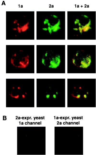
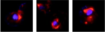
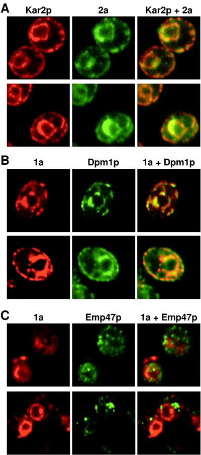
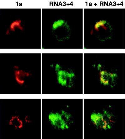
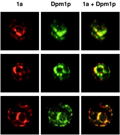
The universal membrane association of positive-strand RNA virus RNA replication complexes is implicated in their function, but the intracellular membranes used vary among viruses. Brome mosaic virus (BMV) encodes two mutually interacting RNA replication proteins: 1a, which contains RNA capping and helicase-like domains, and the polymerase-like 2a protein. In cells from the natural plant hosts of BMV, 1a and 2a colocalize on the endoplasmic reticulum (ER). 1a and 2a also direct BMV RNA replication and subgenomic mRNA synthesis in the yeast Saccharomyces cerevisiae, but whether the distribution of 1a, 2a, and active replication complexes in yeast duplicates that in plant cells has not been determined. For yeast expressing 1a and 2a and replicating BMV genomic RNA3, we used double-label confocal immunofluorescence to define the localization of 1a, 2a, and viral RNA and to explore the determinants of replication complex targeting. As in plant cells, 1a and 2a colocalized on and were retained on the yeast ER, with no detectable accumulation in the Golgi apparatus. 1a and 2a were distributed over most of the ER surface, with strongest accumulation on the perinuclear ER. In vivo labeling with bromo-UTP showed that the sites of 1a and 2a accumulation were the sites of nascent viral RNA synthesis. In situ hybridization showed that completed viral RNA products accumulated predominantly in the immediate vicinity of replication complexes but that some, possibly more mature cells also accumulated substantial viral RNA in the surrounding cytoplasm distal to replication complexes. Additionally, we find that 1a localizes to the ER when expressed in the absence of other viral factors. These results show that BMV RNA replication in yeast duplicates the normal localization of replication complexes, reveal the intracellular distribution of RNA replication products, and show that 1a is at least partly responsible for the ER localization and retention of the RNA replication complex.


Figures

FIG. 1
FIG. 1
BMV 1a and 2a colocalize in 1a+2a+RNA3 yeast cells. (A) 1a+2a+RNA3 yeast cells were processed for indirect, double-label immunofluorescence using rabbit anti-1a and mouse anti-2a antisera followed by anti-rabbit antibodies conjugated to Texas red and anti-mouse antibodies conjugated to Alexa 488. Rows show 1a (left), 2a (middle), and their superposition (right) for a 0.5-μm optical section of a representative, independent cell. Each image is 9 μm per side. (B) Sample negative controls for antibody specificity. Yeast expressing (expr.) 1a only or 2a only were processed for double-label immunofluorescence using both anti-1a and anti-2a primary and secondary antibodies as described above. The images shown illustrate the absence of 1a immunofluorescence signal from yeast expressing 2a only and the absence of 2a immunofluorescence signal from yeast expressing 1a only. See text for additional controls.
FIG. 2
FIG. 2
BMV 1a accumulates in perinuclear and cytoplasmic regions of 1a+2a+RNA3 yeast cells. 1a+2a+RNA3 yeast cells were processed for 1a immunofluorescence (red) and stained for DNA with To-Pro-3 (blue). Each image (9 μm per side) shows the superimposed 1a and DNA fluorescence patterns from independent, representative cells.
FIG. 3
FIG. 3
1a and 2a accumulate on the ER but not the Golgi apparatus in 1a+2a+RNA3 yeast cells. Each row shows immunofluorescence images for a 0.5-μm optical section of representative, independent BMV 1a+2a+RNA3 yeast cells. The cells were processed for indirect, double-label immunofluorescence using primary antisera against BMV 2a and yeast ER protein Kar2p (A), BMV 1a and yeast ER protein Dpm1p (B), and BMV 1a and yeast Golgi protein Emp47p (C). Images of each individual label and their superposition are shown. Each image is 9 μm per side.
FIG. 4
FIG. 4
(A) Colocalization of newly synthesized BMV RNA with 1a. 1a+2a+RNA3 yeast cells were permeabilized, incubated 10 min with BrUTP, and then fixed and processed for indirect, double-label immunofluorescence with primary antisera that recognize 1a and BrU incorporated into nucleic acid. Rows show 1a (left), incorporated BrUTP (middle), and their superposition (right) for a 0.5 μm optical section of a representative, independent cell. (B) Strong, cytoplasmic BrUTP incorporation is dependent on the presence of a functional BMV RNA replication template. 1a+2a yeast cells were permeabilized, incubated, fixed, and processed as for panel A for indirect immunofluorescence with primary antisera that recognize 1a and BrU incorporated into nucleic acid. Each image is 9 μm per side.
FIG. 5
FIG. 5
BMV RNA3 and RNA4 accumulate predominantly but not exclusively near 1a. 1a+2a+RNA3 yeast cells were fixed and subjected to in situ hybridization with four digoxigenin-labeled oligodeoxynucleotide probes (see Materials and Methods) complementary to the positive strand of the coat protein gene in RNA3 and its subgenomic mRNA, RNA4. After in situ hybridization, the cells were processed for indirect, double-label immunofluorescence using primary antisera against 1a and digoxigenin. Rows show 1a (left), RNA3 and RNA4 (middle), and their superposition (right) for a 0.5-μm optical section of a representative, independent cell. Each image is 9 μm per side.
FIG. 6
FIG. 6
1a colocalizes with ER markers in the absence of 2a or RNA3. Yeast cells expressing 1a but not 2a or RNA3 were processed for indirect, double-label immunofluorescence using primary antisera against 1a and yeast ER protein Dpm1p. Rows show 1a (left), Dpm1p (middle), and their superposition (right) for a 0.5-μm optical section of a representative, independent cell. Each image is 9 μm per side.

References

    1. Ahlquist P, Luckow V, Kaesberg P. Complete nucleotide sequence of brome mosaic virus RNA3. J Mol Biol. 1981;153:23–38. - PubMed
    1. Ahlquist P, Strauss E G, Rice C M, Strauss J H, Haseloff J, Zimmern D. Sindbis virus proteins nsP1 and nsP2 contain homology to nonstructural proteins from several RNA plant viruses. J Virol. 1985;53:536–542. - PMC - PubMed
    1. Ahola T, Ahlquist P. Putative RNA capping activities encoded by brome mosaic virus: methylation and covalent binding of guanylate by replicase protein. J Virol. 1999;73:10061–10069. - PMC - PubMed
    1. Allison R, Thompson C, Ahlquist P. Regeneration of a functional RNA virus genome by recombination between deletion mutants and requirement for cowpea chlorotic mottle virus 3a and coat genes for systemic infection. Proc Natl Acad Sci USA. 1990;87:1820–1824. - PMC - PubMed
    1. Ausubel F M, Brent R, Kingston R E, Moore D D, Seidman J G, Smith J A, Struhl K, editors. Current protocols in molecular biology. New York, N.Y: John Wiley & Sons; 1987.

Publication types

LinkOut - more resources