. 1999 Dec;73(12):10310-9.
doi: 10.1128/JVI.73.12.10310-10319.1999.

Determinants of CD4 independence for a human immunodeficiency virus type 1 variant map outside regions required for coreceptor specificity

Affiliations

Determinants of CD4 independence for a human immunodeficiency virus type 1 variant map outside regions required for coreceptor specificity

C C LaBranche et al. J Virol. 1999 Dec.

Abstract

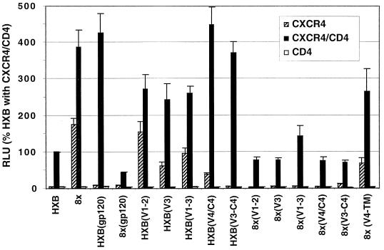
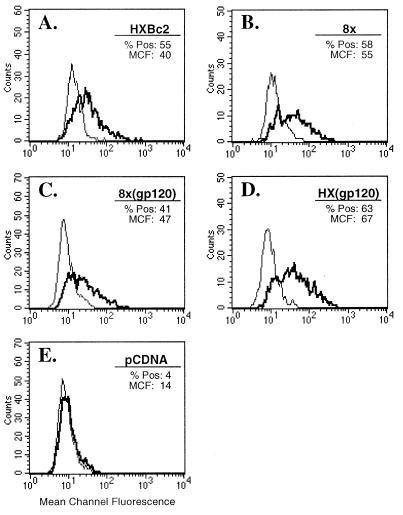
Although infection by human immunodeficiency virus (HIV) typically requires an interaction between the viral envelope glycoprotein (Env), CD4, and a chemokine receptor, CD4-independent isolates of HIV and simian immunodeficiency virus have been described. The structural basis and underlying mechanisms for this phenotype are unknown. We have derived a variant of HIV-1/IIIB, termed IIIBx, that acquired the ability to utilize CXCR4 without CD4. This virus infected CD4-negative T and B cells and fused with murine 3T3 cells that expressed human CXCR4 alone. A functional IIIBx env clone exhibited several mutations compared to the CD4-dependent HXBc2 env, including the striking loss of five glycosylation sites. By constructing env chimeras with HXBc2, the determinants for CD4 independence were shown to map outside the V1/V2 and V3 hypervariable loops, which determine chemokine receptor specificity, and at least partly within an area on the gp120 core that has been implicated in forming a conserved chemokine receptor binding site. We also identified a point mutation in the C4 domain that could render the IIIBx env clone completely CD4 dependent. Mutations in the transmembrane protein (TM) were also required for CD4 independence. Remarkably, when the V3 loop of a CCR5-tropic Env was substituted for the IIIBx Env, the resulting chimera was found to utilize CCR5 but remained CD4 independent. These findings show that Env determinants for chemokine receptor specificity are distinct from those that mediate CD4-independent use of that receptor for cell fusion and provide functional evidence for multiple steps in the interaction of Env with chemokine receptors. Combined with our observation that the conserved chemokine receptor binding site on gp120 is more exposed on the IIIBx gp120 (T. L. Hoffman, C. C. LaBranche, W. Zhang, G. Canziani, J. Robinson, I. Chaiken, J. A. Hoxie, and R. W. Doms, Proc. Natl. Acad. Sci. USA 96:6359-6364, 1999), the findings from this study suggest novel approaches to derive and design Envs with exposed chemokine receptor binding sites for vaccine purposes.


Figures

FIG. 1
FIG. 1
Derivation and characterization of CD4-independent HIV-1. (A) Viral replication in CD4-negative T cells. SupT1 and CD4-negative BC7 cells were inoculated with equal amounts of HIV-1/IIIB or IIIBx, and viral replication was determined by RT activity in culture supernatants. (B) Inhibition of IIIBx by anti-CXCR4 MAb. BC7 cells were inoculated with IIIBx in the presence or absence of 12G5, and RT activity determined at the indicated time points. (C) IIIBx-induced fusion on murine cells expressing CXCR4. IIIBx-infected BC7 cells were cocultured for 24 h with murine 3T3 cells or 3T3 cells that expressed human CXCR4 and stained for syncytium formation as described previously (21).
FIG. 2
FIG. 2
Evaluation of a IIIBx env clone in fusion assays. env genes indicated were cloned into pSP73, transfected into QT6 cells, and evaluated in fusion assays on QT6 cells expressing CD4 plus CXCR4, CXCR4 alone, or CD4 alone (55). Results are expressed as RLU (mean + SEM) normalized to the activity of 8x on CXCR4+ CD4+ cells. Also shown are fusion activities for 8x and HXBc2 Envs that contain a D368R mutation in gp120, which ablates the CD4 binding site (44).
FIG. 3
FIG. 3
Generation of a replication-competent virus with a IIIBx Env and evaluation of its TM size. (A) The 8x env was inserted into pNL4-3, and a virus stock was generated after transfection into BC7 cells. Equal amounts of the resulting virus (designated NL43/8x) and HIV-1/IIIB were inoculated onto SupT1 and BC7 cells, and RT levels were monitored over time. (B) Viral lysates from HIV-1/IIIB-infected SupT1 cells (IIIB), IIIBx-infected BC7 cells (IIIBx), and NL43/8x-infected BC7 cells (8x) were evaluated by Western blotting using an anti-TM MAb, D12. “Mock” viral lysate was prepared from the supernatant of uninfected BC7 cells. Consistent with sequence analysis shown in Fig. 4, both IIIBx and NL43/8x have a truncated TM.
FIG. 4
FIG. 4
Sequence analysis for IIIBx env clones 8x and S10 in comparison to HXBc2. Shaded regions indicate mutations that are also found in other clones from HIV-1/IIIB. Predicted N-linked glycosylation sites are indicated (formula image
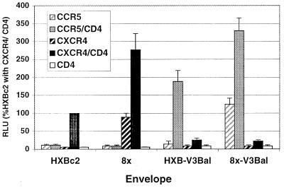
), as are the positions of variable loops, the gp120/gp41 cleavage site, and the TM membrane-spanning domain (msd). 8x contains a frameshift mutation at position 706 resulting in a prematurely truncated TM cytoplasmic tail. S10 contains a deletion of 50 nt, which also leads to frameshift and a prematurely truncated TM cytoplasmic tail.
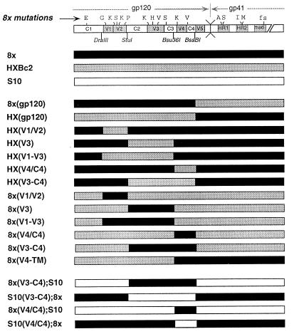
FIG. 5
FIG. 5
Construction of chimeric Env proteins for fusion assays. Diagrams representing HXBc2, 8x, and S10 env genes are shown along with chimeras constructed by using the indicated restriction sites. Mutations present in 8x are indicated above the top schematic. Chimeras were cloned into pSP73 and evaluated in cell fusion assays described in the legend to Fig. 6.
FIG. 6
FIG. 6
Evaluation of chimeric Env proteins in fusion assays. Chimeric Env proteins between HXBc2 and 8x shown in Fig. 5 were evaluated in fusion assays on QT6 target cells expressing CXCR4 alone, CXCR4 and CD4, or CD4 alone. Results are expressed as luciferase activity (RLU) relative to that of HXBc2 on CXCR4+ CD4+ cells. Bars indicate the mean RLU for at least five experiments + SEM.
FIG. 7
FIG. 7
Surface expression of CD4-dependent and -independent Env proteins on transfected cells. QT6 cells were transiently transfected with the Env proteins indicated or a vector-only control (pCDNA), and surface expression was quantitated by FACS. Shown for each Env protein is a histogram for cells labeled with NHS (thin line) or serum from an HIV-1-infected patient (bold line). The percent cells positive (% Pos) and the mean channel of fluorescence intensity (MCF) are indicated for cells stained with anti-HIV serum. Thresholds for positivity were defined so that <5% of cells stained with NHS were considered reactive.
FIG. 8
FIG. 8
Mapping determinants for a CD4-dependent clone of IIIBx. Fusion activity is shown for the CD4-dependent S10 clone of IIIBx and S10-8x chimeras indicated in Fig. 5. In addition, activity is shown for an S10 Env in which the G431E mutation in the C4 domain was corrected (S10-431G) and for an 8x Env that contained this mutation (8x-431E). Results are expressed as the percentage of S10 luciferase activity on target cells that coexpressed CXCR4 and CD4.
FIG. 9
FIG. 9
Construction of a CCR5-tropic, CD4-independent Env. 8x and HXB2 Env proteins containing V3 loop from the CCR5-tropic Env, HIV-1/BaL, were constructed, and their fusion activities were compared to those of parental 8x and HXBc2 Envs on target cells that expressed CCR5 or CXCR4 with or without CD4. Fusion activity is expressed as the percentage of luciferase activity of HXBc2 on target cells that expressed both CXCR4 and CD4. Bars indicate mean + SEM.
FIG. 10
FIG. 10
Locations of HIV-1/IIIBx mutations on the gp120 crystal structure. (A) A space-filling model of the HIV-1/HXB2 gp120 core crystal structure is shown (white) in conjunction with a ribbon diagram for CD4 (yellow) (34). Amino acid sites at which mutations produced a 50% decrease or increase gp120 binding to CCR5 are shown in red (52). Of the six mutations in 8x that could be mapped onto the gp120 core, three (shown in blue) are located immediately adjacent to this putative chemokine receptor binding site. (B) A ribbon diagram of the gp120-CD4 complex is depicted in a slightly different orientation to indicate the position of the G431E mutation, which was sufficient to abrogate CD4-independent but not -dependent fusion of the 8x clone.

References

    1. Alexander W A, Moss B, Fuerst T R. Regulated expression of foreign genes in vaccinia virus under the control of bacteriophage T7 RNA polymerase and the Escherichia coli lac repressor. J Virol. 1992;66:2934–2942. - PMC - PubMed
    1. Alkhatib G, Combadiere C, Broder C C, Feng Y, Kennedy P E, Murphy P M, Berger E A. CC CKR5: a RANTES, MIP-1α, MIP-1β receptor as a fusion cofactor for macrophage-tropic HIV-1. Science. 1996;272:1955–1958. - PubMed
    1. Bandres J C, Wang Q F, O’Leary J, Baleaux F, Amara A, Hoxie J A, Zolla-Pazner S, Gorny M K. Human immunodeficiency virus (HIV) envelope binds to CXCR4 independently of CD4, and binding can be enhanced by interaction with soluble CD4 or by HIV envelope deglycosylation. J Virol. 1998;72:2500–2504. - PMC - PubMed
    1. Berger E A. HIV entry and tropism: the chemokine receptor connection. AIDS. 1997;11(Suppl. A):S3–S16. - PubMed
    1. Cao J, Bergeron L, Helseth E, Thali M, Repke H, Sodroski J. Effects of amino acid changes in the extracellular domain of the human immunodeficiency virus type 1 gp41 envelope glycoprotein. J Virol. 1993;67:2747–2755. - PMC - PubMed

Publication types

MeSH terms

Associated data