. 1999 Dec;73(12):10359-70.
doi: 10.1128/JVI.73.12.10359-10370.1999.

Induction and prevention of apoptosis in human HEp-2 cells by herpes simplex virus type 1

Affiliations

Induction and prevention of apoptosis in human HEp-2 cells by herpes simplex virus type 1

M Aubert et al. J Virol. 1999 Dec.

Abstract

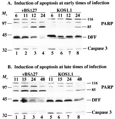
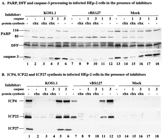
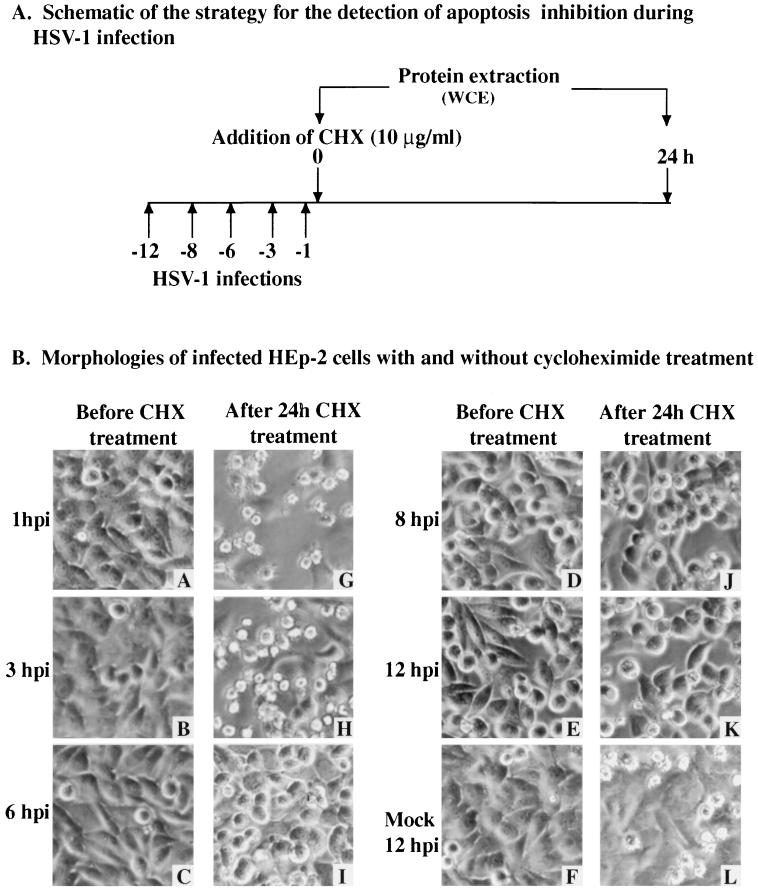
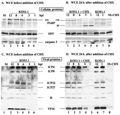
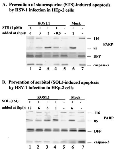
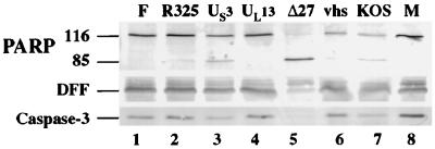
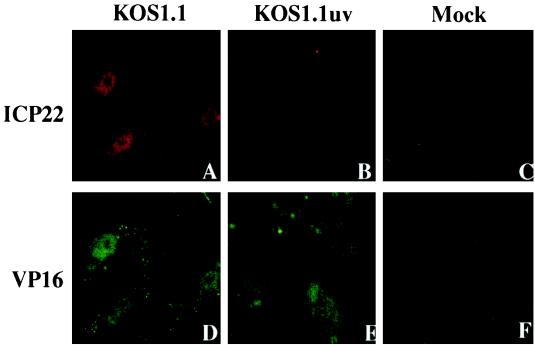
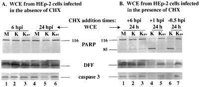
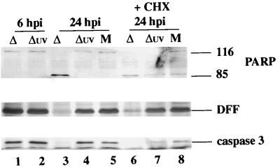
Cultured human epithelial cells infected with an ICP27 deletion strain of herpes simplex virus type 1 (HSV-1) show characteristic features of apoptotic cells including cell shrinkage, nuclear condensation, and DNA fragmentation. These cells do not show such apoptotic features when infected with a wild-type virus unless the infections are performed in the presence of a protein synthesis inhibitor. Thus, both types of virus induce apoptosis, but the ICP27-null virus is unable to prevent this process from killing the cells. In this report, we show that this ICP27-deficient virus induced apoptosis in human HEp-2 cells through a pathway which involved the activation of caspase-3 and the processing of the death substrates DNA fragmentation factor and poly(ADP-ribose) polymerase. The induction of apoptosis by wild-type HSV-1 occurred prior to 6 h postinfection (hpi), and de novo viral protein synthesis was not required to induce the process. The ability of the virus to inhibit apoptosis was shown to be effective between 3 to 6 hpi. Wild-type HSV-1 infection was also able to block the apoptosis induced in cells by the addition of cycloheximide, staurosporine, and sorbitol. While U(S)3- and ICP22-deficient viruses showed a partial prevention of apoptosis, deletion of either the U(L)13 or vhs gene products did not affect the ability of HSV-1 to prevent apoptosis in infected cells. Finally, we demonstrate that in UV-inactivated viruses, viral binding and entry were not sufficient to induce apoptosis. Taken together, these results suggest that either gene expression or another RNA metabolic event likely plays a role in the induction of apoptosis in HSV-1-infected human cells.


Figures

FIG. 1
FIG. 1
Detection of PARP, DFF, and caspase-3 processing in infected HEp-2 cells at early times from 6 to 24 hpi (A) or late times from 11 to 48 hpi (B). Whole cell extracts prepared at various times postinfection from vBSΔ27- or KOS1.1-infected cells were separated in a denaturing gel, transferred to nitrocellulose, and reacted with anti-PARP, anti-DFF, or anti-caspase-3 antibodies as described in Materials and Methods. “116” and “85” denote full-length (116,000-molecular-weight) and processed (85,000-molecular-weight) PARP, respectively. The locations of prestained molecular weight markers are indicated on the left.
FIG. 2
FIG. 2
Detection of PARP, DFF, and caspase-3 (A) and ICP4, ICP22, and ICP27 (B) in infected HEp-2 cells. Total cell extracts were prepared from mock-infected cells or cells infected with KOS1.1 or vBSΔ27 in the absence (−) and presence of the protein synthesis inhibitor CHX (10 μg/ml) or in the absence (−) and presence of caspase-1 or caspase-3 (1 or 3; 50 μg/ml) as described in Materials and Methods. Immunoblot analyses were done with the anti-PARP, anti-DFF, and anti-caspase-3 antibodies (A) or anti-ICP4 (1114), anti-ICP22 (RGST22), and anti-ICP27 (1113) antibodies (B). Bar at the left mark the distribution of the modified forms of ICP22. Minor background bands due to the anti-ICP22 antibody were observed in all of the CHX-treated cells.
FIG. 3
FIG. 3
Schematic of infections (A) and morphologies (B) of KOS1.1-infected HEp-2 cells with and without CHX treatment. A to L, phase-contrast images of mock-infected cells at 12 hpi (F) and KOS1.1-infected cells at 1, 3, 6, 8, and 12 hpi (A to E) and of mock-infected cells 24 h after the addition of CHX at 12 hpi (L) and KOS1.1-infected cells 24 h after the addition of CHX at 1, 3, 6, 8 and 12 hpi (G to K). Magnification, ×60. WCE, whole cell extract.
FIG. 4
FIG. 4
Detection of cellular (A and B) and viral (C and D) protein accumulation in KOS1.1-infected HEp-2 cells with and without CHX treatment. Whole cell extracts (WCE) prepared at 1, 3, 6, 8, or 12 hpi or at 24 h after the addition of CHX (10 μg/ml) at 1, 3, 6, 8, or 12 hpi from KOS1.1-infected cells were used for immunoblot analyses with anti-PARP, anti-DFF, and anti-caspase-3 antibodies (A and B) or with anti-ICP4, anti-ICP0, anti-ICP22, anti-ICP27 (IE proteins), anti-TK (E protein), and anti-VP16 (L protein) antibodies (C and D). M, mock-infected cells at 12 hpi; + CHX, addition of CHX for 24 h. In lanes 6 and 7 of panels B and D, the infected cells were incubated for an additional 24 h in the absence of CHX. “116” and “85” denote full-length (116,000-molecular-weight) and processed (85,000-molecular-weight) PARP, respectively.
FIG. 5
FIG. 5
Detection of PARP, DFF, and caspase-3 processing in KOS1.1-infected HEp-2 cells treated with staurosporine (A) or sorbitol (B). Whole cell extracts prepared from mock- or KOS1.1 (K)-infected cells untreated (−) or treated with staurosporine or sorbitol at 1, 3, 6, or 12 hpi were used for immunoblotting analyses with anti-PARP, anti-DFF, and anti-caspase-3 antibodies as described in Materials and Methods. “116” and “85” denote full-length (116,000-molecular-weight) and processed (85,000-molecular-weight) PARP, respectively.
FIG. 6
FIG. 6
Detection of PARP, DFF, and caspase-3 processing in HEp-2 cells infected with wild-type and mutant HSV-1. Whole cell extracts prepared from mock-infected cells (M) or cells infected with HSV-1 (F), vBSΔ27 (Δ27), R7041 (US3 deletion), R325 (ICP22 deletion), R7356 (UL13 deletion), vhs-ΔSma (vhs deletion), or KOS1.1 at 24 hpi were used for immunoblot analyses with anti-PARP, anti-DFF, and anti-caspase-3 antibodies as described in Materials and Methods. “116” and “85” denote full-length (116,000-molecular-weight) and processed (85,000-molecular-weight) PARP, respectively.
FIG. 7
FIG. 7
Indirect immunofluorescence of infected cells double labeled with antibodies specific for ICP22 (A to E) and VP16 (D to F). Vero cells were mock infected or infected (50 PFU/cell) with KOS1.1 or UV-inactivated KOS1.1 (KOS1.1uv) for 2 h and then subjected to formaldehyde-acetone fixation followed by immunostaining with anti-ICP22 (rabbit) and anti-VP16 (mouse) antibodies as described in Materials and Methods.
FIG. 8
FIG. 8
Detection of PARP, DFF, and caspase-3 processing in KOS1.1-infected HEp-2 cells untreated (A) or treated (B) with CHX. Whole cell extracts (WCE) were prepared from mock-infected cells (M) or cells infected with KOS1.1 (K) or UV-inactivated KOS1.1 (Kuv) virus at 6 or 24 hpi in the absence of CHX (A) and at 24 h after the addition of CHX (B) at either 1 hpi, 6 hpi, or 30 min before infection (−0.5 hpi). Immunoblotting analyses were performed with anti-PARP, anti-DFF, and anti-caspase-3 antibodies. “116” and “85” are full-length (116,000-molecular-weight) and processed (85,000-molecular-weight) PARP, respectively.
FIG. 9
FIG. 9
Detection of PARP, DFF, and caspase-3 processing in vBSΔ27-infected HEp-2 cells. vBSΔ27 was inactivated by exposure to UV light as described in Materials and Methods. Whole cell extracts (WCE) prepared from mock-infected cells (M) or cells infected with vBSΔ27 (Δ) and UV-inactivated vBSΔ27 (Δuv) at 6 and 24 hpi in the absence of or at 24 hpi in the presence (+) of CHX were used for immunoblot analyses with anti-PARP, anti-DFF, and anti-caspase-3 antibodies. “116” and “85” denote full-length (116,000-molecular-weight) and processed (85,000-molecular-weight) PARP, respectively.

References

    1. Alfonso C L, Neilan J G, Kutish G F, Rock D L. An African swine fever virus Bcl-2 homolog, 5-HL, suppresses apoptotic cell death. J Virol. 1996;70:4858–4863. - PMC - PubMed
    1. Alnemri E S, Livingston D J, Nicholson D W, Salvesen G, Thornberry N A, Wong W W, Yuan J. Human ICE/CED-3 protease nomenclature. Cell. 1996;87:171. . (Letter.) - PubMed
    1. Asano S, Honda T, Goshima F, Watanabe D, Miyake Y, Sugiura Y, Nishiyama Y. US3 protein kinase of herpes simplex virus type 2 plays a role in protecting corneal epithelial cells from apoptosis in infected mice. J Gen Virol. 1999;80:51–56. - PubMed
    1. Aubert, M., and J. A. Blaho. Unpublished results.
    1. Aubert M, Blaho J A. The herpes simplex virus type 1 regulatory protein ICP27 is required for the prevention of apoptosis in infected human cells. J Virol. 1999;73:2803–2813. - PMC - PubMed

Publication types

MeSH terms

Substances