. 1999 Dec;73(12):10377-86.
doi: 10.1128/JVI.73.12.10377-10386.1999.

In vivo regulation of hepatitis B virus replication by peroxisome proliferators

Affiliations

In vivo regulation of hepatitis B virus replication by peroxisome proliferators

L G Guidotti et al. J Virol. 1999 Dec.

Abstract

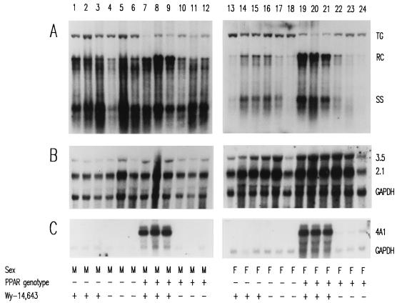
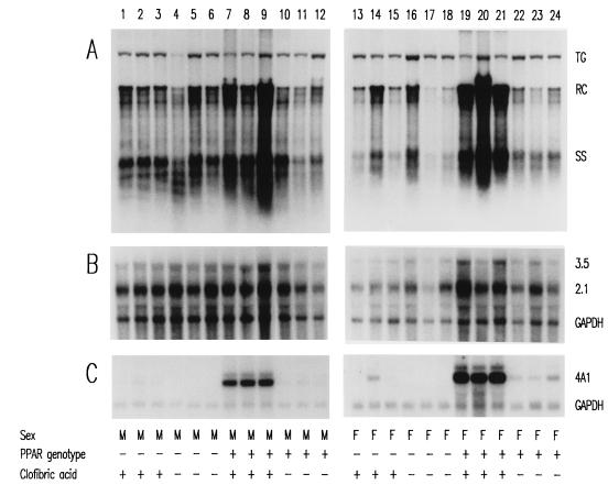
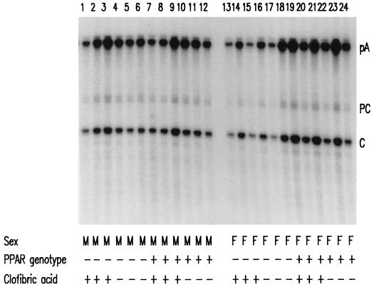
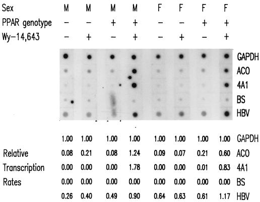
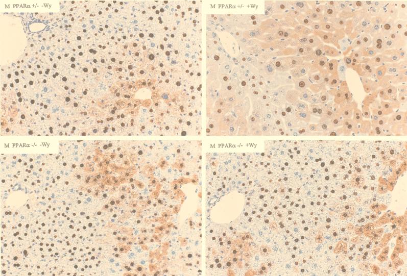
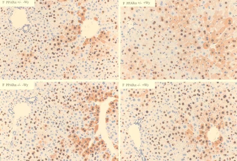
The role of the peroxisome proliferator-activated receptor alpha (PPARalpha) in regulating hepatitis B virus (HBV) transcription and replication in vivo was investigated in an HBV transgenic mouse model. Treatment of HBV transgenic mice with the peroxisome proliferators Wy-14,643 and clofibric acid resulted in a less than twofold increase in HBV transcription rates and steady-state levels of HBV RNAs in the livers of these mice. In male mice, this increase in transcription was associated with a 2- to 3-fold increase in replication intermediates, whereas in female mice it was associated with a 7- to 14-fold increase in replication intermediates. The observed increases in transcription and replication were dependent on PPARalpha. HBV transgenic mice lacking this nuclear hormone receptor showed similar levels of HBV transcripts and replication intermediates as untreated HBV transgenic mice expressing PPARalpha but failed to demonstrate alterations in either RNA or DNA synthesis in response to peroxisome proliferators. Therefore, it appears that very modest alterations in transcription can, under certain circumstances, result in relatively large increases in HBV replication in HBV transgenic mice.


Figures

FIG. 1
FIG. 1
DNA (Southern) and RNA (Northern) filter hybridization analysis of HBV DNA replication intermediates (A) and transcripts (B) in livers of HBV transgenic mice. Groups of three mice of each gender and genotype were fed rodent chow either with or without Wy-14,643 for 7 days before their livers were analyzed. Induction of the transcript detected by the rat cytochrome P450 4A1 cDNA (4A1) was used as a control to demonstrate the PPARα-dependent activation of transcription by the peroxisome proliferator from a responsive gene. The GAPDH transcript was used as an internal control for quantitation of the HBV 3.5- and 2.1-kb RNAs (3.5 and 2.1). The HBV transgene was used as an internal control for quantitation of the HBV replication intermediates. The probes used were HBVayw genomic DNA (A), HBVayw genomic DNA plus GAPDH cDNA (B) and cytochrome P450 4A1 cDNA plus GAPDH cDNA (C). TG, HBV transgene; RC, HBV relaxed circular replication intermediates; SS, HBV single-stranded replication intermediates; M, male HBV transgenic mouse; F, female HBV transgenic mouse; PPAR genotype −, PPARα knockout (−/−) mouse; PPAR genotype +, PPARα heterozygous (+/−) mouse; Wy-14,643 +, HBV transgenic mouse fed 0.1% (wt/wt) Wy-14,643; Wy-14,643 −, HBV transgenic mouse fed normal rodent chow.
FIG. 2
FIG. 2
DNA (Southern) and RNA (Northern) filter hybridization analysis of HBV DNA replication intermediates (A) and transcripts (B) in livers of HBV transgenic mice. Groups of three mice of each gender and genotype were fed rodent chow either with or without clofibric acid for 14 days before their livers were analyzed. Induction of the transcript detected by the rat cytochrome P450 4A1 cDNA (4A1) was used as a control to demonstrate the PPARα-dependent activation of transcription by the peroxisome proliferator from a responsive gene. The GAPDH transcript was used as an internal control for the quantitation of the HBV 3.5- and 2.1-kb RNAs (3.5 and 2.1). The HBV transgene was used as an internal control for quantitation of the HBV replication intermediates. The probes used were HBVayw genomic DNA (A), HBVayw genomic DNA plus GAPDH cDNA (B), and cytochrome P450 4A1 cDNA plus GAPDH cDNA (C). TG, HBV transgene; RC, HBV relaxed circular replication intermediates; SS, HBV single-stranded replication intermediates; M, male HBV transgenic mouse; F, female HBV transgenic mouse; PPAR genotype −, PPARα knockout (−/−) mouse; PPAR genotype +, PPARα heterozygous (+/−) mouse; clofibric acid +, HBV transgenic mouse fed 0.5% (wt/wt) clofibric acid; clofibric acid −, HBV transgenic mouse fed normal rodent chow.
FIG. 3
FIG. 3
RNase protection analysis mapping the transcription initiation sites of precore (PC) and pregenomic (C) transcripts from livers of HBV transgenic mice. The 3′ ends of all HBV transcripts corresponding to the polyadenylation sites (pA) of these RNAs also generated a protected fragment in this analysis. Groups of three mice of each gender and genotype were fed rodent chow either with or without clofibric acid for 14 days before their livers were analyzed. The riboprobe used included the HBVayw sequence spanning nucleotide coordinates 1990 to 1658. M, male HBV transgenic mouse; F, female HBV transgenic mouse; PPAR genotype −, PPARα knockout (−/−) mouse; PPAR genotype +, PPARα heterozygous (+/−) mouse; clofibric acid +, HBV transgenic mouse fed 0.5% (wt/wt) clofibric acid; clofibric acid −, HBV transgenic mouse fed normal rodent chow.
FIG. 4
FIG. 4
Nuclear run-on analysis of HBV transcripts in livers of HBV transgenic mice. A mouse of each gender and genotype was fed rodent chow either with or without Wy-14,643 for 7 days before their livers were analyzed. Induction of the transcription rates of the RNAs detected by the rat ACO and cytochrome P450 4A1 (4A1) cDNAs were used as controls to demonstrate the PPARα-dependent increase in transcription rates induced by the peroxisome proliferator from these responsive genes. The rate of transcription from the GAPDH gene was used as an internal control for quantitation of the relative transcription rates of the other genes analyzed. The rate of transcription of the GAPDH gene was designated 1.00. The nonspecific background rate of transcription detected with the pBluescript SK(−) plasmid (BS) control was designated 0.00. The rates of transcription for the HBVayw (HBV), ACO, and 4A1 genes were estimated relative to the GAPDH gene after subtracting the nonspecific background rate of transcription. The probes hybridized to each strip of filter were 32P-labeled transcripts isolated from in vitro-labeled mouse liver nuclei. The DNA on each strip of filter is indicated at the right. M, male HBV transgenic mouse; F, female HBV transgenic mouse; PPAR genotype −, PPARα knockout (−/−) mouse; PPAR genotype +, PPARα heterozygous (+/−) mouse; Wy-14,643 +, HBV transgenic mouse fed 0.1% (wt/wt) Wy-14,643; Wy-14,643 −, HBV transgenic mouse fed normal rodent chow.
FIG. 5
FIG. 5
Immunohistochemical staining of HBcAg in the livers of male HBV transgenic mice (original magnification, ×200). Nuclear staining of HBcAg is observed throughout the liver, whereas cytoplasmic staining is located primarily in the centrolobular hepatocytes in the livers of untreated PPARα +/− HBV transgenic mice (upper left). Treatment of PPARα +/− HBV transgenic mice with Wy-14,643 (upper right) results in hepatocyte hypertrophy and HBcAg staining that extends to the periportal hepatocytes. Livers from Wy-14,643-treated (lower right) or untreated (lower left) PPARα −/− HBV transgenic mice display the same distribution of HBcAg staining as untreated PPARα +/− HBV transgenic mice.
FIG. 6
FIG. 6
Immunohistochemical staining of HBcAg in the livers of female HBV transgenic mice (original magnification, ×200). Nuclear staining of HBcAg is observed throughout the liver, whereas cytoplasmic staining is located primarily in the centrolobular hepatocytes in the livers of untreated PPARα +/− HBV transgenic mice (upper left). Treatment of PPARα +/− HBV transgenic mice with Wy-14,643 (upper right) results in hepatocyte hypertrophy and HBcAg staining that extends to the periportal hepatocytes. Livers from Wy-14,643 treated (lower right) or untreated (lower left) PPARα −/− HBV transgenic mice display the same distribution of HBcAg staining as untreated PPARα +/− HBV transgenic mice.

Similar articles

Cited by

References

    1. Block T M, Lu X Y, Mehta A, Park J, Blumberg B S, Dwek R. Role of glycan processing in hepatitis B virus envelope protein trafficking. Adv Exp Med Biol. 1998;435:207–216. - PubMed
    1. Buckwold V E, Chen M, Ou J H. Interaction of transcription factors RFX1 and MIBP1 with the gamma motif of the negative regulatory element of the hepatitis B virus core promoter. Virology. 1997;227:515–518. - PubMed
    1. Chang C, Jeng K, Hu C, Lo S J, Su T, Ting L-P, Chou C-K, Han S, Pfaff E, Salfeld J, Schaller H. Production of hepatitis B virus in vitro by transient expression of cloned HBV DNA in a hepatoma cell line. EMBO J. 1987;6:675–680. - PMC - PubMed
    1. Chen I-H, Huang C-J, Ting L-P. Overlapping initiator and TATA box functions in the basal core promoter of hepatitis B virus. J Virol. 1995;69:3647–3657. - PMC - PubMed
    1. Chen M, Ou J H. Cell type-dependent regulation of the activity of the negative regulatory element of the hepatitis B virus core promoter. Virology. 1995;214:198–206. - PubMed

Publication types

MeSH terms

LinkOut - more resources