

High viral load in the cerebrospinal fluid and brain correlates with severity of simian immunodeficiency virus encephalitis
- PMID: 10559366
- PMCID: PMC113103
- DOI: 10.1128/JVI.73.12.10480-10488.1999
High viral load in the cerebrospinal fluid and brain correlates with severity of simian immunodeficiency virus encephalitis
Abstract
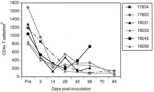
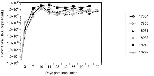
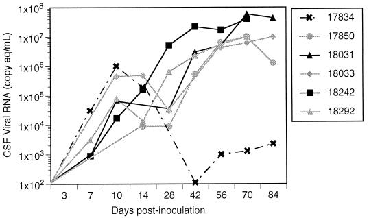
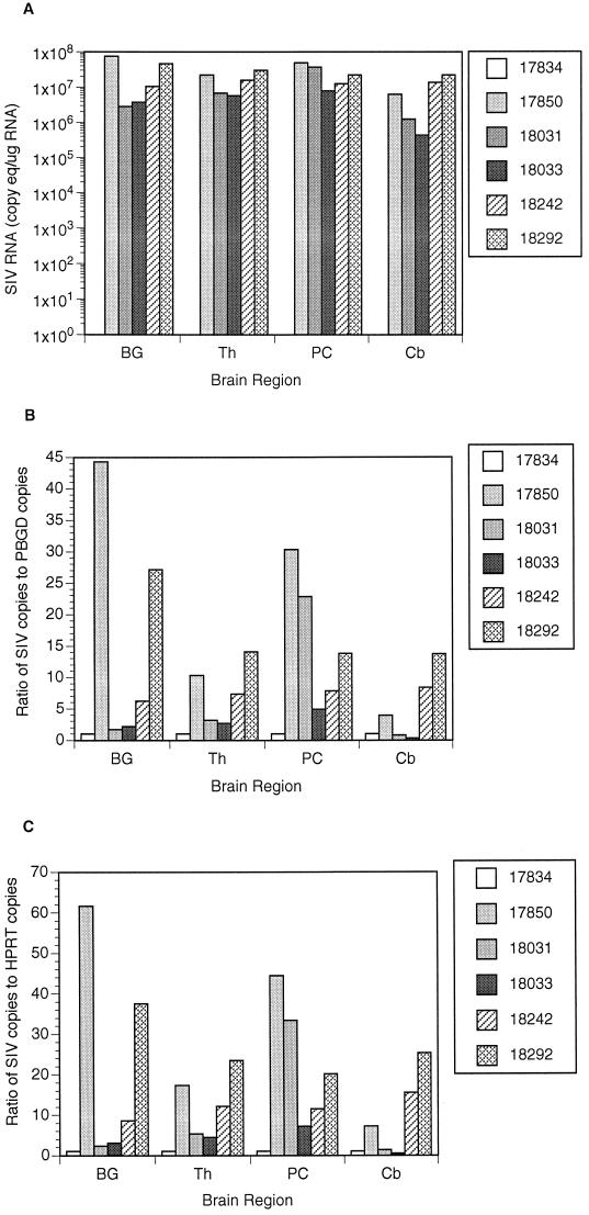
AIDS dementia and encephalitis are complications of AIDS occurring most frequently in patients who are immunosuppressed. The simian immunodeficiency virus (SIV) model used in this study was designed to reproducibly induce AIDS in macaques in order to examine the effects of a neurovirulent virus in this context. Pigtailed macaques (Macaca nemestrina) were coinoculated with an immunosuppressive virus (SIV/DeltaB670) and a neurovirulent molecularly cloned virus (SIV/17E-Fr), and more than 90% of the animals developed moderate to severe encephalitis within 6 months of inoculation. Viral load in plasma and cerebrospinal fluid (CSF) was examined longitudinally to onset of AIDS, and viral load was measured in brain tissue at necropsy to examine the relationship of systemic and central nervous system (CNS) viral replication to the development of encephalitis. In all animals, plasma viral load peaked at 10 to 14 days postinfection and remained high throughout infection with no correlation found between plasma viremia and SIV encephalitis. In contrast, persistent high levels of CSF viral RNA after the acute phase of infection correlated with the development of encephalitis. Although high levels of viral RNA were found in the CSF of all macaques (six of six) during the acute phase, this high level was maintained only in macaques developing SIV encephalitis (five of six). Furthermore, the level of both viral RNA and antigen in the brain correlated with the severity of the CNS lesions. The single animal in this group that did not have CNS lesions had no detectable viral RNA in any of the regions of the brain. The results substantiate the use of CSF viral load measurements in the postacute phase of SIV infection as a marker for encephalitis and CNS viral replication.
Figures
References
-
- Anderson M G, Hauer D, Sharma D P, Joag S V, Narayan O, Zink M C, Clements J E. Analysis of envelope changes acquired by SIVmac239 during neuroadaption in rhesus macaques. Virology. 1993;195:616–626. - PubMed
-
- Baskin G B, Murphey-Corb M, Martin L N, Soike K F, Hu F-S, Kuebler D. Lentivirus-induced pulmonary lesions in rhesus monkeys (Macaca mulatta) infected with simian immunodeficiency virus. Vet Pathol. 1991;28:506–513. - PubMed
-
- Brew B J, Pemberton L, Cunningham P, Law M G. Levels of human immunodeficiency virus type 1 RNA in cerebrospinal fluid correlate with AIDS dementia stage. J Infect Dis. 1997;175:963–966. - PubMed
Publication types
MeSH terms
Substances
Grants and funding
LinkOut - more resources
Full Text Sources
Other Literature Sources
