. 1999 Nov 15;19(22):9986-95.
doi: 10.1523/JNEUROSCI.19-22-09986.1999.

Identification, isolation, and promoter-defined separation of mitotic oligodendrocyte progenitor cells from the adult human subcortical white matter

Affiliations

Identification, isolation, and promoter-defined separation of mitotic oligodendrocyte progenitor cells from the adult human subcortical white matter

N S Roy et al. J Neurosci. .

Abstract

Previous studies have suggested the persistence of oligodendrocyte progenitor cells in the adult mammalian subcortical white matter. To identify oligodendrocyte progenitors in the adult human subcortical white matter, we transfected dissociates of capsular white matter with plasmid DNA bearing the gene for green fluorescence protein (hGFP), placed under the control of the human early promoter (P2) for the oligodendrocytic protein cyclic nucleotide phosphodiesterase (P/hCNP2). Within 4 d after transfection with P/hCNP2:hGFP, a discrete population of small, bipolar cells were noted to express GFP. These cells were A2B5-positive (A2B5(+)), incorporated bromodeoxyuridine in vitro, and constituted <0.5% of all cells. Using fluorescence-activated cell sorting (FACS), the P/hCNP2-driven GFP(+) cells were then isolated and enriched to near-purity. In the weeks after FACS, most P/hCNP2:hGFP-sorted cells matured as morphologically and antigenically characteristic oligodendrocytes. Thus, the human subcortical white matter harbors mitotically competent progenitor cells, which give rise primarily to oligodendrocytes in vitro. By using fluorescent transgenes of GFP expressed under the control of an early oligodendrocytic promoter, these oligodendrocyte progenitor cells may be extracted and purified from adult human white matter in sufficient numbers for implantation and cell-based therapy.


Figures

Fig. 1.
Fig. 1.
The human P/CNP2:hGFP vector. Humanized GFP (Levy et al., 1996), a mutant form of red-shifted GFP optimized for expression in human cells, was placed under the control of the human CNP2 promoter (P/hCNP2) (Gravel et al., 1996). To construct P/hCNP2:hGFP, an XbaI-XbaI fragment encompassing exon 0 and part of intron 1 was isolated from the SK/hgCNP plasmid, which contains the complete sequence of the human CNP gene (Gravel et al., 1996), and was then linked to the hGFP gene fused to the SV40 polyadenylation signal.
Fig. 2.
Fig. 2.
Adult human white matter harbors oligodendrocyte progenitors. Immunocytochemistry of white matter dissociates for a panel of cell type-selective antigens revealed a diverse representation of phenotypes before sorting. A–C, A typical bipolar cell, double-labeled for A2B5 (red) and BrdU (yellow), fixed after 4 DIV. D–F, A cluster of postmitotic O4+ cells (D, E) and an overtly less mature BrdU-incorporating O4+/BrdU+ cell (F), all fixed after 7 DIV. G–I, Representative examples of the diverse phenotypes present in the adult white matter. These included cells expressing CNP (G), GFAP (H), and TuJ1 (I) immunoreactivities, which respectively identify oligodendrocytes, astrocytes, and neurons; each cell type was found in the proportion noted in Results. Scale bar, 40 μm.
Fig. 3.
Fig. 3.
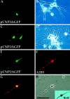
P/hCNP2:hGFP identifies a population of bipolar, A2B5+ cells. GFP expression was observed within 4–5 d after transfection. The P/hCNP2:hGFP+ cells typically first appeared as small, bipolar cells. A–F, P/hCNP2:hGFP-expressing cells (A, C, E) and their corresponding phase contrast micrographs (B, D, H). E, F, Immunocytochemistry identified the P/hCNP2:hGFP+ bipolar cells as A2B5+; G indicates double-labeling of the two. Inset in H shows that this cell incorporated BrdU. Scale bar, 30 μm.
Fig. 4.
Fig. 4.
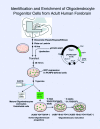
Culture, isolation, and enrichment of oligodendrocyte progenitors. Adult human subcortical white matter, derived from surgical samples of frontal and temporal lobe, was dissected and enzymatically dissociated using papain and DNase and then cultured and transfected with either P/hCNP2:hGFP or control plasmids (P/CMV:hGFP and P/hCNP2:lacZ).
Fig. 5.
Fig. 5.
Isolation of P/hCNP2:hGFP+cells by FACS. A and B shows a representative sort of a human white matter sample, derived from the frontal lobe of a 42-year-old woman during repair of an intracranial aneurysm. This plot shows 50,000 cells (sorting events) with their GFP fluorescence intensity (FL1), plotted against their forward scatter (FSC, a measure of cell size). A indicates the plot obtained from a nonfluorescent P/hCNP2:lacZ-transfected control, whereas B indicates the corresponding result from a matched culture transfected with P/hCNP2:hGFP. The boxed area (R1 and R2) includes those P/hCNP2:hGFP+ cells recognized and separated on the basis of their fluorescence emission. The many cells thereby recognized in the P/hCNP2:hGFP-transfected sample (B) contrasts to the rare cells so identified in the nonfluorescent P/hCNP2:lacZ-transfected control (A). C and D show phase and fluorescence images of GFP+ cells 2 hr after sorting. Scale bar, 20 μm.
Fig. 6.
Fig. 6.
P/hCNP2:hGFP-sorted cells divide and express oligodendrocytic markers. A–C, A bipolar A2B5+/BrdU+ cell 48 hr after FACS. D–F, Within 3 weeks, the bipolar cells matured into fibrous, O4+ cells. These cells often incorporated BrdU, indicating their in vitro origin from replicating A2B5+ cells. G–I, A multipolar oligodendrocyte expressing CNP, still expressing GFP 3 weeks after FACS. Scale bar, 20 μm.
Fig. 7.
Fig. 7.
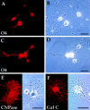
FACS-sorted P/hCNP2:hGFP+ cells mature primarily as oligodendrocytes. A,B, P/hCNP2:hGFP-sorted cells express O4 (red) and begin process elaboration within 4 d after FACS. C, D, By 2 weeks after FACS, these cells generally develop multipolar morphologies.Red, O4-immunoreactive cells. E,F, Progenitor derived-cells matured further over the following weeks, developing oligodendrocytic morphologies and both CNP protein (E) and galactocerebroside (F) expression by 4 weeks in vitro. Scale bar, 30 μm.
Fig. 8.
Fig. 8.
White matter precursor cells may constitute a pool of multipotential progenitors. By 1 week after FACS, some P/hCNP2:hGFP-sorted cells were noted to mature into either TuJ1+ neurons (A) or GFAP+ astrocytes (B). Both the TuJ1+ (red in A) and GFAP+ (red in B) cells were confirmed visually as expressing P/hCNP2:hGFP (green). No such neuronal differentiation of CNP2:hGFP-identified cells was ever noted in unsorted plates, within which these cells generally matured as oligodendrocytes and much less so as astrocytes. This suggests that P/hCNP2-defined progenitors may retain some degree of multilineage potential, which may be selectively exercised in the low-density, homogeneous environment of a sorted cell pool in which paracrine influences on differentiation are minimized.

Similar articles

Cited by

References

    1. Armstrong RC, Dorn HH, Kufta CV, Friedman E, Dubois-Dalcq ME. Pre-oligodendrocytes from adult human CNS. J Neurosci. 1992;12:1538–1547. - PMC - PubMed
    1. Bansal R, Lemmon V, Gard A, Ranscht B, Pfeiffer S. Multiple and novel specificities of monoclonal antibodies O1, O4, and R-mAb, in the analysis of oligodendrocyte development. J Neurosci Res. 1989;24:548–557. - PubMed
    1. Bernier L, Alvarez F, Norgard E, Schembri J, Sabatini D, Colman D. Molecular cloning of a 2′,3′-cyclic nucleotide phosphodiesterase. J Neurosci. 1987;7:2703–2710. - PMC - PubMed
    1. Blakemore W, Franklin R, Noble M. Glial cell transplantation and the repair of demyelinating lesions. In: Jessen K, Richardson W, editors. Glial cell development: basic principles and clinical relevance. BIOS Scientific; Oxford: 1996. pp. 209–220.
    1. Butt AM, Ransom BR. Morphology of astrocytes and oligodendrocytes during development in the intact rat optic nerve. J Comp Neurol. 1993;338:141–158. - PubMed

Publication types

Substances