. 1999 Nov 15;19(22):10036-43.
doi: 10.1523/JNEUROSCI.19-22-10036.1999.

Embryonic neurons adapt to the inhibitory proteoglycan aggrecan by increasing integrin expression

Affiliations

Embryonic neurons adapt to the inhibitory proteoglycan aggrecan by increasing integrin expression

M L Condic et al. J Neurosci. .

Abstract

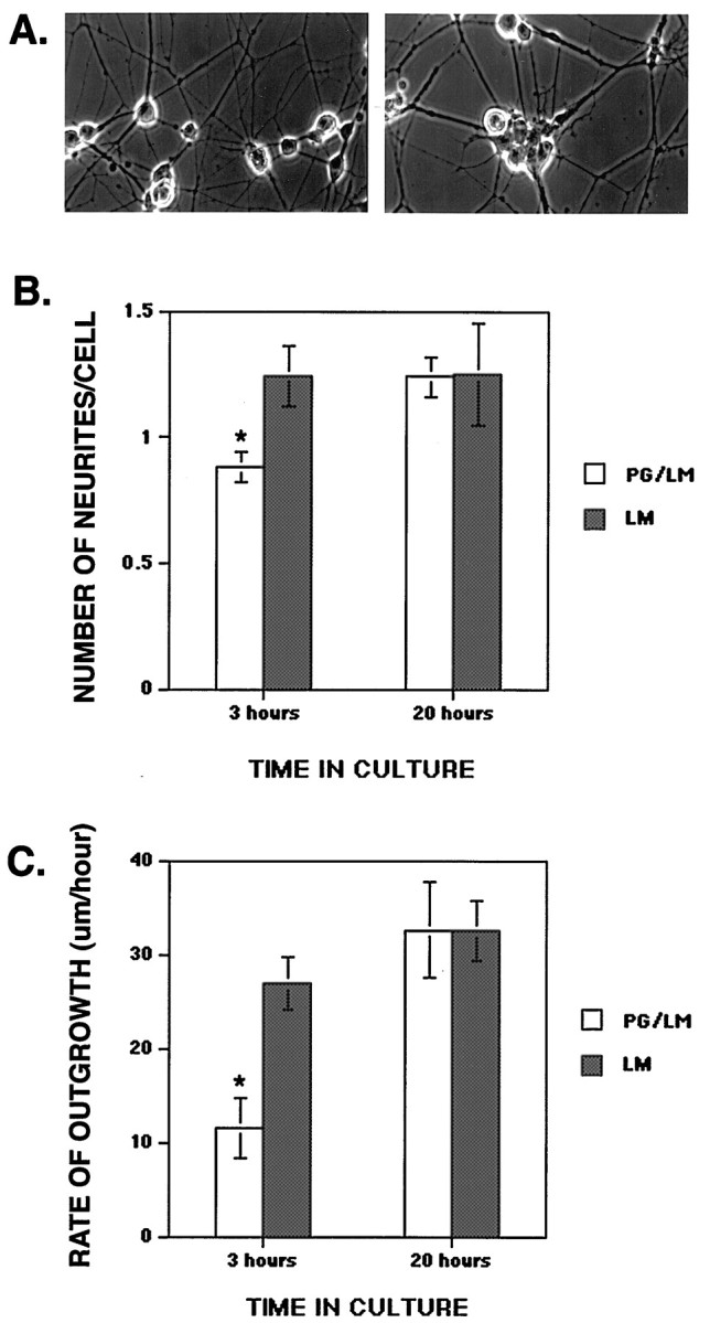
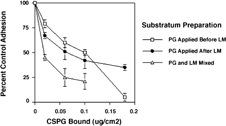
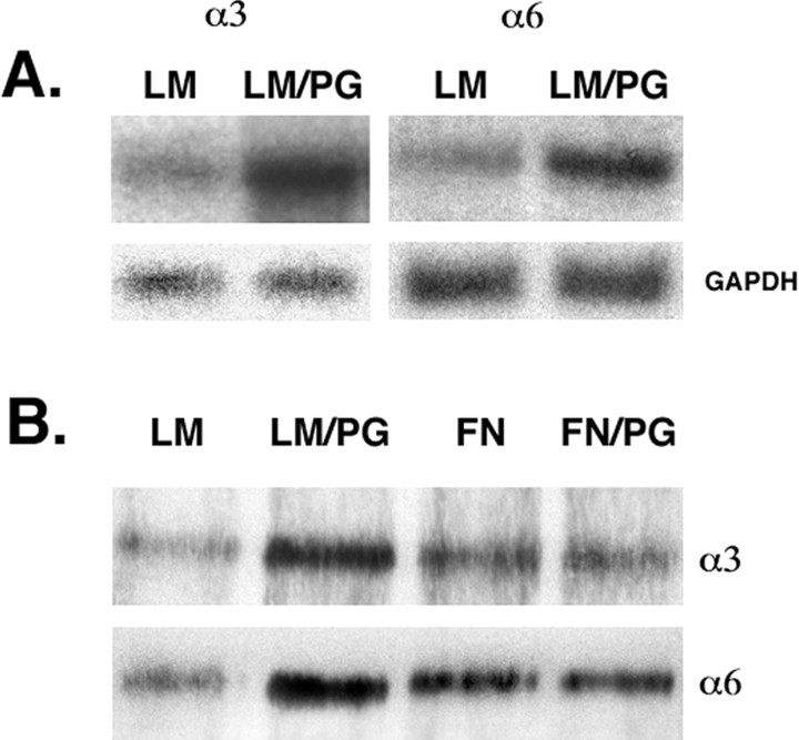
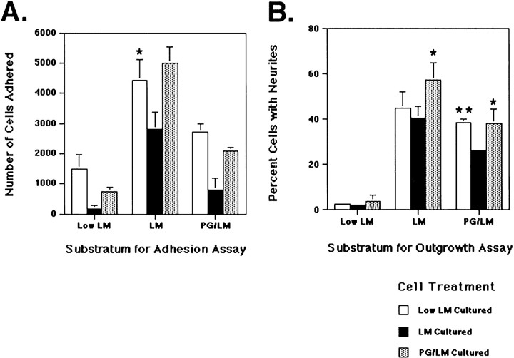
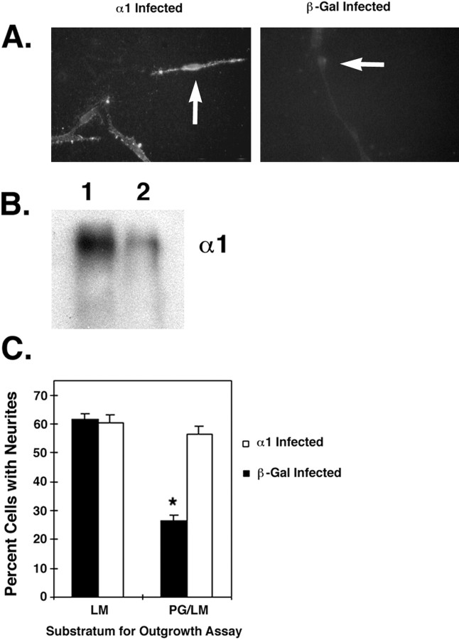
The primary mediators of cell migration during development, wound healing and metastasis, are receptors of the integrin family. In the developing and regenerating nervous system, chondroitin sulfate proteoglycans (CSPGs) inhibit the integrin-dependent migration of neuronal growth cones. Here we report that embryonic sensory neurons cultured on the growth-promoting molecule laminin in combination with the inhibitory CSPG aggrecan rapidly adapt to inhibition. Adaptation is associated with a two- to threefold increase in the levels of RNA and surface protein for two laminin receptors, integrin alpha6beta1 and alpha3beta1, indicating that integrin expression is regulated by aggrecan. Increased integrin expression is associated both with increases in neuronal cell adhesion/outgrowth and with decreases in the ability of aggrecan to inhibit cell adhesion. Directly increasing integrin expression by adenoviral infection is sufficient to eliminate the inhibitory effects of aggrecan, indicating that upregulation of integrin receptors may promote neuronal regeneration in the presence of inhibitory matrix components.


Figures

Fig. 1.
Fig. 1.
Neurons cultured on substrata containing a low concentration of the inhibitory proteoglycan aggrecan adapt to inhibition and extend neurites. A, Neurons were cultured on substrata containing either laminin alone (left) or laminin in combination with aggrecan (right). After 16–20 hr in culture, neurons have extended axons on both substrata. Outgrowth was less profuse and axons were more fasciculated on substrata containing aggrecan relative to outgrowth on laminin alone.B, The initiation of neurites is delayed on substrata containing aggrecan. At 3 hr, significantly fewer cells (p < 0.002, t test) have initiated neurites on substrata containing aggrecan relative to cells on laminin alone. By 6 hr in culture, the number of neurites initiated on aggrecan-containing substrata has greatly increased, suggesting that the neurons have adapted to inhibition. Means + SEMs of three experiments, each measuring at least 174 neurons/condition, are given.C, The rate of axon extension on laminin at 3 hr in culture is reduced by the presence of aggrecan (p < 0.001, t test), but by 20 hr in culture the rate of outgrowth on the two substrata is identical. Means + SEMs of three experiments, each measuring at least 14 neurons/condition, are given.
Fig. 2.
Fig. 2.
The adhesion of neurons to substrata containing constant amounts of bound laminin (300 ng laminin/cm2) and increasing amounts of bound aggrecan is affected by the order of application of the two proteins to the substrata. Aggrecan mixed with laminin in solution (shaded triangles) is more inhibitory than aggrecan applied before or after adsorption of laminin to the substrata (Mixedpoints are statistically different from either Before orAfter at p < 0.05, ttest), suggesting that conformational changes in laminin may underlie some of the inhibitory effect of aggrecan. At the highest concentrations tested, aggrecan is more inhibitory when applied before laminin, supporting the idea that interactions of soluble laminin with aggrecan result in greater inhibition. Adhesion is given as a percentage of adhesion to control (laminin only) substrata. Means + SEMs from three experiments are given. When error bars do not appear, they are smaller than the size of the symbol.
Fig. 3.
Fig. 3.
Increased expression of integrin α3 and α6 RNA and cell surface receptor protein in response to aggrecan. Quantifications of the results are given in Table 1. A, Northern analysis of integrin total RNA. The expression of RNA for α3 and α6 integrin is increased in neurons cultured on substrata containing both laminin and aggrecan (LM/PG) relative to those cultured on laminin alone (LM). The blots were stripped and reprobed for GAPDH as a loading control.B, Western analysis of biotinylated cell-surface integrin protein. Neurons cultured on substrata containing laminin and aggrecan (LM/PG) show an increase in the expression of laminin receptors integrin α3 and α6 at the cell surface relative to those cultured on laminin alone (LM), whereas the expression of these receptors does not increase in neurons cultured on substrata containing fibronectin and aggrecan (FN/PG) when compared with those cultured on fibronectin alone (FN).
Fig. 4.
Fig. 4.
Increased expression of laminin receptors at the cell surface is associated with increased neuronal adhesion and outgrowth. Means + SEMs from three experiments are given.A, Neurons expressing high levels of integrin α3 and α6 at the cell surface (Low LM Cultured andPG/LM Cultured) show increased adhesion to laminin and to PG/LM substrata. *Different from LM cultured atp < 0.1 (t test). Values for all other low laminin-cultured and PG/LM-cultured conditions are significantly different from LM cultured at p < 0.05 or better. B, Neurons expressing high levels of integrin α3 and α6 at the cell surface extend neurites more readily on laminin both in the presence and absence of the growth inhibitor aggrecan. *Different from LM cultured at p < 0.1. **Significantly different from LM cultured at p < 0.005. Where error bars do not appear, they are <1%.
Fig. 5.
Fig. 5.
Increased expression of α1-integrin is sufficient to mediate adaptation of neurons to aggrecan.A, Neurons infected with adenoviral constructs expressing a control protein (β-Gal,right) or mouse α1-integrin (left) were stained live with an antibody that specifically recognizes the mouse α1 subunit.Arrows indicate the positions of neuronal cells.B, Overexpression of mouse α1-integrin in chick neurons results in an approximately twofold increase in total α1-integrin expressed at the cell surface (2.25 + 0.2). Infected neurons were cultured on LM substrata, where endogenous integrin expression is low (see Materials and Methods). Cell surface-biotinylated proteins from mouse α1-integrin-infected (lane 1) or control, β-galactosidase-infected (lane 2) neurons were immunoprecipitated with an antisera that recognizes both mouse and chick α1-integrin subunits. Total α1-integrin expressed at the surface is increased after infection with α1-expressing adenovirus, reflecting the contribution of the exogenous mouse protein to total α1 expression. Expression of endogenous α1 (control cells, lane 2) is quite low under these conditions. C, Control or α1-integrin-infected neurons were cultured on substrata containing laminin alone or laminin in combination with aggrecan as in Figure 4. Control neurons expressing β-galactosidase (black bars) are strongly inhibited in the presence of aggrecan after 3 hr in culture (Figs. 1, 4). At this time in culture, endogenous increases in integrin expression have not yet occurred. Neurons with increased laminin-receptor expression (α1-expressing; white bars) are not inhibited by aggrecan, showing outgrowth equivalent to that seen on laminin alone. Means + SEMs from at least three experiments are given. *Significantly different from all other conditions at p < 0.0001 (ttest).

Similar articles

Cited by

References

    1. Balsamo J, Ernst H, Zanin MK, Hoffman S, Lilien J. The interaction of the retina cell surface N-acetylgalactosaminylphosphotransferase with an endogenous proteoglycan ligand results in inhibition of cadherin-mediated adhesion. J Cell Biol. 1995;129:1391–1401. - PMC - PubMed
    1. Barkalow FJ, Schwarzbauer JE. Interactions between fibronectin and chondroitin sulfate are modulated by molecular context. J Biol Chem. 1994;269:3957–3962. - PubMed
    1. Barnea G, Grumet M, Milev P, Silvennoinen O, Levy JB, Sap J, Schlessinger J. Receptor tyrosine phosphatase beta is expressed in the form of proteoglycan and binds to the extracellular matrix protein tenascin. J Biol Chem. 1994;269:14349–14352. - PubMed
    1. Barres BA, Silverstein BE, Corey DP, Chun LL. Immunological, morphological, and electrophysiological variation among retinal ganglion cells purified by panning. Neuron. 1988;1:791–803. - PubMed
    1. Braunewell KH, Pesheva P, McCarthy JB, Furcht LT, Schmitz B, Schachner M. Functional involvement of sciatic nerve-derived versican- and decorin-like molecules and other chondroitin sulphate proteoglycans in ECM-mediated cell adhesion and neurite outgrowth. Eur J Neurosci. 1995;7:805–814. - PubMed

Publication types

MeSH terms

LinkOut - more resources