. 1999 Nov 15;19(22):10044-52.
doi: 10.1523/JNEUROSCI.19-22-10044.1999.

Gaze direction modulates finger movement activation patterns in human cerebral cortex

Affiliations

Gaze direction modulates finger movement activation patterns in human cerebral cortex

J T Baker et al. J Neurosci. .

Abstract

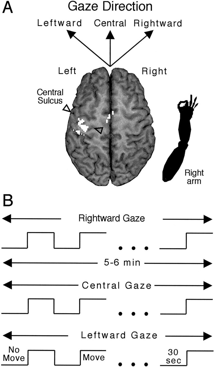
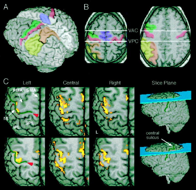
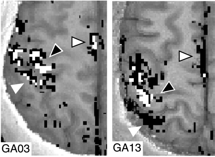
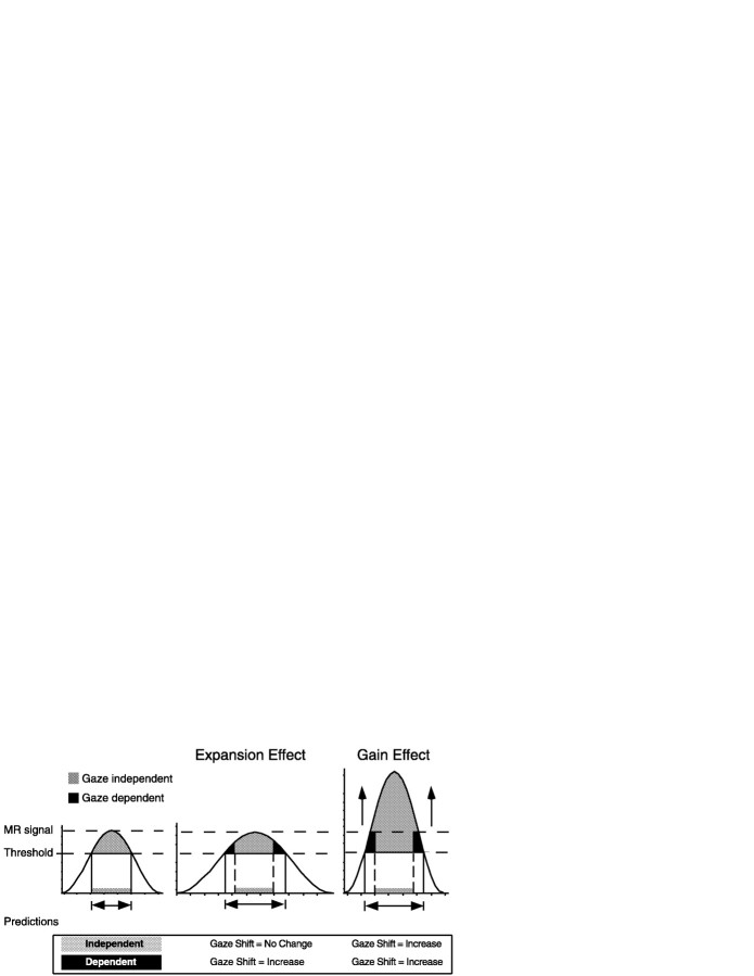
We investigated whether gaze direction modified the pattern of finger movement activation in human cerebral cortex using functional magnetic resonance imaging (MRI). Participants performed a sequential finger-tapping task or made no finger movements while maintaining gaze in the direction of the moving hand (aligned conditions) or away from the location of the moving hand. Functional MR signals, measured in the hemisphere contralateral to the moving hand, revealed finger movement-related activation in primary motor cortex, lateral and medial premotor cortex, and a wide extent of the lateral superior and inferior parietal lobules. In each area, the extent of the finger movement activation increased when static gaze was more aligned with the moving hand compared to when gaze was directed away from the moving hand. These data suggest the existence of large-scale cortical networks related to finger actions and indicate that skeletomotor processing in the cerebral cortex is consistently modified by gaze direction signals.


Figures

Fig. 1.
Fig. 1.
Experimental design. A, Schematic indicating the arm (bottom right) positioned at the right side of a participant, an expected activation in the contralateral hemisphere in motor areas (note area in opaque white in the vicinity of the central sulcus as demarcated by the two outlined triangles), and the three directions of gaze performed separately. B, Time and event lines indicating alternation of no-movement and finger movements for successive 30 sec periods for a total of 5–6 min for each gaze direction. See Materials and Methods for additional details and gaze terminology.
Fig. 2.
Fig. 2.
Cerebral cortical regions assessed and exemplar activation patterns. A, B, Color-coded illustration of the cortical regions assessed for functional MR activation. MI in red, PMA in green, SMA in purple, SPL in orange, and IPL inyellow. See Materials and Methods for additional details of sulcal and gyral landmarks defining each region. VAC, Vertical plane through the anterior commissure perpendicular to a line between the anterior and posterior commissures; VPC,vertical plane through the posterior commissure perpendicular to a line between the anterior and posterior commissures. C, Functional MR labeling in two exemplars, slice obtained from a single participant (slice planes indicated on whole brain volumes at right). The least activation occurred for leftward gaze, whereas that for both central and rightward gaze exceeded that for leftward gaze. The images with overlain label depict mostly portions of the left, contralateral hemisphere (L, left; R, right).Red arrowhead indicates fundus of central sulcus (indicated by green lines).
Fig. 3.
Fig. 3.
Functional activation. The number of activated voxels in each analyzed brain region for each direction of gaze. All areas exhibited the least amount of activation for leftward gaze.
Fig. 4.
Fig. 4.
Spatial distribution for gaze-independent and gaze-dependent voxels. Exemplar activation patterns from two participants (one slice each in left and right panels), illustrating the intermixing of gaze-independent and gaze-dependent voxels across brain regions. Voxels indicated inwhite correspond to gaze-independent, and those inblack correspond to gaze-dependent. White triangle indicates the interparietal sulcus; white triangle with a black outline indicates SMAc;black triangle with white outlineindicates MIc.
Fig. 5.
Fig. 5.
Model of gaze-independent and gaze-dependent voxels. Hypothetical distributions of MR signal intensity for the sampled voxels for different gaze directions. Theleftmost distribution represents MR signal obtained during leftward gaze. The voxels passing the statistical criteria for identification as “activated” would fall between the twohorizontal dashed lines and exhibit the highest MR signal intensity for the entire distribution. The centerand rightmost distributions represent possible response functions that could explain the observed data, with the dashed lines remaining as that for leftward gaze. Additional details in Results.
Fig. 6.
Fig. 6.
Gaze-related MR signal intensity. The percentage increase in movement-related (vs no-movement) in MR signal intensity is illustrated for MIc (A) and SMAc (B) for each of the gaze directions and for the two classes of activated voxels; gaze-dependent and gaze-independent. No differences in MR signal were observed for the gaze-independent voxels in either MIc or SMAc. By contrast, MR signal obtained from labeled voxels in MIc and SMAc in increased for the gaze-dependent voxels when participants looked in a sector of visual space that yielded more active voxels.

References

    1. Andersen RA. Coordinate transformations and motor planning in posterior parietal cortex. In: Gazzaniga M, editor. The cognitive neurosciences. MIT; Cambridge: 1995. pp. 519–532.
    1. Andersen RA, Asanuma C, Essick G, Siegel RM. Corticocortical connections of anatomically and physiologically defined subdivisions within the inferior parietal lobule. J Comp Neurol. 1990;296:65–113. - PubMed
    1. Bandettini PA, Jesmanowicz A, Wong EC, Hyde JS. Processing strategies for time-course data sets in functional MRI of the human brain. Magn Reson Med. 1993;30:161–173. - PubMed
    1. Batista AP, Buneo CA, Snyder LH, Andersen RA. Reach plans in eye-centered coordinates. Science. 1999;285:257–260. - PubMed
    1. Boussaoud D. Primate premotor cortex: modulation of preparatory neuronal activity by gaze. J Neurophysiol. 1995;73:886–890. - PubMed

Publication types

LinkOut - more resources