. 1999 Nov 15;19(22):10053-64.
doi: 10.1523/JNEUROSCI.19-22-10053.1999.

Assessment of inhibition and epileptiform activity in the septal dentate gyrus of freely behaving rats during the first week after kainate treatment

Affiliations

Assessment of inhibition and epileptiform activity in the septal dentate gyrus of freely behaving rats during the first week after kainate treatment

J L Hellier et al. J Neurosci. .

Abstract

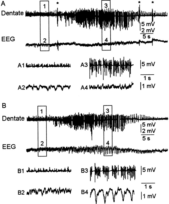
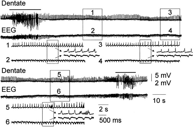
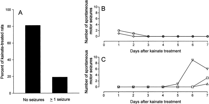
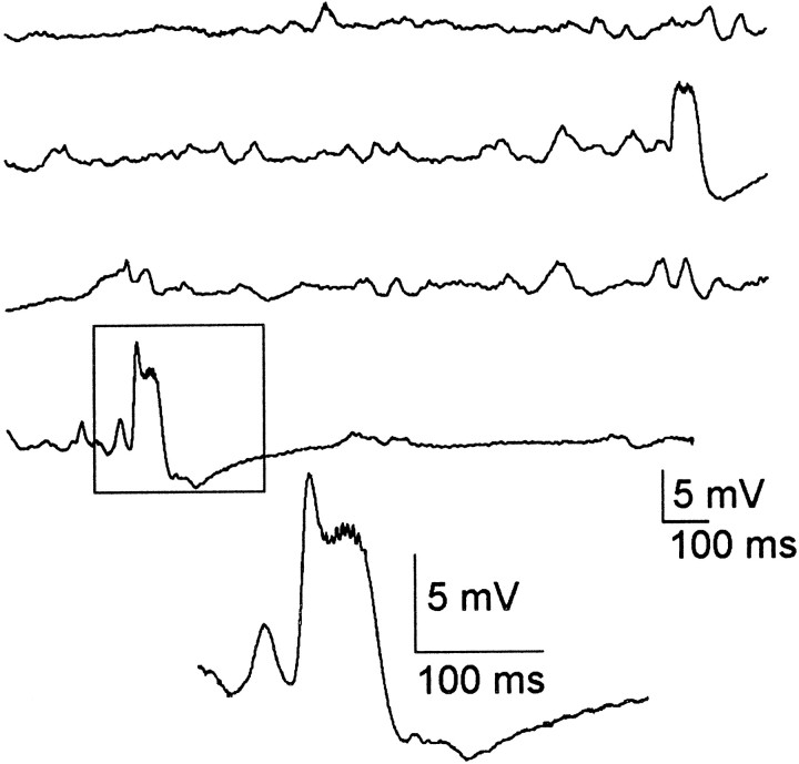
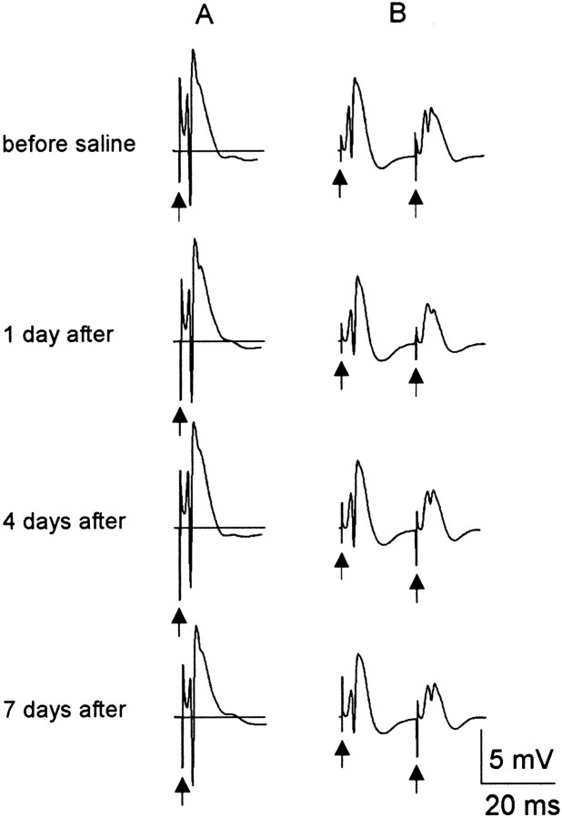
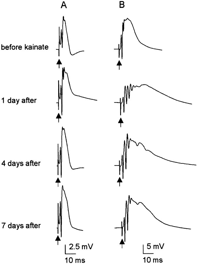
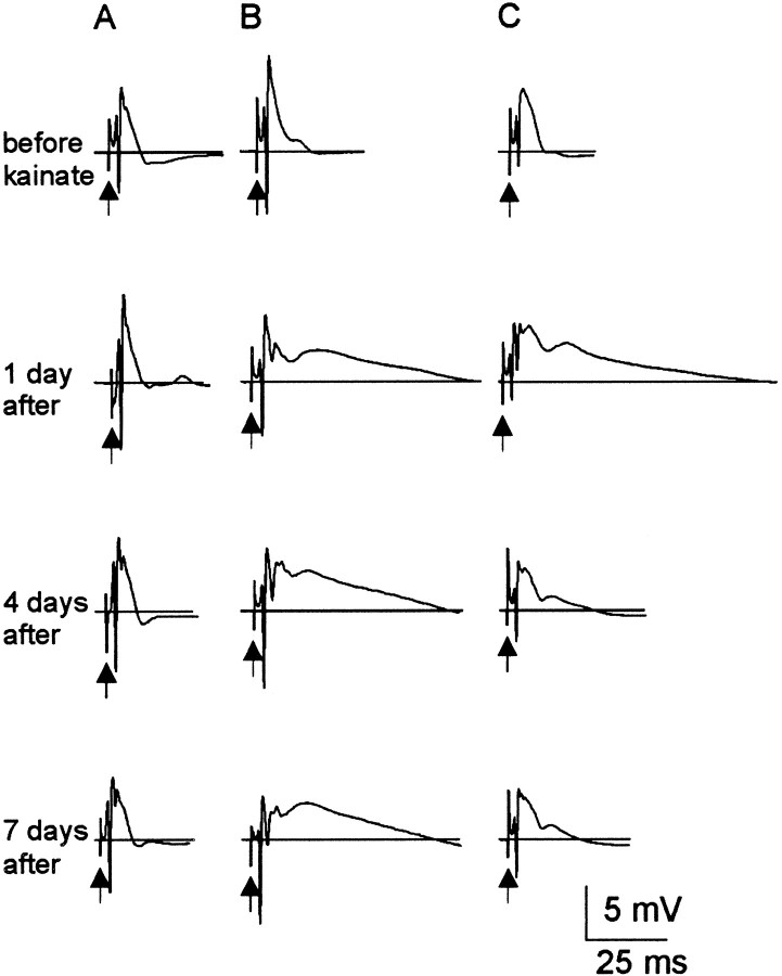
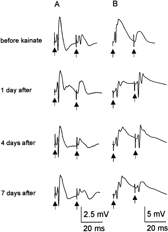
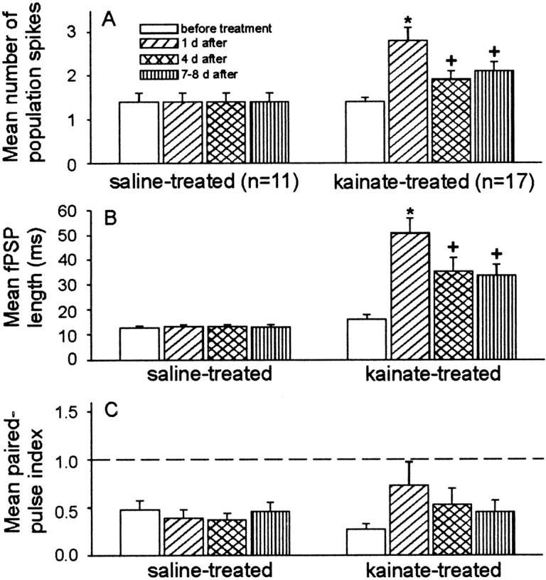
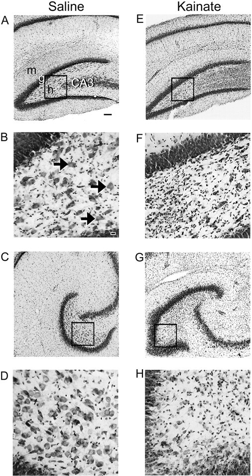
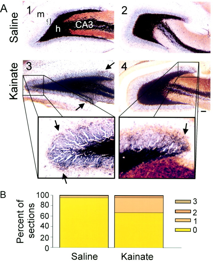
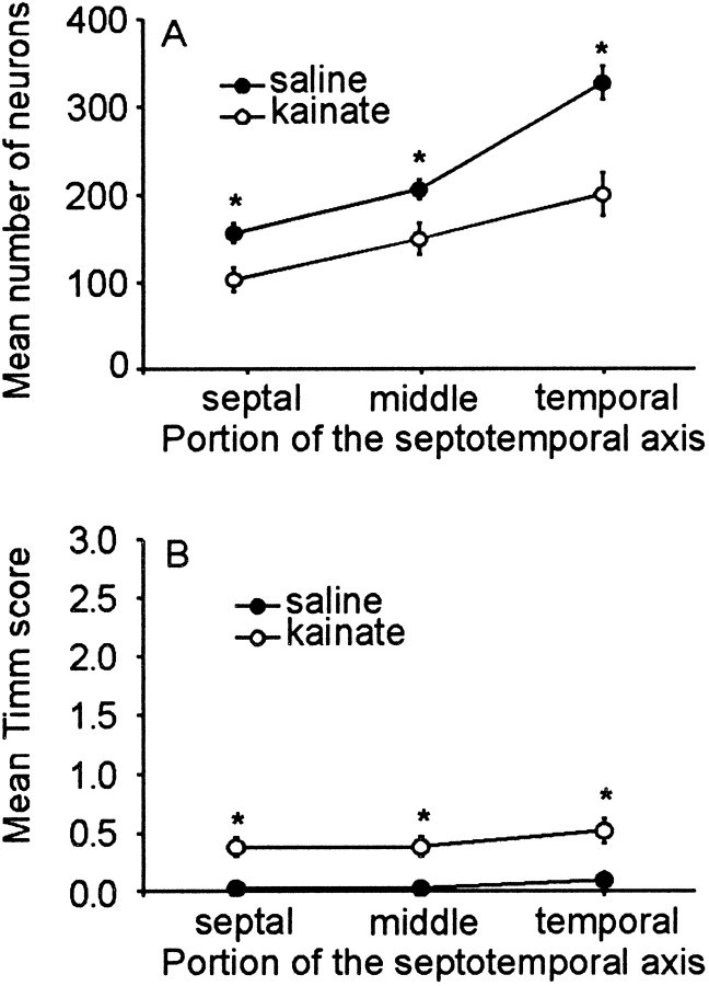
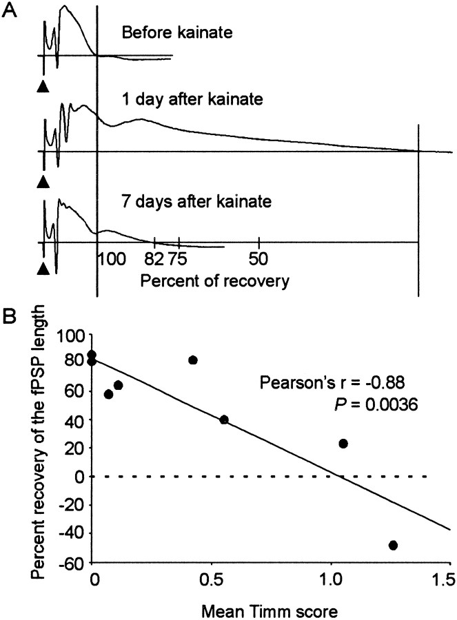
Mossy fiber reorganization has been hypothesized to restore inhibition months after kainate-induced status epilepticus. The time course of recovery of inhibition after kainate treatment, however, is not well established. We tested the hypothesis that if inhibition is decreased after kainate treatment, it is restored within the first week when little or no mossy fiber reorganization has occurred. Chronic in vivo recordings of the septal dentate gyrus were performed in rats before and 1, 4, and 7-8 d after kainate (multiple injections of 5 mg/kg, i.p.; n = 17) or saline (n = 11) treatment. Single and paired-pulse stimuli were used to assess synaptic inhibition. The first day after kainate treatment, only a fraction of rats showed multiple population spikes (35%), prolonged field postsynaptic potentials (76%), and loss of paired-pulse inhibition (29%) to perforant path stimulation. Thus, inhibition was reduced in only some of the kainate-treated rats. By 7-8 d after treatment, nearly all kainate-treated rats showed partial or full recovery in these response characteristics. Histological analysis indicated that kainate-treated rats had a significant decrease in the number of hilar neurons compared to controls, but Timm staining showed little to no mossy fiber reorganization. These results suggest that a decrease in synaptic inhibition in the septal dentate gyrus is not a prerequisite for epileptogenesis and that most of the recovery of inhibition occurs before robust Timm staining in the inner molecular layer.


Figures

Fig. 1.
Fig. 1.
Granule cell layer (Dentate) and surface (EEG) recordings of nonconvulsive and convulsive seizures during kainate treatment. A, Nonconvulsive seizure during kainate treatment. In the expanded traces (A1–A4) seizure activity is shown in the dentate electrode (A3), but it is absent in the EEG trace (A4). B, Stage IV motor seizure during kainate treatment. Note the epileptiform activity in the expanded dentate and EEG recordings (B3 andB4, respectively). All dentate traces are shown at the same gain (calibration, 5 mV), but the EEG traces are at two gains. Thenumbered boxes in the toptwo traces in each panel are expanded in the bottom traces. Asterisks represent truncated motor artifacts induced by “wet dog shakes.”
Fig. 2.
Fig. 2.
Electrographic status epilepticus during kainate treatment. The top and bottom panels of the dentate and EEG traces are continuous. Stage IV motor seizures that were recorded at both the beginning and end of the traces are marked with a bar. Between the motor seizures, nonconvulsive ictal activity with a frequency of 2.5–10 Hz was observed in both the dentate and EEG recordings. The dentate and EEG traces are different gain (note the 5 vs 2 mV calibrations). The numbered boxes in the top traces are shown below at a faster time scale (2 sec), and the arrows point to traces with the fastest time scale (500 msec).
Fig. 3.
Fig. 3.
During the first week after kainate treatment, few spontaneous motor seizures were observed in kainate-treated rats.A, Continuous video monitoring (24 hr) showed that 81% of kainate-treated rats did not have any spontaneous motor seizures during the first week after treatment. The remaining 19% of rats, however, were observed to have ≥1 behavioral seizures. The seizure frequencies of these rats are shown as a function of time after kainate treatment. B, Two of five rats experienced motor seizures within the first 27 hr after treatment. C, The remaining three rats had their first motor seizure at 5–7 d after treatment.
Fig. 4.
Fig. 4.
Spontaneous interictal events. In the granule cell layer of the septal dentate gyrus in kainate-treated rats, spontaneous interictal events were observed from the first day after treatment until killing. These events varied in frequency and duration among kainate-treated rats. The boxed event is expanded below.
Fig. 5.
Fig. 5.
A representative response to perforant path stimuli during the first week after saline treatment in a single rat. For this and all following figures, the traces are an average of 10 evoked events, and arrows point to truncated stimulus artifacts. A, In all control rats, single stimuli produced responses with one or two population spikes and uniform lengths of field PSP throughout the testing paradigm. B,Paired-pulse stimulation produced inhibition in the response to the test stimulus compared to the conditioning stimulus.
Fig. 6.
Fig. 6.
Number of population spikes produced from perforant path stimulation during the first week after kainate treatment. Each group of traces is a response from a single kainate-treated rat recorded in serial order. A, In 47% of treated rats, the number of population spikes 1–8 d after treatment was not significantly different from controls. B, In the remaining 53% of treated rats, few population spikes were observed 1–4 d after treatment (i.e., ≥3 population spikes). By 7–8 d after treatment, all rats had ≤3 population spikes.
Fig. 7.
Fig. 7.
Representative examples of prolonged field PSPs in the septal dentate gyrus during the first week after kainate treatment in a single rat from each of the groups. A, In 4 of 17 treated rats, single stimuli produced responses with the same length of field PSP throughout the testing period (i.e., 1–8 d after treatment).B, Single stimuli produced prolonged field PSPs with little or no recovery from 1–8 d after treatment in 7 of 17 treated rats. C, Partial to full recovery of the length of the field PSP was observed in the remaining 6 of 17 treated rats.
Fig. 8.
Fig. 8.
Paired-pulse responses in kainate-treated rats during the first week after treatment. Each group of traces is a recording from a single rat in chronological order. A,In 65% of treated rats, paired-pulse inhibition was enhanced throughout the testing period after kainate treatment. Compare the paired-pulse responses before treatment to 1 and 4 d after treatment. Although a population spike was present in the second response before kainate treatment, it was no longer present in the second response at 1 and 4 d after treatment. This result suggests that an increase in inhibition occurs in some animals after kainate treatment. B, Paired-pulse stimuli produced facilitated responses 1–4 d after treatment in the remaining 35% of rats. By 7–8 d, however, four of the six kainate-treated rats with a facilitated response 1 d after treatment had a paired-pulse index <1. Thus, partial recovery of paired-pulse inhibition occurred in these animals (i.e., although the paired-pulse index was <1, a population spike was present in the second response at 7 d, but not before treatment).
Fig. 9.
Fig. 9.
The mean number of population spikes and duration of the field PSP of kainate-treated rats, but not of paired-pulse inhibition, were significantly different from controls.A, Kainate-treated rats had significantly more population spikes 1 d after treatment compared to before, 4, and 7–8 d after treatment. By 4–8 d after treatment, a partial recovery of the number of population spikes was observed. B,Similarly, kainate-treated rats had significantly longer field PSPs at 1 d after treatment compared to before, 4, and 7–8 d after treatment. However, a partial to full recovery of the field PSP duration was observed 7–8 d after treatment. C, Both kainate-treated and control rats showed paired-pulse inhibition before and 1, 4, and 7–8 d after treatment. At 1 d after treatment, kainate-treated rats had an increase in the mean paired-pulse index; however, this difference was not significant. Theasterisks represent significant differences over all data points (p < 0.05; Student–Newman–Keuls). The plus symbols indicate significant differences from controls and 1 d after treatment (i.e., asterisks).
Fig. 10.
Fig. 10.
Region used for counting neurons and analyzing Timm stain. The hilus was determined by: (1) drawing a line along the inner margin of the granule cell layer from the tip of the inner blade to the tip of the outer blade and (2) extending these lines to the proximal end of the CA3 pyramidal cell layer (white line). Abnormal Timm staining was scored as the amount of dark reaction product in the inner molecular layer and through the granule cell layer (black line).
Fig. 11.
Fig. 11.
Cresyl violet-stained hippocampal sections of saline- and kainate-injected rats (A–D vsE–H) 7 d after treatment. Sections from the septal one-third of the hippocampus (A, B, E, F) show partial loss of hilar neurons in a kainate-treated rat (E, F) versus severe loss at the temporal one-third (G, H). Compare the larger number of neurons in the hilus and the more tightly packed CA3 pyramidal cell layer in the control rat relative to the prominent gliosis in the hilar region and the loss of CA3 pyramidal cells in the kainate-treated rat. Thearrows point to neurons located in the hilus, and theboxed regions are magnified in the bottom panels. m, Molecular layer; g, granule cell layer; h, hilus; CA3, CA3 pyramidal cell layer. Scale bars: A, C,E, G, 100 μm; B,D, F, H, 20 μm.
Fig. 12.
Fig. 12.
Timm- and cresyl violet-stained hippocampal sections of saline- and kainate-injected rats 7 d after treatment. Sections from a control rat at the septal (A1) and temporal one-thirds (A2) of the hippocampus lack dark reaction product in the inner molecular layer of the dentate gyrus. In a kainate-treated rat, the septal (A3) and temporal sections (A4) have slight but detectable Timm staining in the granule cell layer and molecular layer (arrows indicate dark reaction product in the inner molecular layer). Scale bar, 100 μm. B, In both controls and kainate-treated rats, most hippocampal sections (99 and 96%, respectively) had a Timm score of 0 or 1 (Tauck and Nadler, 1985).
Fig. 13.
Fig. 13.
Septotemporal distribution of hilar neurons and Timm staining in control (n = 6, filled circles) and kainate-treated rats (n = 8,open circles). A, Saline-treated rats had more cresyl violet-stained neurons in the temporal pole (100% septotemporal distance) compared to the septal pole (0% septotemporal distance). However, there were significantly fewer neurons in all three regions of the hippocampi in kainate-treated rats (p < 0.005; Student's ttest). B, Similarly, the difference in the mean Timm score between controls and kainate-treated rats was small but significant (p < 0.0005; Student'st test). Error bars represent SEM, and some means are smaller than the data point symbol. The asterisksrepresent significant differences between data point pairs.
Fig. 14.
Fig. 14.
Recovery of the duration of the field PSP.A, Graphical representation of how the amount of recovery in the duration of the field PSP was measured. The original field PSP duration is shown in the top trace. This length was subtracted from the field PSP duration at 1 and 7 d after treatment. The duration of the field PSP was prolonged 1 d after treatment. The bottom trace shows that at 7 d after treatment, the abnormal length of the field PSP recovered by 82%. B, At 7 d after treatment, the relationship between the percentage of recovery and the mean Timm score showed a significant negative correlation (Pearson's r = −0.88; p = 0.0036).

Similar articles

Cited by

References

    1. Babb TL, Kupfer WR, Pretorius JK, Crandall PH, Levesque MF. Synaptic reorganization by mossy fibers in human epileptic fascia dentata. Neuroscience. 1991;42:351–363. - PubMed
    1. Bekenstein JW, Lothman EW. Dormancy of inhibitory interneurons in a model of temporal lobe epilepsy. Science. 1993;259:97–100. - PubMed
    1. Ben-Ari Y. Limbic seizures and brain damage produced by kainic acid: Mechanisms and relevance to human temporal lobe epilepsy. Neuroscience. 1985;14:375–403. - PubMed
    1. Buckmaster PS, Dudek FE. Network properties of the dentate gyrus in epileptic rats with hilar neuron loss and granule cell axon reorganization. J Neurophysiol. 1997a;77:2685–2696. - PubMed
    1. Buckmaster PS, Dudek FE. Neuron loss, granule cell reorganization, and functional changes in the dentate gyrus of epileptic kainate-treated rats. J Comp Neurol. 1997b;385:385–404. - PubMed

Publication types

MeSH terms