. 1999 Nov 15;19(22):10107-15.
doi: 10.1523/JNEUROSCI.19-22-10107.1999.

Null mutation of c-fos causes exacerbation of methamphetamine-induced neurotoxicity

Affiliations

Null mutation of c-fos causes exacerbation of methamphetamine-induced neurotoxicity

X Deng et al. J Neurosci. .

Abstract

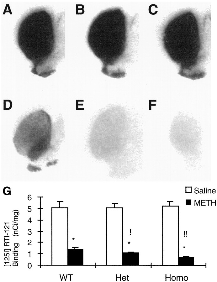
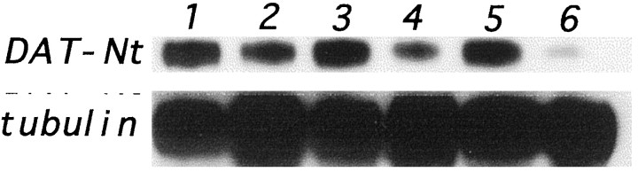
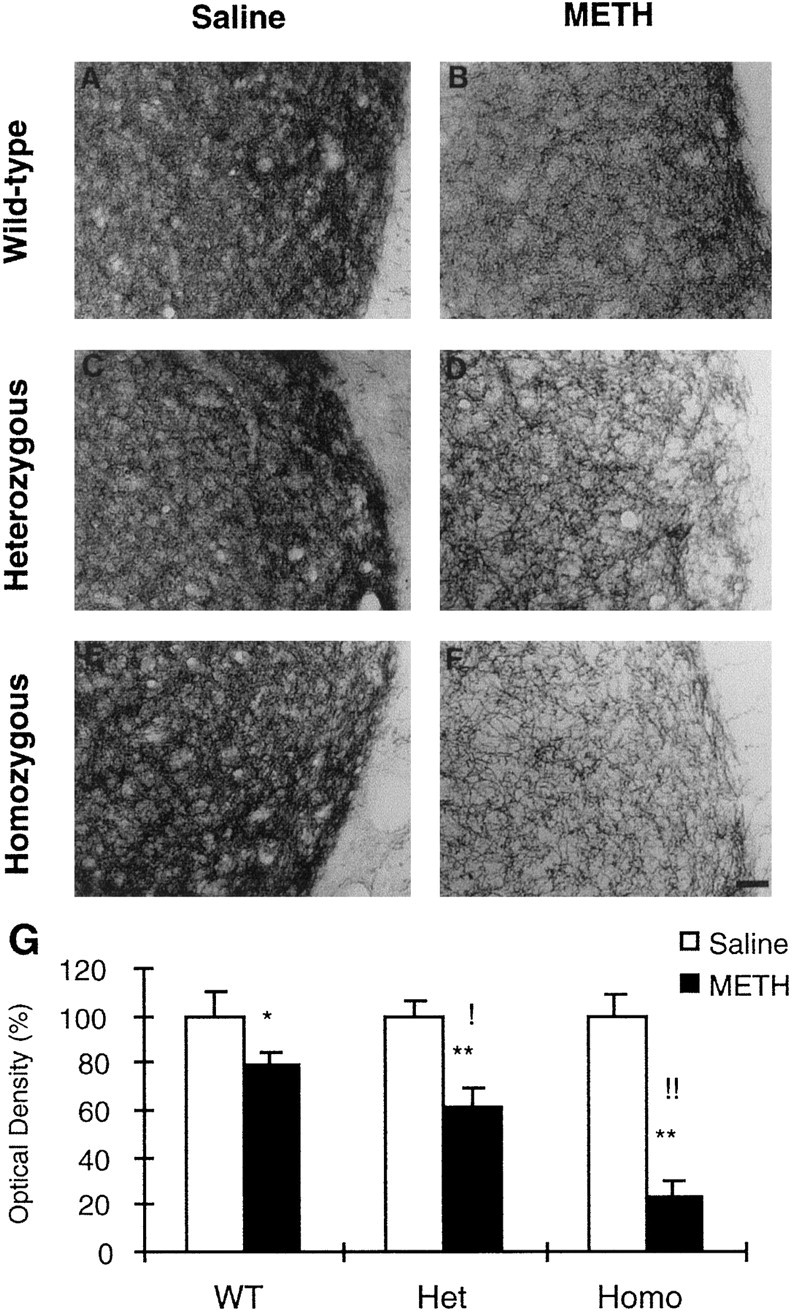
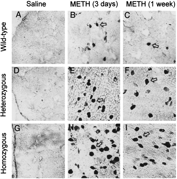
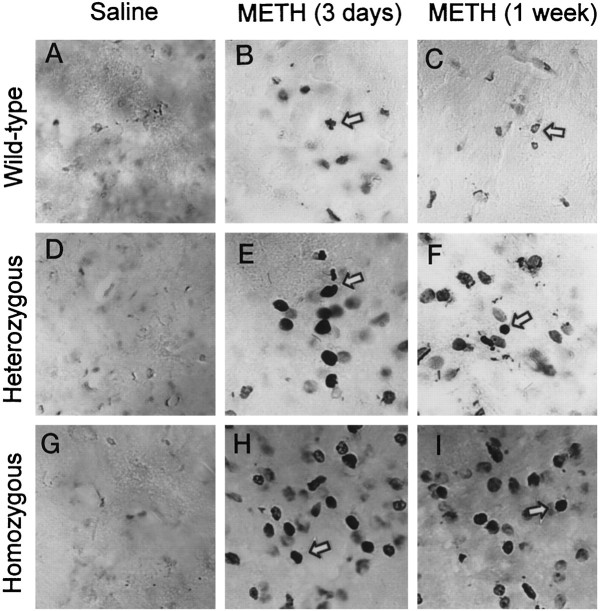
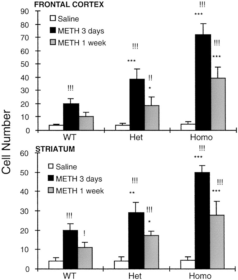
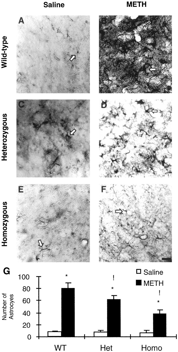
Methamphetamine neurotoxicity has been demonstrated in rodents and nonhuman primates. These neurotoxic effects may be associated with mechanisms involved in oxidative stress and the activation of immediate early genes (IEG). It is not clear, however, whether these IEG responses are involved in a methamphetamine-induced toxic cascade or in protective mechanisms against the deleterious effects of the drug. As a first step toward clarifying this issue further, the present study was thus undertaken to assess the toxic effects of methamphetamine in heterozygous and homozygous c-fos knock-out as well as wild-type mice. Administration of methamphetamine caused significant reduction in [(125)I]RTI-121-labeled dopamine uptake sites, dopamine transporter protein, and tyrosine hydroxylase-like immunohistochemistry in the striata of wild-type mice. These decreases were significantly exacerbated in heterozygous and homozygous c-fos knock-out mice, with the homozygous showing greater loss of striatal dopaminergic markers. Moreover, in comparison with wild-type animals, both genotypes of c-fos knock-out mice showed more DNA fragmentation, measured by the number of terminal deoxynucleotidyl transferase-mediated dUTP nick-end-labeled nondopaminergic cells in their cortices and striata. In contrast, wild-type mice treated with methamphetamine demonstrated a greater number of glial fibrillary acidic protein-positive cells than did c-fos knock-out mice. These data suggest that c-fos induction in response to toxic doses of methamphetamine might be involved in protective mechanisms against this drug-induced neurotoxicity.


Figures

Fig. 1.
Fig. 1.
A–F, Effects of METH on [125I]RTI-121–labeled striatal DA uptake sites in WT (A, D), heterozygous (B, E), and homozygous (C, F) c-fos mutant mice. Animals received saline (A–C) or METH (D–F) as described in Materials and Methods. They were killed 1 week after drug treatment. [125I]RTI-121–binding density is similar in the three saline-treated genotypes (A–C). METH administration caused marked reduction of DA uptake sites (D–F), with the greatest decreases occurring in c-fos −/− mice (F). G, The results of the statistical analyses of the quantitative data obtained from the image analyses. Values represent means ± SEM of five to eight animals per group. Key to statistics: *p < 0.0001 in comparison with saline-treated mice of similar genotypes; !p < 0.05, and !!p < 0.0001 in comparison with METH-treated WT mice.
Fig. 2.
Fig. 2.
Representative immunoblot of the effects of METH on DAT protein concentration. Lanes 1, 3, 5, From saline-injected +/+, +/−, and −/− c-fos mutant mice, respectively.Lanes 2, 4, 6, From METH-injected +/+, +/−, and −/− c-fos knock-out mice, respectively. There were significant changes in DAT concentration consisting of 44.6 ± 4.7, 57.9 ± 6.8, and 78.8 ± 5.3% decreases in +/+, −/−, and −/− c-fos knock-out mice, respectively. α-Tubulin is also shown and reveals similar loading for all groups (6 mice per group). Nt, Antibody against N terminus of the protein.
Fig. 3.
Fig. 3.
Effects of METH on TH-like immunoreactivity in mice. A–F, Animals were treated with either saline (A, C, E) or METH (B, D, F) 1 week after drug treatment as described in Materials and Methods. The intensity of staining is comparable in the saline-treated mice of the three genotypes (A, C, E). METH administration caused a visually obvious reduction of TH staining (B, D, F), which was more severe in homozygous c-fos knock-out mice (F). G, The results of the statistics for the semiquantitative data obtained using image analysis are shown. Values represent means ± SEM of five to eight animals (6 sections per animal) per group. Key to statistics: *p < 0.001, and **p < 0.0001 in comparison with saline-treated mice of similar genotypes; !p < 0.001, and !!p < 0.0001 in comparison with METH-treated WT mice. Scale bar, 100 μm.
Fig. 4.
Fig. 4.
Representative photomicrographs of TUNEL-stained frontal cortices of mice. Very few positive cells appeared in the frontal cortices of saline-treated mice of the three genotypes (A, D, G). METH caused marked increases in TUNEL-positive cells at 3 d (B, E, H) and 1 week (C, F, I) after drug treatment. Thearrows point to typical positive cells. These photomicrographs were generated by using a Carl Zeiss Laser Scanning Confocal System with Axiovert 135–inverted microscopy. The objective lens was 40×. Quantitative data are provided below (see Fig.6).
Fig. 5.
Fig. 5.
Representative photomicrographs of TUNEL-stained striata of mice. Very few positive cells could be seen in the striata of saline-treated mice (A, D, G). As in the cortex, METH caused marked increases in TUNEL-positive cells at 3 d (B, E, H) and 1 week (C, F, I). Thearrows point to typical positive cells. The photomicrographs were generated as described in Figure 4. Quantitative data are provided below (see Fig. 6).
Fig. 6.
Fig. 6.
METH caused greater increases in TUNEL-positive cells in the frontal cortex and striatum of c-fos knock-out than in WT mice. The animals were treated and the brains were processed as described in Materials and Methods. Values represent means ± SEM of five to eight mice per group. Key to statistics: *p < 0.05, **p < 0.001, and ***p < 0.0001 in comparison with METH-treated WT mice killed at similar time points; !p < 0.05, !!p < 0.001, and !!!p < 0.0001 in comparison with saline-treated mice of similar genotypes.
Fig. 7.
Fig. 7.
A–F, Effects of METH on GFAP-like immunohistochemistry in the striata of +/+ (A, B), +/− (C, D), and −/− (E, F) c-fos knock-out mice. A few small positive astrocytes are seen in the striata of saline-treated mice (A, C, E). METH caused marked increases in the number of astrocytes in the striatum in mice killed 1 week after drug treatment (B, D, F). METH-induced astrocytes were hypertrophic and densely stained in the WT mice (B). However, in the c-fos +/− (D) and −/− (F) mice, METH treatment caused a measurable increase in the number of astrocytes, but these were small in size and weakly stained. Thearrows point to typical positive cells.G, The statistical analyses of the data obtained from the counts of GFAP-positive cells in the striatum. Values represent means ± SEM from five to eight animals (6 sections per mouse) per group. Key to statistics: *p < 0.0001 in comparison with saline-treated mice of similar genotypes; !p < 0.01 in comparison with METH-treated WT mice. Scale bar, 100 μm.
Fig. 8.
Fig. 8.
Effects of METH on core temperature in WT and heterozygous c-fos mutant mice. Core temperature was recorded in animals every 30 min during the administration of METH. Values represent means ± SEM from seven mice per group. There were no statistical differences in the METH-induced temperature elevation between the two genotypes of mice. Saline-treated animals did not exhibit changes in core body temperature over time (data not shown for the sake of clarity). Recorded ambient temperature was between 20.5 and 21.2°C.

References

    1. Albers DS, Sonsalla PK. Methamphetamine-induced hyperthermia and dopaminergic neurotoxicity in mice: pharmacological profile of protective and nonprotective agents. J Pharmacol Exp Ther. 1995;275:1104–1114. - PubMed
    1. Ali SF, Newport GD, Holson RR, Slikker W, Jr, Bowyer JF. Low environmental temperatures or pharmacologic agents that produce hypothermia decrease methamphetamine neurotoxicity in mice. Brain Res. 1994;658:33–38. - PubMed
    1. Asanuma M, Cadet JL. Methamphetamine-induced increase in striatal NF-kappaB DNA-binding activity is attenuated in superoxide dismutase transgenic mice. Brain Res Mol Brain Res. 1998;60:305–309. - PubMed
    1. Asanuma M, Hirata H, Cadet JL. Attenuation of 6-hydroxydopamine-induced dopaminergic nigrostriatal lesions in superoxide dismutase transgenic mice. Neuroscience. 1998;85:907–917. - PubMed
    1. Baba T, Yamamda H, Oguri K, Yoshimura H. Participation of cytochrome P-450 isozymes in N-demethylation, N-hydroxylation and aromatic hydroxylation of methamphetamine. Xenobiotica. 1988;18:475–484. - PubMed

MeSH terms