. 1999 Nov 15;19(22):10153-63.
doi: 10.1523/JNEUROSCI.19-22-10153.1999.

Motor skills training enhances lesion-induced structural plasticity in the motor cortex of adult rats

Affiliations

Motor skills training enhances lesion-induced structural plasticity in the motor cortex of adult rats

T A Jones et al. J Neurosci. .

Abstract

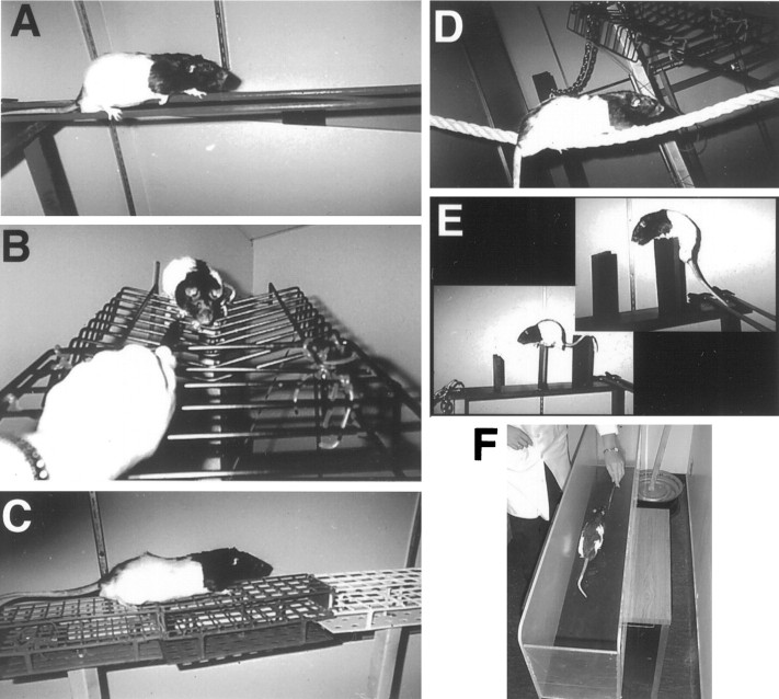
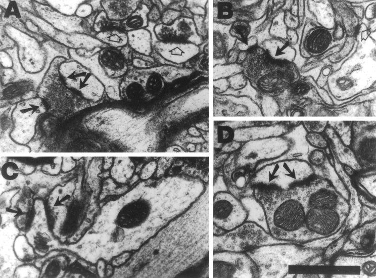
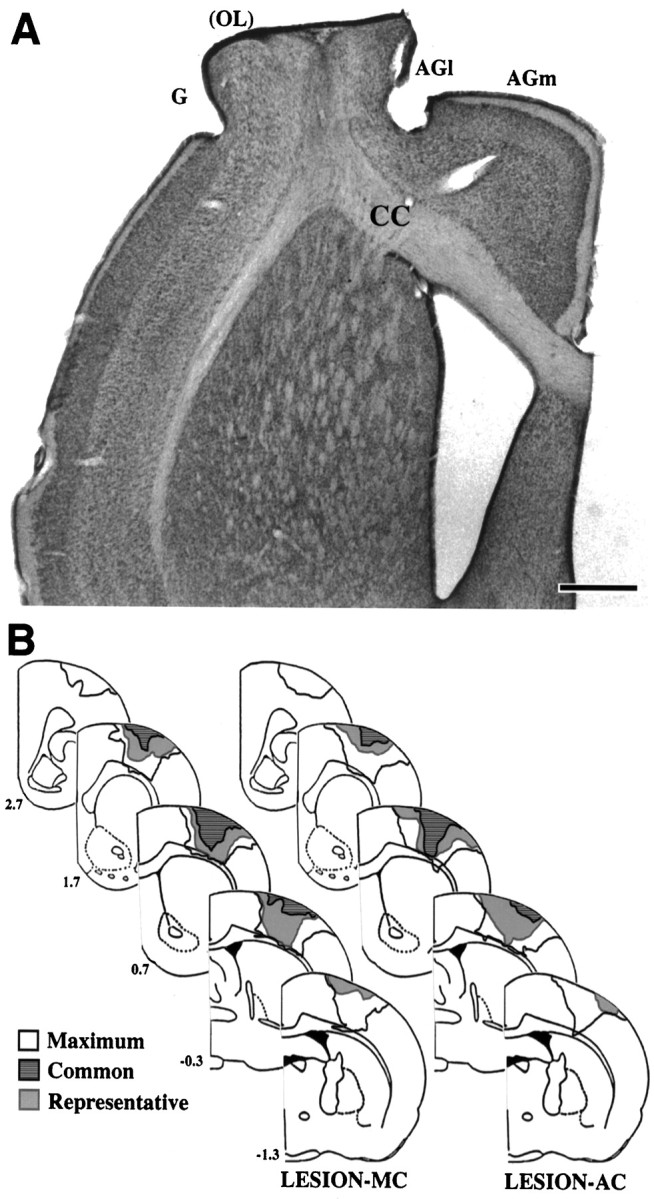
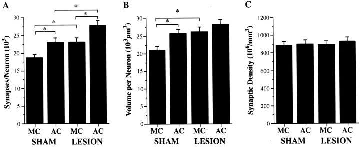
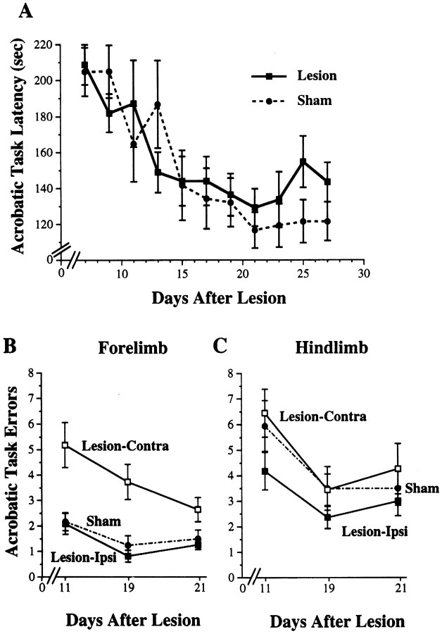
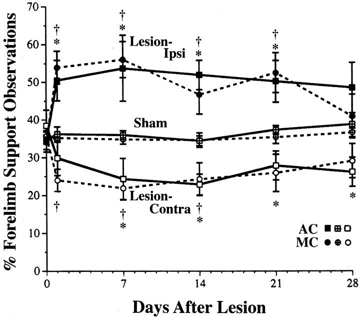
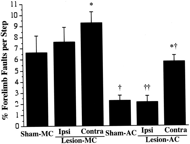
To assess behavioral experience effects on synaptic plasticity after brain damage, the present study examined the effects of complex motor skills training (the acrobatic task) on synaptic changes in layer V of the motor cortex opposite unilateral damage to the forelimb sensorimotor cortex (FLsmc). Adult male rats were given lesions or sham operations followed by 28 d of training on the acrobatic task [acrobat condition (AC)]. As a motor activity control [motor control (MC)], lesion and sham animals were given simple repetitive exercise. Previously, FLsmc lesions and acrobatic training have independently been found to result in increases in synapse to neuron ratios in the intact motor cortex relative to controls, and both of these effects were replicated in the present study. In addition, acrobat training after lesions significantly increased layer V synapses per neuron relative to sham-AC and lesion-MC rats. Thus, the combination of acrobatic training and lesions resulted in an enhanced synaptogenic response. Synapse subtypes were also differentially affected by the conditions. Lesion-MC and sham-AC primarily had increases in the number of synapses per neuron formed by multiple synaptic boutons in comparison to sham-MC. In contrast, lesion-AC had increases in both multiple and single synapses. Multiple synaptic spines and perforated synapses were also differentially affected by training versus lesions. On tests of coordinated forelimb use, lesion-AC rats performed better than lesion-MC rats. In addition to supporting a link between behavioral experience and structural plasticity after brain damage, these findings suggest that adaptive neural plasticity may be enhanced using behavioral manipulations as "therapy."


Figures

Fig. 1.
Fig. 1.
Rats trained on the acrobatic task were required to traverse a series of eight obstacles, which included small parallel rods (A), a ladder with widely spaced rungs (B), a grid platform (C), a rope (D), and barriers (E). As a motor activity control, rats received simple repetitive exercise in a straight runway (F).
Fig. 2.
Fig. 2.
Examples of synapses formed by MSBs (A, B), a synapse formed by an MSS (C), and synapses with perforated postsynaptic densities (A,D) in layer V of the motor cortex. A,B, MSBs consist of a single axonal bouton forming synaptic contacts (arrows) with two or more postsynaptic dendritic processes, i.e., spines or shafts. One of the synapses inA has a perforated postsynaptic density (double arrows). Synapses between individual pairings of boutons and spines, or single synapses, are indicated by open arrows. C, MSSs consist of a single dendritic spine forming synaptic contacts (arrows) with two or more axonal boutons. D, A synapse between a single synaptic spine and single synaptic bouton with a perforated postsynaptic density (arrows). Scale bar, 0.5 μm.
Fig. 3.
Fig. 3.
A, Photomicrograph of a representative FLsmc lesion in a coronal hemisection stained with methylene blue-azure II. Lesions created complete or near complete damage to the overlapping SI and MI representations of the forelimb (OL) as well as extensive damage to the adjacent MI (within lateral agranular cortex, AGl) and SI (within granular cortex, G) cortex. In this example, there is no normal appearing overlap zone remaining.AGm, Medial agranular cortex; CC, corpus callosum. Scale bar, 1 mm. B, Lesions reconstructed based on the extent of remaining non-necrotic tissue. The outer boundaries of all lesions combined (maximum), the region of damage common to all lesions (common), and a representative lesion (representative), for each lesion group are shown. Numbers to the leftindicate coordinates relative to bregma. Structural measurements were made in the motor cortex opposite the lesions.
Fig. 4.
Fig. 4.
Structural effects in layer V of the motor cortex opposite unilateral FLsmc lesions or sham operations in AC and MC rats.A, Acrobatic training after lesions (lesion–AC) significantly enhanced synapse number per neuron increases in comparison to the increases that were found as a result of the training alone (sham–AC) or as a result of the lesion in motor control rats (lesion–MC). B, Volume per neuron (the inverse of neuronal density) was significantly increased in sham–AC and lesion–MC in comparison to sham–MC. Volume per neuron was elevated but not significantly increased in lesion–AC relative to sham–AC and lesion–MC. C, There were no significant differences in the density of layer V synapses for any planned comparison. *p < 0.05.
Fig. 5.
Fig. 5.
Performance on the acrobatic task in lesion–AC and sham–AC rats. A, Latency in seconds to complete the acrobatic task over days of training. There was a significant reduction in latency over days of training (p < 0.0001) but no significant differences between lesion and sham animals on this measure. The data shown are from the first traverse of the acrobatic task per training session. B, Forelimb errors in limb placement on the acrobatic task, measured as the number of foot slips per traverse. The lesions resulted in a significant increase in contralateral forelimb errors in comparison to sham (p < 0.0005). Ipsilateral forelimb errors were not significantly different from sham. C, There were no significant differences between lesion and sham rats in hindlimb footslips.
Fig. 6.
Fig. 6.
Forelimb asymmetries in postural support behaviors during exploratory movements. These data are from observations of the use of the forelimbs for upright postural support against the walls of a transparent cage. FLsmc lesions resulted in the preferential use of the forelimb ipsilateral to the lesion and a reduction in the use of the contralateral forelimb in comparison to shams. There were no significant differences between lesion–AC and lesion–MC in forelimb use asymmetries. Data for lesions are percentage ipsilateral or contralateral forelimb use/total support observations (ipsi + contra + bilateral support). For shams, data for left and right forelimbs are pooled. The percentage of simultaneous use of both forelimbs was not significantly affected by the lesions (see Results). *p < 0.05 lesion–AC versus sham–AC; †p < 0.05 lesion–MC versus sham–MC.
Fig. 7.
Fig. 7.
Performance on the footfault task. This task was performed after the last day of training (on postoperative day 29) as a test of coordinated forelimb placement during locomotion. Acrobatic training in both lesion and sham animals resulted in a reduction in forelimb-placing errors on this task in comparison to lesion and sham motor controls, respectively. Lesion–AC made fewer errors with either forelimb in comparison to lesion–MC, most notably with the ipsilateral forelimb. *p < 0.05 significantly different from sham of the same training condition, †p < 0.005 and ††p < 0.0005 significantly different from motor control of the same lesion condition.

References

    1. Adams FS, Schwarting RK, Huston JP. Behavioral and neurochemical asymmetries following unilateral trephination of the rat skull: is this control operation always appropriate? Physiol Behav. 1994;55:947–952. - PubMed
    1. Anker RL, Cragg BG. Estimation of the number of synapses in a volume of nervous tissue from counts in thin sections by electron microscopy. J Neurocytol. 1974;3:725–735. - PubMed
    1. Barth TM, Jones TA, Schallert T. Functional subdivisions of the rat somatic sensorimotor cortex. Behav Brain Res. 1990;39:73–95. - PubMed
    1. Bendre AA, Swain RA, Wheeler BC, Greenough WT. Augmentation of cerebellar responses to parallel fiber activation following skilled motor acquisition in rats. Soc Neurosci Abstr. 1995;21:444.
    1. Black JE, Isaacs K, Anderson BJ, Alcantara AA, Greenough WT. Learning causes synaptogenesis whereas motor activity causes angiogenesis, in cerebellar cortex of adult rats. Proc Natl Acad Sci USA. 1990;87:5568–5572. - PMC - PubMed

Publication types