

Nerve growth factor and its high-affinity receptor in chronic pancreatitis
- PMID: 10561084
- PMCID: PMC1420914
- DOI: 10.1097/00000658-199911000-00002
Nerve growth factor and its high-affinity receptor in chronic pancreatitis
Abstract
Objective: To study the mechanisms that are involved in nerve growth and contribute to pain generation in chronic pancreatitis (CP).
Summary background data: Chronic pancreatitis is a painful disease associated with characteristic nerve changes, including an increase in nerve number and diameter. The mechanisms that influence nerve growth are not known. Nerve growth factor (NGF) and its high-affinity tyrosine kinase receptor A (TrkA) are involved in neural development and survival and growth of central and peripheral nerves.
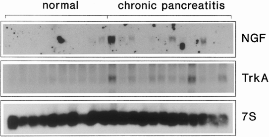
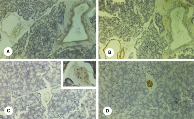
Methods: Nerve growth factor and TrkA were investigated by Northern blot analysis, in situ hybridization, and immunohistochemical staining in the pancreases of 24 patients with CP, and the findings were correlated with clinical parameters.
Results: By Northern blot analysis, NGF and TrkA mRNA expression were increased in 42% (13.1-fold) and 54% (5.5-fold) of the CP samples (p < 0.01), respectively. In situ hybridization revealed that in CP, enhanced NGF mRNA expression was present in metaplastic ductal cells, in degenerating acinar cells, and in acinar cells dedifferentiating into tubular structures. TrkA mRNA was intensely present in the perineurium. Further, enhanced NGF and TrkA mRNA signals were also present in intrapancreatic ganglia cells in CP samples. Immunohistochemistry confirmed the in situ hybridization findings. Analysis of the molecular findings with clinical parameters revealed a significant relation (p < 0.05) between NGF mRNA levels and pancreatic fibrosis (r = 0.64) and acinar cell damage (r = 0.74) and between TrkA mRNA and pain intensity (r = 0.84).
Conclusion: Activation of the NGF/TrkA pathway occurs in CP. It might influence neural morphologic changes and the pain syndrome in this disorder.
Figures


Comment in
-
Unravelling the mystery of pancreatic pain.Ann Surg. 1999 Nov;230(5):625-6. doi: 10.1097/00000658-199911000-00003. Ann Surg. 1999. PMID: 10561085 Free PMC article. No abstract available.
References
-
- Di Magno EP, Layer P, Clain JE. The exocrine pancreas: biology, pathobiology and disease. In: Go VLW, ed. Chronic pancreatitis. New York: Raven Press; 1993: 665–706.
-
- DiMagno EP. A short, eclectic history of exocrine pancreatic insufficiency and chronic pancreatitis. Gastroenterology 1993; 104:1255–1262. - PubMed
-
- Oertel JE, Heffess CS, Oertel YC. Pancreas. In: Sternberg SS, ed. Diagnostic surgical pathology. New York: Raven Press; 1989: 1057–1093.
-
- Kennedy RH, Bockman DE, Uscanga L, Choux R, Grimaud JA, Sales H. Pancreatic extracellular matrix alterations in chronic pancreatitis. Pancreas 1987; 2:61–72. - PubMed
-
- Hunger R, Müller C, Zgraggen K, Friess H, Büchler MW. Cytotoxic cells are activated in cellular infiltrates of alcoholic chronic pancreatitis. Gastroenterology 1997; 112:1656–1663. - PubMed
Publication types
MeSH terms
Substances
LinkOut - more resources
Full Text Sources
Other Literature Sources
Medical
Miscellaneous
