. 1999 Nov 15;147(4):707-14.
doi: 10.1083/jcb.147.4.707.

Protein tyrosine phosphatase alpha (PTPalpha) and contactin form a novel neuronal receptor complex linked to the intracellular tyrosine kinase fyn

Affiliations

Protein tyrosine phosphatase alpha (PTPalpha) and contactin form a novel neuronal receptor complex linked to the intracellular tyrosine kinase fyn

L Zeng et al. J Cell Biol. .

Abstract

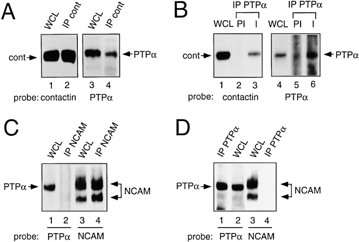
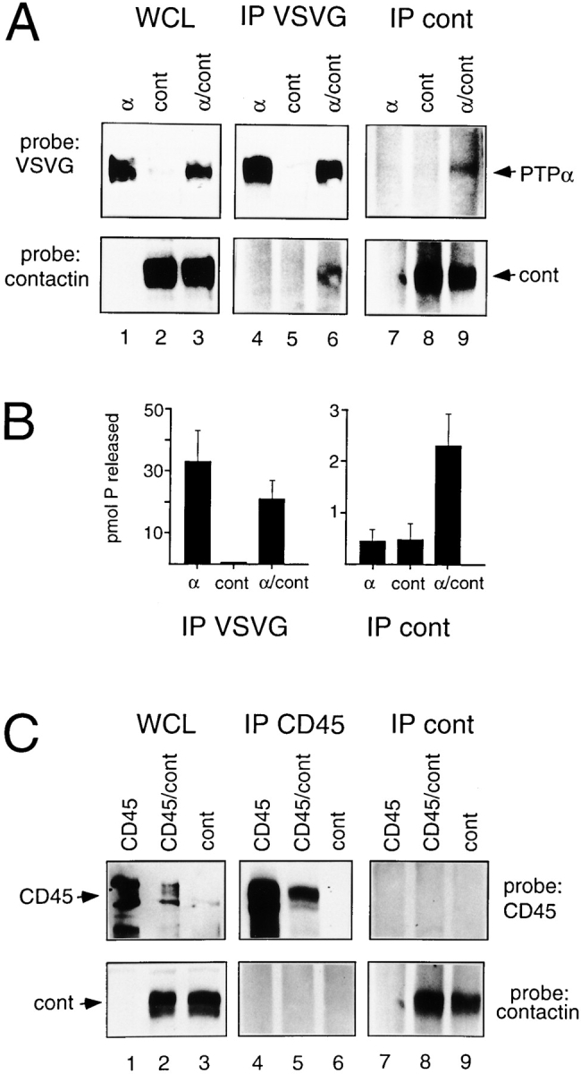
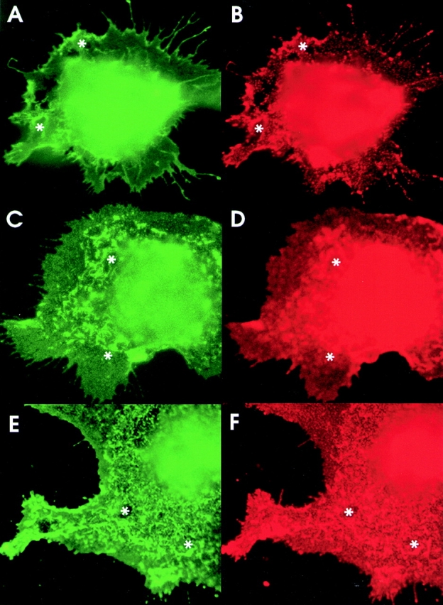
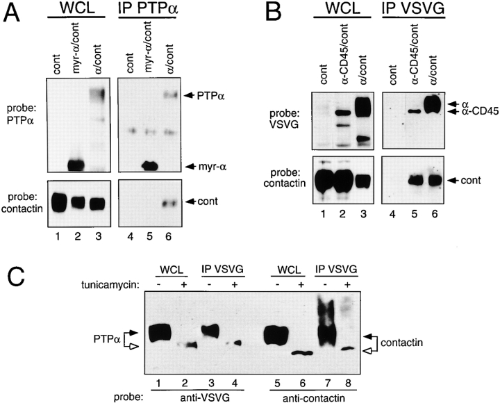
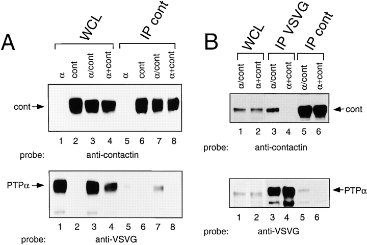
Glycosyl phosphatidylinositol (GPI)-linked receptors and receptor protein tyrosine phosphatases (RPTPs), both play key roles in nervous system development, although the molecular mechanisms are largely unknown. Despite lacking a transmembrane domain, GPI receptors can recruit intracellular src family tyrosine kinases to receptor complexes. Few ligands for the extracellular regions of RPTPs are known, relegating most to the status of orphan receptors. We demonstrate that PTPalpha, an RPTP that dephosphorylates and activates src family kinases, forms a novel membrane-spanning complex with the neuronal GPI-anchored receptor contactin. PTPalpha and contactin associate in a lateral (cis) complex mediated through the extracellular region of PTPalpha. This complex is stable to isolation from brain lysates or transfected cells through immunoprecipitation and to antibody-induced coclustering of PTPalpha and contactin within cells. This is the first demonstration of a receptor PTP in a cis configuration with another cell surface receptor, suggesting an additional mode for regulation of a PTP. The transmembrane and catalytic nature of PTPalpha indicate that it likely forms the transducing element of the complex, and we postulate that the role of contactin is to assemble a phosphorylation-competent system at the cell surface, conferring a dynamic signal transduction capability to the recognition element.


Figures

Figure 1
Figure 1
PTPα associates with contactin in chick brain. Embryonic chick (A and B) or adult mouse (C and D) brain lysates (WCL) and immunoprecipitates made with anticontactin antibody (A), anti–NCAM antibody (C), or affinity-purified anti–PTPα antibody or preimmune (PI) serum (B and D), were probed for the presence of contactin, NCAM, or PTPα as indicated.
Figure 2
Figure 2
Association of ectopically expressed PTPα and contactin. COS-1 cells were transfected with VSVG-PTPα (α), contactin (cont), or VSVG-PTPα and contactin (α/cont) cDNAs. (A) Whole cell lysates (WCL), and anti-VSVG or anticontactin immunoprecipitates (IP) were probed with anti-VSVG or anticontactin antibodies. (B) Anti-VSVG and anticontactin immunoprecipitates were assayed for phosphatase activity as described in Materials and Methods. The bars represent the values of the mean phosphatase activity ± SEM measured in three independent experiments. (C) COS-1 cells were transfected with CD45, contactin (cont), or CD45 and contactin (CD45/cont) cDNAs. Whole cell lysates (WCL), and anti-CD45 or anticontactin immunoprecipitates (IP) were probed with anti-CD45 or anticontactin antibodies.
Figure 3
Figure 3
Ectopically expressed PTPα and contactin colocalize. (A and B) COS-7 cells cotransfected with contactin and VSVG-tagged PTPα cDNAs were fixed and incubated with a mouse anti–VSVG antibody and with a rabbit anticontactin antibody, with detection by FITC-labeled goat-anti–rabbit (A) and RITC-labeled goat anti–mouse antibodies (B). (C–F) In COS-7 cells cotransfected with contactin and PTPα, contactin was induced to form cell surface clusters by incubation with mouse 4D1 antibody (C and D) and PTPα was induced to form cell surface clusters by incubation with mouse anti-VSVG antibody (E and F), in each case followed by FITC-labeled goat anti–mouse antibodies. In the PTPα-induced clustering, contactin was detected with rabbit anticontactin, and in the 4D1-induced clustering, PTPα was detected with rabbit anti-PTPα, followed in both cases by RITC-labeled goat anti–rabbit antibodies. C and F show the distribution of contactin and D and E that of PTPα, with focus levels close to the plane of cell-substratum attachment and with corresponding structures in each panel marked (asterisk).
Figure 4
Figure 4
The extracellular region of PTPα is required for association with contactin. (A) Lysates (WCL) and anti-PTPα immunoprecipitates (IP) from COS-1 cells transiently expressing contactin (cont), myr-PTPα/contactin (myr-α/cont), or PTPα/contactin (α/cont) were probed for PTPα and contactin. (B) Whole cell lysates (WCL) and anti-VSVG immunoprecipitates (IP) of COS cells transiently expressing contactin (cont), VSVG-PTPα-CD45/contactin (α-CD45/cont), or VSVG-PTPα/contactin (α/cont) were probed with anti-VSVG or anticontactin antibodies. (C) COS-1 cells were cotransfected with VSVG-PTPα and contactin cDNAs and treated with 20 μg/ml tunicamycin (lanes 2, 4, 6, and 8) or left untreated (lanes 1, 3, 5, and 7). Whole cell lysates (WCL) and anti-VSVG immunoprecipitates (IP) were probed with anti-VSVG or anticontactin antibodies.
Figure 5
Figure 5
PTPα and contactin do not associate in a trans configuration. (A) COS cells were transfected with VSVG-PTPα (α), contactin (cont), or VSVG-PTPα and contactin cDNAs (α/cont). Whole cell lysates (WCL) (lanes 1–3), a mixed lysate (α+cont) (lane 4) made of equal parts of the VSVG-PTPα–expressing cell lysate, the contactin-expressing cell lysate, and corresponding anticontactin immunoprecipitates (IP) (lanes 5–8) were probed with anticontactin (top) and anti-VSVG (bottom) antibodies. (B) Cells coexpressing VSVG-PTPα and contactin (α/cont) were cultured for 48 h and harvested. Contactin-expressing cells were trypsinized 24 h after transfection, replated onto VSVG-PTPα–expressing cells (α+cont), and harvested after coculture for 24 h. Whole cell lysates (WCL) (lanes 1 and 2), anti-VSVG (lanes 3 and 4) and anticontactin (lanes 5 and 6) immunoprecipitates (IP) were probed with anticontactin (top) or anti-VSVG (bottom) antibodies.

Similar articles

Cited by

References

    1. Beggs H.E., Baragona S.C., Hemperly J.J., Maness P.F. NCAM140 interacts with the focal adhesion kinase p125fak and the SRC related tyrosine kinase p59fyn . J. Biol. Chem. 1997;272:8310–8319 . - PubMed
    1. Bhandari V., Lim K.L., Pallen C.J. Physical and functional interactions between receptor-like protein-tyrosine phosphatase α and p59fyn . J. Biol. Chem. 1998;273:8691–8698 . - PubMed
    1. Brümmendorf T., Wolff J.M., Frank R., Rathjen F.G. Neural cell recognition molecule F11homology with fibronectin type III and immunoglobulin type C domains. Neuron. 1989;2:1351–1361 . - PubMed
    1. Brümmendorf T., Hubert M., Treubert U., Leuschner R., Tàrnok A., Rathjen F.G. The axonal recognition molecule F11 is a multifunctional proteinspecific domains mediate interactions with Ng-CAM and restrictin. Neuron. 1993;10:711–727 . - PubMed
    1. Buj-Bello A., Adu J., Pinon L.G.P., Horton A., Thompson J., Rosenthal A., Chinchetru M., Buchman V.L., Davies A.M. Neurturin responsiveness requires a GPI-linked receptor and the Ret receptor tyrosine kinase. Nature. 1997;387:721–724 . - PubMed

Publication types

MeSH terms

Substances