. 1999 Nov 15;147(4):715-28.
doi: 10.1083/jcb.147.4.715.

The spinal muscular atrophy disease gene product, SMN: A link between snRNP biogenesis and the Cajal (coiled) body

Affiliations

The spinal muscular atrophy disease gene product, SMN: A link between snRNP biogenesis and the Cajal (coiled) body

T Carvalho et al. J Cell Biol. .

Abstract

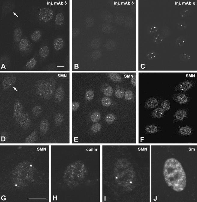
The spliceosomal snRNAs U1, U2, U4, and U5 are synthesized in the nucleus, exported to the cytoplasm to assemble with Sm proteins, and reimported to the nucleus as ribonucleoprotein particles. Recently, two novel proteins involved in biogenesis of small nuclear ribonucleoproteins (snRNPs) were identified, the Spinal muscular atrophy disease gene product (SMN) and its associated protein SIP1. It was previously reported that in HeLa cells, SMN and SIP1 form discrete foci located next to Cajal (coiled) bodies, the so-called "gemini of coiled bodies" or "gems." An intriguing feature of gems is that they do not appear to contain snRNPs. Here we show that gems are present in a variable but small proportion of rapidly proliferating cells in culture. In the vast majority of cultured cells and in all primary neurons analyzed, SMN and SIP1 colocalize precisely with snRNPs in the Cajal body. The presence of SMN and SIP1 in Cajal bodies is confirmed by immunoelectron microscopy and by microinjection of antibodies that interfere with the integrity of the structure. The association of SMN with snRNPs and coilin persists during cell division, but at the end of mitosis there is a lag period between assembly of new Cajal bodies in the nucleus and detection of SMN in these structures, suggesting that SMN is targeted to preformed Cajal bodies. Finally, treatment of cells with leptomycin B (a drug that blocks export of U snRNAs to the cytoplasm and consequently import of new snRNPs into the nucleus) is shown to deplete snRNPs (but not SMN or SIP1) from the Cajal body. This suggests that snRNPs flow through the Cajal body during their biogenesis pathway.


Figures

Figure 1
Figure 1
Localization of SMN in human cell lines. (A) HeLa cells were fixed in formaldehyde, permeabilized with Triton X-100, and immunostained with anti–SMN monoclonal antibody 2B1. Note the cytoplasmic labeling with additional nuclear foci. Staining of the cytoplasm is more difficult to detect when cells are extracted with detergent before fixation (B–G). (B and D) Phase contrast images of HeLa cells with superimposed SMN labeling produced by either monoclonal antibody 2B1 (B, green foci) or serum 95020 (D, red foci). (C and E) The corresponding cells double labeled with anticoilin antibodies. The arrow and the arrowhead in C indicate gems; one is adjacent to a Cajal body (arrow), whereas the other is independent (arrowhead). The yellow color observed in E demonstrates a precise colocalization of SMN (red staining) and coilin (green staining). (F) Phase contrast image of an Huh7 cell with superimposed SMN staining (red focus); (G) the same cell double labeled for SMN (red staining) and coilin (green staining); the yellow color indicates colocalization of both proteins in the same focus. (H–K) HeLa cell triple labeled for SMN (H), coilin (I), and Sm (J) proteins. This experiment was performed using mouse anti–SMN monoclonal antibody 2B1 (detected with Texas red), rabbit anticoilin serum 204.3 (detected with Cy5), and human anti–Sm autoimmune serum C45 (detected with FITC). Superimposition of the three images (K) reveals that SMN colocalizes with Sm in Cajal bodies (arrows), but not in gems (red foci). Bars, 10 μm.
Figure 2
Figure 2
(continues on facing page). Localization of SMN by immunoelectron microscopy. Huh7 cells were embedded in Lowicryl, sectioned, and immunogold labeled with antibodies directed against coilin (A, rabbit serum 204.3) or SMN (B, mouse monoclonal antibody 2B1). (C and D) 0.1 μm. Sections double labeled using anticoilin (5-nm gold particles) and anti–SMN (10-nm gold particles) antibodies. Bars: (A and B) 0.2 μm; (C and D) 0.1 μm.
Figure 3
Figure 3
Localization of SMN in primary neurons. (A–F) Squash preparations from rat trigeminal ganglia were double labeled with antibodies directed against either SMN (A, green staining) and coilin (B, red staining) or U2snRNP B″ (D, green staining) and coilin (E, red staining). (C and F) Superimpositions of red and green images. The yellow color indicates that SMN and B″ proteins are localized in Cajal bodies (arrows), but not in the perinucleolar rings (arrowheads). (G–I) Primary Schwann cells from rat sciatic nerve were kept in culture and stimulated to proliferate by addition of forskolin and pituitary extract. The cells were double labeled with antibodies directed against SMN (green staining) and coilin (red staining). Although most cells reveal colocalization of the two proteins (G), some (∼15%) contain gems that are located either independent of or in close proximity to a Cajal body (H and I, arrows). Bars, 10 μm.
Figure 5
Figure 5
Cajal bodies represent a platform for colocalization of SMN and snRNPs. (A–F) HeLa cells were microinjected in the nucleus with purified anticoilin monoclonal antibodies and allowed to incubate for 24 h. (A) The cells were incubated with fluorescein-conjugated anti–mouse IgG to detect the injected antibody. The arrow points to a noninjected cell; note that all injected cells are devoid of Cajal bodies. (D) Double labeling with anti–SMN antibody does not reveal any intranuclear staining; however, the noninjected cell contains SMN localized in Cajal bodies (D, arrow). (B) A field of cells that were not injected. Note that most of these cells contain SMN in Cajal bodies (E). (C) Microinjection of monoclonal antibody 5P10-pi, which does not affect the structure of Cajal bodies. (F) In these cells, SMN localizes in Cajal bodies. (G–J) WI-38 cells were double labeled with either anti–SMN (G) and anticoilin (H) antibodies, or anti–SMN (I) and anti–Sm (J) antibodies. Note that gems are present in cells devoid of Cajal bodies (G and H) and that gems are not enriched in snRNPs (I and J). Bars, 10 μm.
Figure 4
Figure 4
Localization of SMN during mitosis. HeLa cells were fixed in formaldehyde, permeabilized with Triton X-100, and double labeled with anti–SMN monoclonal antibody 2B1 (green staining) and anticoilin antibody (red staining). Metaphase (A), anaphase (B), and telophase (C) cells contain foci in the cytoplasm that appear yellow, demonstrating perfect colocalization of SMN and coilin proteins. In contrast, in early G1 cells (D), SMN foci persist in the cytoplasm (D, arrowhead), while Cajal bodies are already detected in the nucleus (D, arrows).
Figure 6
Figure 6
SIP1 colocalizes with snRNPs in the Cajal body. Squash preparations from rat trigeminal ganglia neurons (A–C) and HeLa cells (D–F) were triple labeled with anti–SIP1 (A and D), anti–Sm (B and E), and anticoilin (C and F) antibodies. These experiments were performed using mouse anti–SIP1 monoclonal antibody 2E17 (detected with Texas red), rabbit anticoilin serum 204.3 (detected with Cy5), and human anti–Sm autoimmune serum C45 (detected with FITC). Bars, 10 μm.
Figure 7
Figure 7
Leptomycin B alters the intranuclear distribution of snRNPs. HeLa cells were either untreated (-leptomycin B) or incubated with 30 nM leptomycin B for 3 h (3h leptomycin B). Untreated cells were labeled for snRNPs using Y12 monoclonal antibody (which recognizes the B, B′, and D peptides of the Sm family), antibody R1131 (which recognizes the m3G cap of the U snRNAs), 4G3 monoclonal antibody (which recognizes the U2 snRNP-specific protein B″), and an antisense riboprobe complementary to U2 snRNA. Arrows denote high concentration of snRNPs in the Cajal body. After 3 h of leptomycin B treatment, the cells were double labeled for snRNPs and coilin. Arrows indicate the position of Cajal bodies. Note that snRNPs are no longer highly enriched in the Cajal body. Bar, 10 μm.
Figure 8
Figure 8
Leptomycin B depletes snRNPs from the Cajal body. Untreated and leptomycin B–treated cells were randomly selected for image segmentation and quantification. The ICM algorithm ( Fwu and Djuriç 1996) was applied to segment images into four classes (A). The average U2 snRNA labeling intensity of Cajal bodies (m2) and nucleoplasm (m1) was calculated for each image and their ratio (r = m2/m1) was estimated (B).
Figure 9
Figure 9
Leptomycin B affects the dynamics of the Cajal body. HeLa cells were treated with 30 nM leptomycin B for 3 h and double labeled using anticoilin (A) and antifibrillarin (B) antibodies. Note that coilin colocalizes with fibrillarin in intranucleolar foci (A, arrows). The graph depicted in C illustrates the effect of leptomycin B on the average number of Cajal bodies per cell nucleus (∼150 cells were counted at each time point; means ± SEM are indicated). (E and G) Early G1 cells stained with anticoilin antibodies; (D and F) corresponding phase contrast images. The cells depicted in D and E were untreated, whereas the cells in F and G were treated with leptomycin B for 3 h. In the absence of leptomycin B, Cajal bodies are seen in >80% of early G1 cells (H). In contrast, >95% of the cells that complete mitosis in the presence of leptomycin B fail to assemble new Cajal bodies (H).
Figure 10
Figure 10
Leptomycin B affects the dynamics of gems. (A) Huh7 cells were treated with 30 nM leptomycin B for 3 h and double labeled using anticoilin (red staining) and anti–SMN (green staining) antibodies. A depicts a superimposition of eight optical sections through the cell nucleus (consecutive sections are separated by 0.5 μm). (B) HeLa cells were either untreated or incubated with 30 nM leptomycin B for 1, 3, 8, or 10 h. The cells were double labeled with anticoilin and anti–SMN antibodies. At each time point, ∼100 cells containing Cajal bodies were randomly selected. The graph depicts the proportion of these cells that contained gems. Note that absence of SMN from Cajal bodies was very rarely observed (<1%). Bar, 10 μm.

Similar articles

Cited by

References

    1. Abbott J., Marzluff W.F., Gall J.G. The stem loop binding protein (SLBP1) is present in coiled bodies of the Xenopus germinal vesicle. Mol. Biol. Cell. 1999;10:487–499 . - PMC - PubMed
    1. Almeida F., Saffrich R., Ansorge W., Carmo-Fonseca M. Microinjection of anti-coilin antibodies affects the structure of coiled bodies. J. Cell Biol. 1998;142:899–912 . - PMC - PubMed
    1. Bauer D.W., Murphy C., Wu Z., Wu C.-H.H., Gall J.G. In vitro assembly of coiled bodies in Xenopus egg extracts. Mol. Biol. Cell. 1994;5:633–644 . - PMC - PubMed
    1. Bellini M., Gall J.G. Coilin can form a complex with the U7 small nuclear ribonucleoprotein. Mol. Biol. Cell. 1998;9:2987–3001 . - PMC - PubMed
    1. Bertrandy S., Burlet P., Clermont O., Huber C., Fondrat C., Thierry-Mieg D., Munnich A., Lefebvre S. The RNA-binding properties of SMNdeletion analysis of the zebrafish orthologue defines domains conserved in evolution. Hum. Mol. Genet. 1999;8:775–782 . - PubMed

Publication types

MeSH terms