. 1999 Nov 15;147(4):761-74.
doi: 10.1083/jcb.147.4.761.

PEX12 interacts with PEX5 and PEX10 and acts downstream of receptor docking in peroxisomal matrix protein import

Affiliations

PEX12 interacts with PEX5 and PEX10 and acts downstream of receptor docking in peroxisomal matrix protein import

C C Chang et al. J Cell Biol. .

Abstract

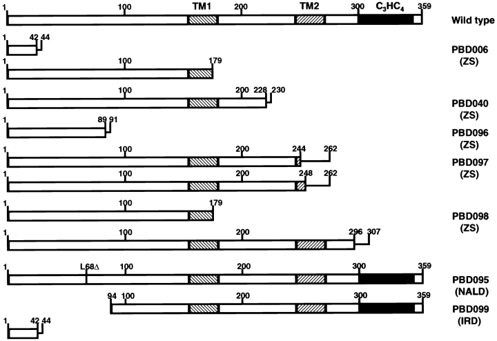
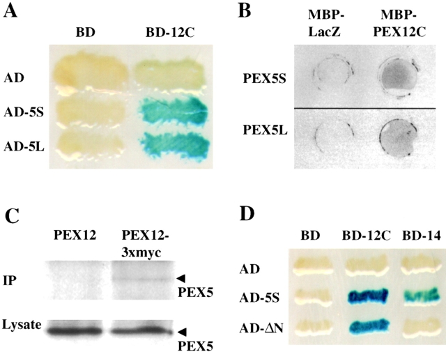
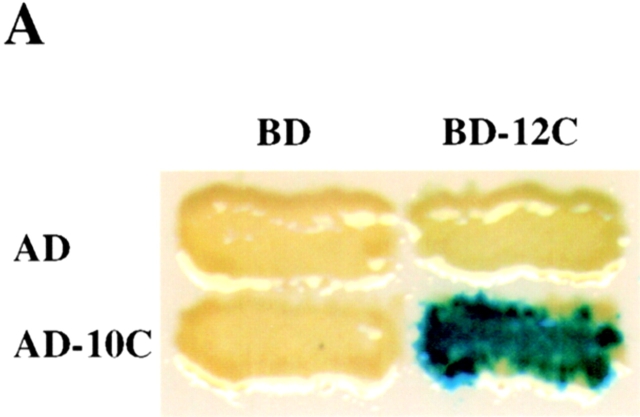
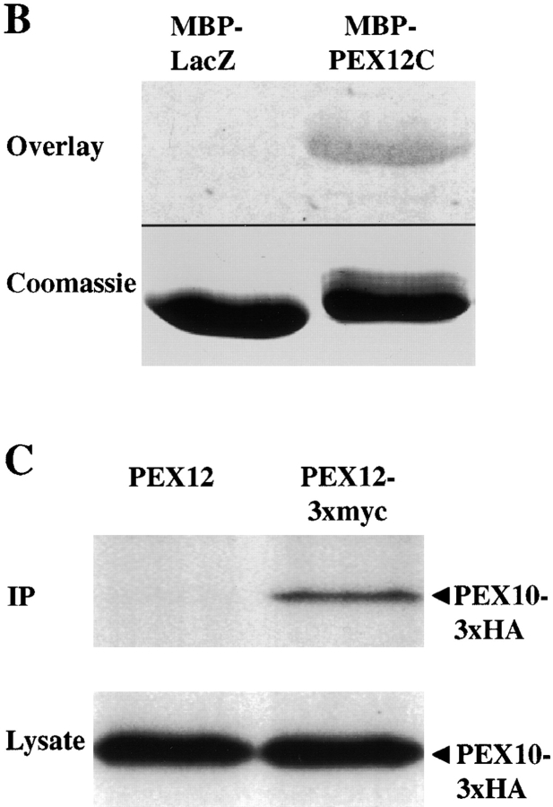
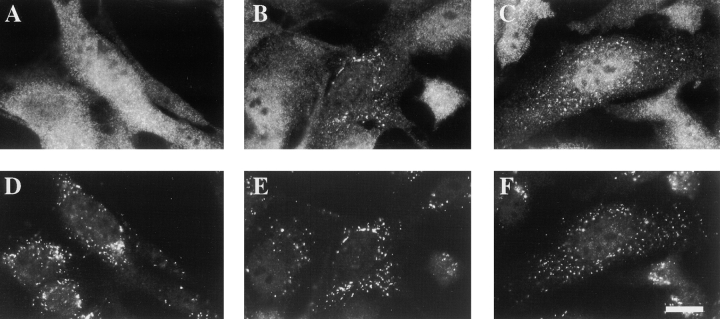
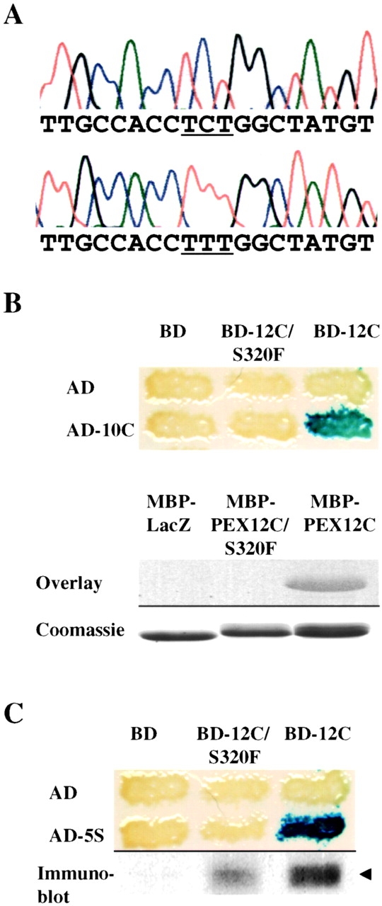
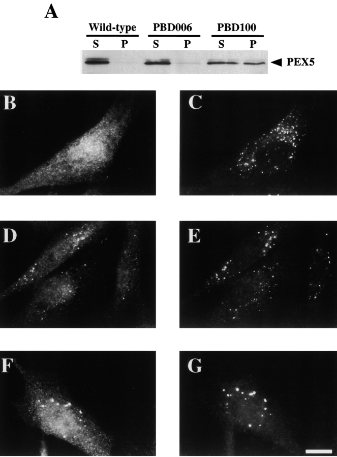
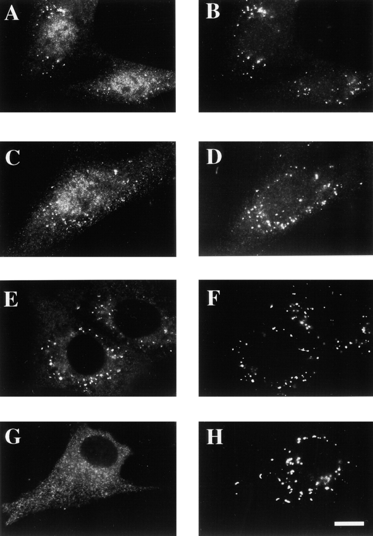
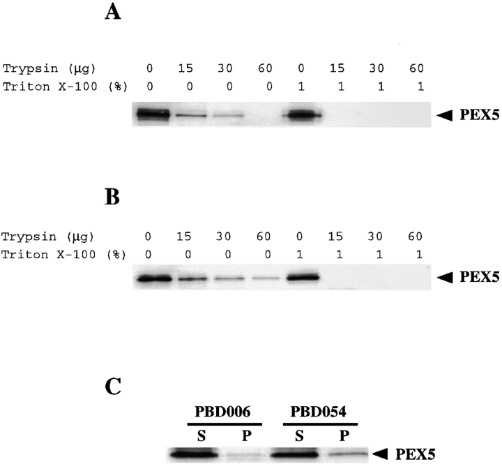
Peroxisomal matrix protein import requires PEX12, an integral peroxisomal membrane protein with a zinc ring domain at its carboxy terminus. Mutations in human PEX12 result in Zellweger syndrome, a lethal neurological disorder, and implicate the zinc ring domain in PEX12 function. Using two-hybrid studies, blot overlay assays, and coimmunoprecipitation experiments, we observed that the zinc-binding domain of PEX12 binds both PEX5, the PTS1 receptor, and PEX10, another integral peroxisomal membrane protein required for peroxisomal matrix protein import. Furthermore, we identified a patient with a missense mutation in the PEX12 zinc-binding domain, S320F, and observed that this mutation reduces the binding of PEX12 to PEX5 and PEX10. Overexpression of either PEX5 or PEX10 can suppress this PEX12 mutation, providing genetic evidence that these interactions are biologically relevant. PEX5 is a predominantly cytoplasmic protein and previous PEX5-binding proteins have been implicated in docking PEX5 to the peroxisome surface. However, we find that loss of PEX12 or PEX10 does not reduce the association of PEX5 with peroxisomes, demonstrating that these peroxins are not required for receptor docking. These and other results lead us to propose that PEX12 and PEX10 play direct roles in peroxisomal matrix protein import downstream of the receptor docking event.


Figures

Figure 1
Figure 1
The deduced PEX12 products of seven PBD patients. The diagram shows the predicted protein product of each PEX12 allele. The zinc ring domain is indicated by a black box and each of two transmembrane domains is indicated by the cross-hatched boxes. Straight lines show the length of additional amino acids that are appended as a result of frameshifting mutations. ZS, Zellweger syndrome; NALD, neonatal adrenoleukodystrophy; and IRD, infantile Refsum disease.
Figure 2
Figure 2
PEX12 interacts with PEX5. (A) Results of two-hybrid studies between PEX12 and PEX5. Two-hybrid reporter strains expressing the indicated fusion proteins were transferred to a nitrocellulose filter, submerged in liquid nitrogen to lyse the cells, and assayed for β-galactosidase activity. AD, GAL4 activation domain; and BD, GAL4-binding domain. (B) Filter binding experiments with PEX12 and PEX5. Equal amounts of MBP-LacZα and MBP-PEX12C were spotted on membranes and probed with [35S]PEX5S (upper panel) or [35S]PEX5L (lower panel). (C) PEX5 coimmunoprecipitates with PEX12/3xmyc. Lysates were prepared from fibroblasts that had been transfected with either pcDNA3-PEX12 or pcDNA3-PEX12/3xmyc. After immunoprecipitation with anti–myc antibodies, the immunoprecipitates were analyzed by immunoblot with anti-PEX5 antibodies (upper panel). In addition, equal amounts of the crude lysate before immunoprecipitation were assayed for PEX5 levels by immunoblot (lower panel). (D) PEX12 interacts with the PTS1-binding domain of PEX5. Two-hybrid reporter strains expressing the indicated fusion proteins were transferred to a nitrocellulose filter, submerged in liquid nitrogen to lyse the cells, and assayed for β-galactosidase activity.
Figure 3
Figure 3
The COOH-terminal domain of PEX12 interacts with the COOH-terminal domain of PEX10 in the yeast two-hybrid system. Two-hybrid reporter strains expressing the indicated fusion proteins were transferred to a nitrocellulose filter, submerged in liquid nitrogen to lyse the cells, and assayed for β-galactosidase activity. AD, GAL4 activation domain; and BD, GAL4-binding domain. (B) PEX10 binds immobilized PEX12C in vitro. 10 μg of purified MBP-LacZα and purified MBP-PEX12C were resolved by SDS-PAGE, transferred to membranes, and probed with 35S-labeled PEX10 (upper panel). A duplicate gel was stained before transfer with Coomassie blue (lower panel). (C) Coimmunoprecipitation of PEX10 with PEX12. Lysates from cells cotransfected with either pcDNA3-PEX12 and pcDNA3-PEX10/3xHA (left lane) or pcDNA3-PEX12/3xmyc and pcDNA3-PEX10/3xHA (right lane) were immunoprecipitated with anti–myc antibodies and analyzed by immunoblot with anti–HA antibodies. Aliquots of the crude lysates before immunoprecipitation were also assayed for PEX10/3xHA levels by immunoblot.
Figure 3
Figure 3
The COOH-terminal domain of PEX12 interacts with the COOH-terminal domain of PEX10 in the yeast two-hybrid system. Two-hybrid reporter strains expressing the indicated fusion proteins were transferred to a nitrocellulose filter, submerged in liquid nitrogen to lyse the cells, and assayed for β-galactosidase activity. AD, GAL4 activation domain; and BD, GAL4-binding domain. (B) PEX10 binds immobilized PEX12C in vitro. 10 μg of purified MBP-LacZα and purified MBP-PEX12C were resolved by SDS-PAGE, transferred to membranes, and probed with 35S-labeled PEX10 (upper panel). A duplicate gel was stained before transfer with Coomassie blue (lower panel). (C) Coimmunoprecipitation of PEX10 with PEX12. Lysates from cells cotransfected with either pcDNA3-PEX12 and pcDNA3-PEX10/3xHA (left lane) or pcDNA3-PEX12/3xmyc and pcDNA3-PEX10/3xHA (right lane) were immunoprecipitated with anti–myc antibodies and analyzed by immunoblot with anti–HA antibodies. Aliquots of the crude lysates before immunoprecipitation were also assayed for PEX10/3xHA levels by immunoblot.
Figure 4
Figure 4
Peroxisomal protein import in PBD054 cells is restored by expression of PEX10 or PEX12. PBD054 cells were transfected with pcDNA3 (A and D), pcDNA3-PEX10 (B and E), or pcDNA3-PEX12 (C and F). 2 d after transfection, the cells were fixed, permeabilized with Triton X-100, and processed for double indirect immunofluorescence using anticatalase antibodies (A–C) and anti–PMP70 antibodies (D–F). Bar, 25 μm.
Figure 5
Figure 5
PBD054 is mutated in PEX12. (A) Sequence chromatographs of PEX12 genomic DNA showing the wild-type sequence (top) and PBD054 sequence (bottom) in the region corresponding to nucleotides 950–968 of the PEX12 ORF. Note the C to T transition mutation, S320F, which changes the serine codon TCT to the phenylalanine codon TTT (underlined). (B) The S320F mutation attenuates the interaction between PEX12 and PEX10. Two-hybrid reporter strains expressing the indicated fusion proteins were assayed for β-galactosidase activity (top). To assess the effects of this mutation by blot overlay assay, purified MBP-LacZα, MBP-PEX12C/S320F, and MBP-PEX12C were separated by SDS-PAGE, transferred to membranes, and probed with 35S-labeled PEX10 (upper half of lower panel). These proteins were also separated by SDS-PAGE and stained with Coomassie blue (lower half of lower panel). (C) The S320F mutation attenuates the PEX12-PEX5 interaction in the yeast two-hybrid assay. Two-hybrid reporter strains expressing the listed proteins were assayed for β-galactosidase activity (top). The three strains expressing AD-5S were also lysed and assayed for BD-PEX12 levels by immunoblot with anti–PEX12 antibodies.
Figure 6
Figure 6
PEX12 and PEX10 are not required for PEX5 docking. (A) Postnuclear supernatants were separated into cytosolic supernatants (S) and organelle pellets (P). Equal proportions of the two fractions from each cell line were assayed for PEX5 levels by immunoblot. Immunofluorescence experiments were performed with wild-type (B and C), PEX12-deficient PBD006 (D and E), and PEX10-deficient PBD100 (F and G) cells using anti–PEX5 antibodies (B, D, and F) and anti–PMP70 antibodies (C, E, and G). Bar, 25 μm.
Figure 7
Figure 7
Distribution of PEX5 in PBD006 and PBD054 cells. PBD006 (A, B, E, and F) and PBD054 (C, D, G, and H) cells were fixed, permeabilized with either Triton X-100 (A–D) or digitonin (E–H), and processed for double indirect immunofluorescence using anti–PEX5 antibodies (A, C, E, and G) and anti–PMP70 antibodies (B, D, F, and H). Bar, 25 μm.
Figure 8
Figure 8
Protease protection analysis of peroxisomal PEX5 in PBD006 and PBD054 cells. Organelle fractions were prepared from PBD006 (A) and PBD054 (B) cells and incubated with protease in the absence or presence of detergent, and then assayed for PEX5 by immunoblot binding. (C) Postnuclear supernatants from PBD006 and PBD054 cells were separated into cytosolic supernatants (S) and organelle pellets (P). Equal proportions of each were assayed for PEX5 levels by immunoblot.

Similar articles

Cited by

References

    1. Adams A., Gottschling D.E., Kaiser C.A., Stearns T. Methods in Yeast Genetics 1997. Cold Spring Harbor Laboratory Press; Cold Spring Harbor: pp. 115–116
    1. Albertini M., Rehling P., Erdmann R., Girzalsky W., Kiel J.A., Veenhuis M., Kunau W.-H. Pex14p, a peroxisomal membrane protein binding both receptors of the two PTS-dependent import pathways. Cell. 1997;89:83–92 . - PubMed
    1. Baes M., Gressens P., Baumgart E., Carmeliet P., Casteels M., Fransen M., Evrard P., Fahimi D., Declercq P.E., Collen D., van Veldhoven P.P., Mannaerts G.P. A mouse model for Zellweger syndrome. Nat. Genet. 1997;17:49–57 . - PubMed
    1. Borden K.L. RING fingers and B-boxeszinc-binding protein-protein interaction domains. Biochem. Cell Biol. 1998;76:351–358 . - PubMed
    1. Braverman N., Dodt G., Gould S.J., Valle D. Disorders of peroxisome biogenesis. Hum. Mol. Genet. 1995;4:1791–1798 . - PubMed

Publication types

MeSH terms