. 1999 Nov 15;147(4):791-808.
doi: 10.1083/jcb.147.4.791.

The COOH-terminal domain of Myo2p, a yeast myosin V, has a direct role in secretory vesicle targeting

Affiliations

The COOH-terminal domain of Myo2p, a yeast myosin V, has a direct role in secretory vesicle targeting

D Schott et al. J Cell Biol. .

Abstract

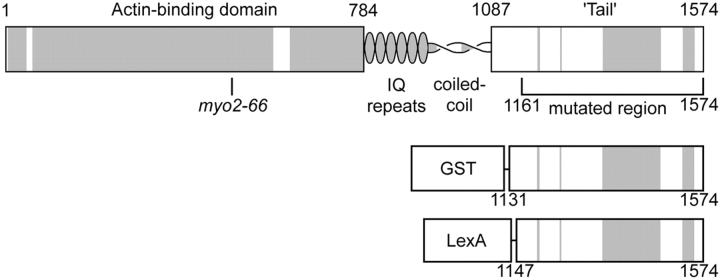
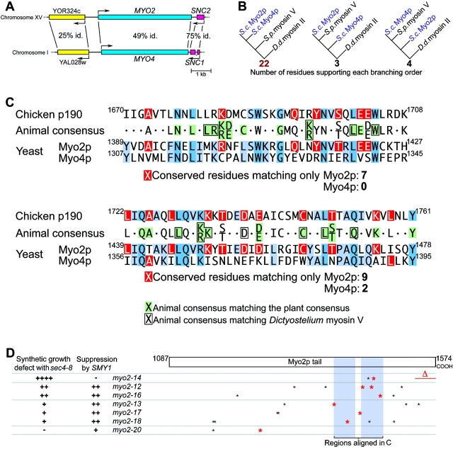
MYO2 encodes a type V myosin heavy chain needed for the targeting of vacuoles and secretory vesicles to the growing bud of yeast. Here we describe new myo2 alleles containing conditional lethal mutations in the COOH-terminal tail domain. Within 5 min of shifting to the restrictive temperature, the polarized distribution of secretory vesicles is abolished without affecting the distribution of actin or the mutant Myo2p, showing that the tail has a direct role in vesicle targeting. We also show that the actin cable-dependent translocation of Myo2p to growth sites does not require secretory vesicle cargo. Although a fusion protein containing the Myo2p tail also concentrates at growth sites, this accumulation depends on the polarized delivery of secretory vesicles, implying that the Myo2p tail binds to secretory vesicles. Most of the new mutations alter a region of the Myo2p tail conserved with vertebrate myosin Vs but divergent from Myo4p, the myosin V involved in mRNA transport, and genetic data suggest that the tail interacts with Smy1p, a kinesin homologue, and Sec4p, a vesicle-associated Rab protein. The data support a model in which the Myo2p tail tethers secretory vesicles, and the motor transports them down polarized actin cables to the site of exocytosis.


Figures

Figure 1
Figure 1
Myo2p. Predicted domain structure showing the region mutagenized and the fusion constructs used. Shaded regions are conserved between Myo2p and nonfungal myosin Vs.
Figure 3
Figure 3
Myo2p and Sec4p distributions in tpm1-2 tpm2Δ MYO2+ (ABY988) and tpm1-2 tpm2Δ myo2-16 (ABY1128) yeast grown at 25°C (upper panels), shifted to 34.5°C for 10 min (middle panels), and then shifted back to 25°C for 2 min (lower panels). Percentages of small-budded cells showing a polarized distribution are shown.
Figure 2
Figure 2
Effect of myo2 mutations on the distribution of Sec4p and Myo2p. (A and B) ABY551 (MYO2+), ABY553 (myo2-16), and ABY555 (myo2-66), grown in SD at 25°C, and shifted from 25 to 35°C for 5 min. (A) Sec4p staining, with percentages of small buds showing a polarized distribution. (B) Myo2p staining, with percentages ± SEM, n = 2 experiments. (C) Myo2-66p is not rapidly degraded at the restrictive temperature. Western blot of ABY551 and ABY555 lysates. (D) Sec4p and Myo2p staining of ABY531 (MYO2) and ABY532 (myo2-12). Bars, 5 μm.
Figure 4
Figure 4
(A) Act1p (actin) immunofluorescence of ABY551 (MYO2+), ABY553 (myo2-16), and ABY555 (myo2-66) grown in SD at 25°C, shifted from 25 to 35°C for 5 min, or shifted for 90 min. Bar, 5 μm. (B and C) Yeast double-labeled for F-actin with rhodamine-phalloidin and for Myo2p. Bars, 1 μm. (B) Myo2p is concentrated where actin cables converge in ABY537 (myo2-17), ABY538 (myo2-18), and ABY530 (myo2-20), shifted to 37°C for 75 min. Images have been aligned using other cells in the same field and the faint cytoplasmic background in both fluorescence channels. Arrows mark the point of brightest Myo2p staining. (C) Gallery of selected wild-type cells in which the images have been overexposed to make faint structures more visible. Long arrows indicate faint structures that appear to contain both Myo2p and F-actin. Diagonal arrows in the lower right panel indicate bright Myo2p and F-actin structures that do not coincide.
Figure 5
Figure 5
(A) Expression of Myo2p tail fusion protein is lethal. Three transformants each of ABY531 with pEG (GSTp), pEGM (GST–Myo2p tail), pCL (LexAp), or pCLM (LexA–Myo2p tail) were streaked onto raffinose or raffinose plus 0.2 or 2% galactose. (B) myo2 tail mutants are hypersensitive to expression of tail fusion protein. ABY531 (MYO2), ABY532 (myo2-12), ABY533 (myo2-13), ABY534 (myo2-14), ABY536 (myo2-16), ABY537 (myo2-17), ABY538 (myo2-18), and ABY530 (myo2-20) bearing pEGM (GST–Myo2p tail) or pEG (GSTp) were streaked onto raffinose medium containing or lacking leucine and incubated at 30°C. Media lacking leucine select for cells with high plasmid copy number. Results are identical for at least four independent transformants for each combination of strain and plasmid.
Figure 6
Figure 6
The polarized distribution of LexA–Myo2p tail requires functional endogenous MYO2. (A) Immunofluorescence of LexA in ABY551 (MYO2) and ABY553 (myo2-16) bearing pCLM, grown in raffinose medium with 2% galactose added for 60 min at 25°C, then shifted to 34.5°C for 5 min. Arrows indicate small buds lacking LexA staining. Images overrepresent the proportion of polarized cells severalfold. Bar, 5 μm. (B) Percentages of cells showing polarized LexA staining. ABY551 (MYO2+) and ABY553 (myo2-16) bearing pCLM or pCL were grown in raffinose medium, then supplemented with 2% galactose for 90 min at 20°C or for 85 min at 20°C, then 34.5°C for 5 min. For each condition, 200 random cells in four separate trials were scored for staining concentrated at presumptive growth sites. After shift to 34.5°C, myo2-16 cells are less often polarized for LexAp staining than wild-type (P ≤ 0.05, two-tailed t test).
Figure 7
Figure 7
The polarized distribution of LexA–Myo2p tail requires functional SEC2. (A) LexAp, Myo2p, and Sec4p in NY13 (wt, upper panels), NY130 (sec2-41, middle panels), and NY3 (sec1-1, lower panels) bearing pCLM, grown in raffinose, then supplemented with 2% galactose for 60 min at 28°C and then shifted to 35°C for 5 min. Bar, 5 μm. (B) Percentages of cells showing polarized LexAp staining. After shift to 35°C, sec2-41 cells are less often polarized for LexAp staining than wild-type (P ≤ 0.05, two-tailed t test), whereas sec1-1 cells are more often polarized for LexAp staining than wild-type (P ≤ 0.05).
Figure 8
Figure 8
Myo2p has features conserved with animal myosin Vs but lost from Myo4p. (A) Map of genomic regions near MYO2 and MYO4. Percentage of amino acid identity is shown (P ≤ 10−21 for all three BLASTP 2.0 scores). (B) Evolutionary relationship among the motor domains of Myo2p, Myo4p, and their closest relative, Schizosaccharomyces myosin V (CAA22641), using Dictyostelium myosin II as an outgroup. 22/29 corresponds to P2) ≤ 10−6 and a bootstrap value of 99.7%. (C) Comparison of unambiguously alignable regions of p190, Myo2p, and Myo4p, showing consensus sequence for animal myosinVs, and matches of the animal consensus with the plant consensus (green) and Dictyostelium myosin V (boxed) sequences. Residues in the consensus sequence matching only Myo2p or only Myo4p are shown in red and other matches among the sequences are shown in blue. The animal consensus represents residues that match in at least six of the seven animal myosin Vs. (D) Summary of genetic interactions and positions of myo2ts mutations relative to the regions aligned in C. Nonconservative mutations in residues that are conserved with animals are shown in red.

References

    1. Adams A.E.M., Pringle J.R. Relationship of actin and tubulin distribution to bud growth in wild-type and morphogenetic-mutant Saccharomyces cerevisiae . J. Cell Biol. 1984;98:934–945 . - PMC - PubMed
    1. Altschul S.F., Madden T.M., Schäffer A.A., Zhang J., Zhang Z., Miller W., Lipman D.J. Gapped BLAST and PSI-BLASTa new generation of protein database search programs. Nucleic Acids Res. 1997;25:3389–3402 . - PMC - PubMed
    1. Ausubel, F.M., R. Brent, R.E. Kingston, D.D. Moore, J.G. Seidman, J.A. Smith, and K. Struhl, editors. 1996. Current Protocols in Molecular Biology. John Wiley & Sons, Inc., New York. 1,906 pp.
    1. Ayscough K.R., Stryker J., Pokala N., Sanders M., Crews P., Drubin D.G. High rates of actin filament turnover in budding yeast and roles for actin in establishment and maintenance of cell polarity revealed using the actin inhibitor latrunculin-A. J. Cell Biol. 1997;137:399–416 . - PMC - PubMed
    1. Bailey, T., and C. Elkan. 1994. Fitting a mixture model by expectation maximization to discover motifs in biopolymers. Proceedings of the Second International Conference on Intelligent Systems for Molecular Biology. AAAI Press, Menlo Park, CA. 28–36. - PubMed

Publication types

MeSH terms