. 1999 Nov 15;147(4):809-22.
doi: 10.1083/jcb.147.4.809.

The pro-apoptotic proteins, Bid and Bax, cause a limited permeabilization of the mitochondrial outer membrane that is enhanced by cytosol

Affiliations

The pro-apoptotic proteins, Bid and Bax, cause a limited permeabilization of the mitochondrial outer membrane that is enhanced by cytosol

R M Kluck et al. J Cell Biol. .

Abstract

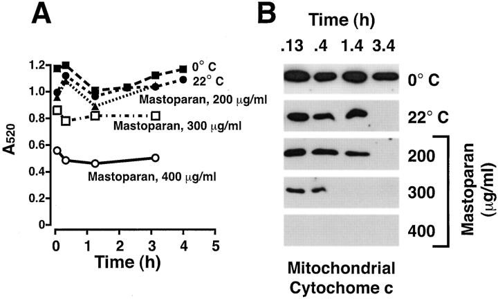
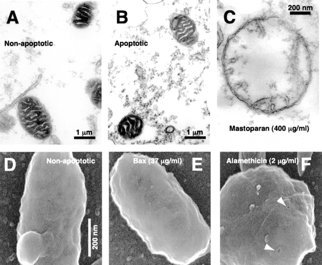
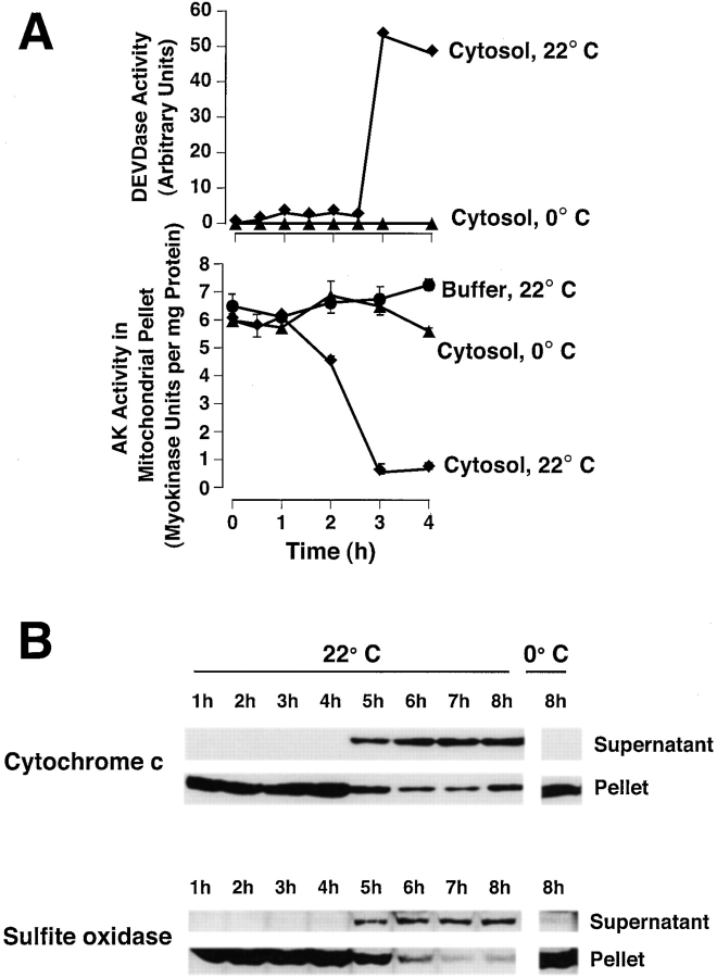
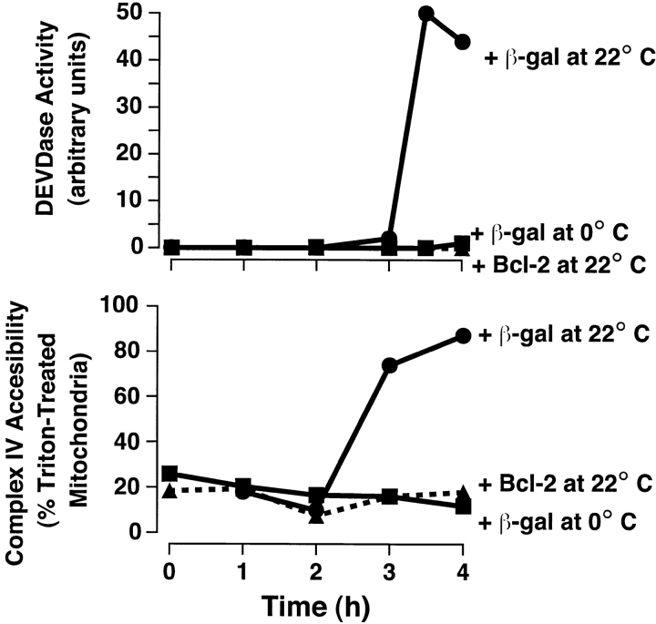
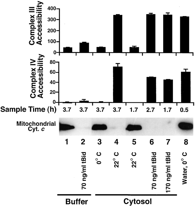
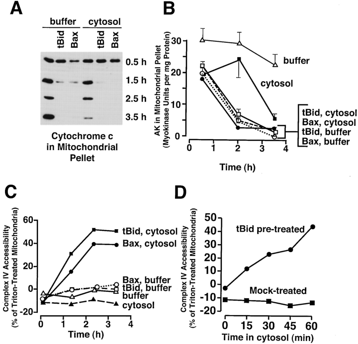
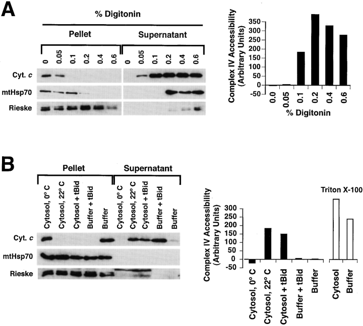
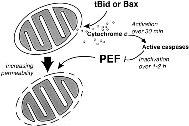
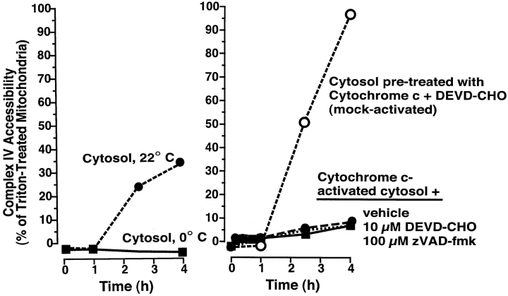
During apoptosis, an important pathway leading to caspase activation involves the release of cytochrome c from the intermembrane space of mitochondria. Using a cell-free system based on Xenopus egg extracts, we examined changes in the outer mitochondrial membrane accompanying cytochrome c efflux. The pro-apoptotic proteins, Bid and Bax, as well as factors present in Xenopus egg cytosol, each induced cytochrome c release when incubated with isolated mitochondria. These factors caused a permeabilization of the outer membrane that allowed the corelease of multiple intermembrane space proteins: cytochrome c, adenylate kinase and sulfite oxidase. The efflux process is thus nonspecific. None of the cytochrome c-releasing factors caused detectable mitochondrial swelling, arguing that matrix swelling is not required for outer membrane permeability in this system. Bid and Bax caused complete release of cytochrome c but only a limited permeabilization of the outer membrane, as measured by the accessibility of inner membrane-associated respiratory complexes III and IV to exogenously added cytochrome c. However, outer membrane permeability was strikingly increased by a macromolecular cytosolic factor, termed PEF (permeability enhancing factor). We hypothesize that PEF activity could help determine whether cells can recover from mitochondrial cytochrome c release.


Figures

Figure 1
Figure 1
Absence of gross mitochondrial swelling with apoptotic cytochrome c release as indicated by light scatter. (A) Xenopus egg mitochondria were incubated in Xenopus egg cytosol at either 0°C or 22°C for 4 h. As positive controls for swelling, mastoparan was added at 200, 300, and 400 μg/ml. At the times indicated, light scatter of mitochondria was assayed by diluting 40 μl of extract with 260 μl buffer B and measuring the average A520 over 1 min. (B) Mitochondria were recovered from the samples measured in A and assessed for cytochrome c translocation by Western blot.
Figure 2
Figure 2
Apoptotic mitochondria exhibit normal size and morphology on electron microscopy. (A–C) Crude extracts were incubated at 0°C as a non-apoptotic control (A) or incubated at 22°C to induce spontaneous cytochrome c release (B). Immediately after cytochrome c release in the sample taken at 2.75 h, aliquots were fixed and examined by transmission electron microscopy. (C) A swollen mitochondrion taken from a further sample treated with mastoparan (400 μg/ml). (D–F) FEISEM of reconstituted extracts incubated at 0°C for 3 h, 40 min (D, non-apoptotic) or 22°C in the presence of 35 μg/ml BaxΔTM for 1 h, 40 min (E). (F) Alamethicin addition (2 μg/ml, 40 min) caused both cytochrome c release and the formation of 10–13-nm pores (arrows). Bar, (D–F) 200 nm.
Figure 3
Figure 3
Adenylate kinase and sulfite oxidase loss from mitochondria, like loss of cytochrome c, precedes DEVDase activation. Xenopus mitochondria were incubated in either Xenopus cytosol or buffer, at either 0 or 22°C for 4 h. At the times indicated, 100-μl aliquots were centrifuged and the mitochondrial pellets washed twice, lysed in 1% Triton X-100, then assayed for adenylate kinase activity (A). Data are means and SD of triplicates. (B) At the times indicated, aliquots (2 μl) were assessed for DEVDase activity to determine the time of cytochrome c release. Data are representative of 6 experiments. (C) Rat liver mitochondria (5 mg/ml) were incubated in Xenopus egg cytosol at either 0 or 22°C for 8 h. At the indicated times, aliquots were assessed for cytochrome c and sulfite oxidase translocation by Western blot. Data are representative of three experiments.
Figure 4
Figure 4
Outer mitochondrial membrane permeability to cytochrome c is bidirectional in the Xenopus cell-free system. Xenopus mitochondria were incubated in cytosol at 0 or 22°C for 4 h in the presence of Sf-9 cell lysates (2% vol/vol) containing either β-galactosidase or Bcl-2 expressed after baculovirus infection. At the times indicated, 2-μl aliquots were assayed for either DEVDase activity (top) or complex IV accessibility (bottom).
Figure 6
Figure 6
Cytosol (PEF) increases both complex III and IV accessibility after tBid-induced cytochrome c release. Mitochondria were incubated with or without tBid in either buffer B or cytosol, at 0 or 22°C. At suitable times (1.7, 2.7, or 3.7 h), mitochondria were pelleted and analyzed by Western blot for cytochrome c translocation (bottom) or washed and resuspended in buffer E for measurement of complex III (top) and IV (middle) accessibility (arbitrary units). For comparison between assays, mitochondria were lysed by incubation in water for 30 min on ice. These osmotically lysed mitochondria retain their cytochrome c due to the low salt content (see Materials and Methods). Data are mean and SD for duplicate measurements and are representative of three experiments. Note: The time course of permeabilization by PEF is slower in Fig. 6 than Fig. 5; this merely reflects a normal variation between Xenopus extracts in time of onset of apoptotic changes ( Newmeyer et al. 1994; Farschon et al. 1997).
Figure 5
Figure 5
In the absence of cytosol, Bid and Bax release cytochrome c and adenylate kinase, but do not develop high complex IV accessibility. Xenopus egg mitochondria were incubated in buffer B or cytosol alone, or with added tBid (170 ng/ml) or Bax (37 μg/ml) for 4 h at 22°C. At the indicated times, aliquots were measured for (A) mitochondrial cytochrome c content, (B) mitochondrial adenylate kinase activity, and (C) complex IV accessibility. Data are single measurements except those for adenylate kinase (mean and SD of triplicates), and are representative of two experiments. (D) PEF acts subsequent to tBid, and tBid effects on the outer membrane are sustained. Xenopus mitochondria were first incubated at 22°C in buffer B with or without tBid (170 ng/ml) for 2 h to allow tBid to cause complete cytochrome c release (not shown). Mitochondria were then washed in buffer B and resuspended in fresh cytosol, and aliquots were tested every 15 min for complex IV accessibility.
Figure 7
Figure 7
tBid and PEF preserve the integrity of the mitochondrial matrix compartment, as measured by retention of a soluble mitochondrial matrix protein, mtHsp70. (A) mtHsp70 is released when mitochondria are treated with intermediate concentrations of digitonin. Xenopus egg mitochondria were treated with increasing concentrations of digitonin to selectively permeabilize the outer, then the inner mitochondrial membranes. After two hours incubation, the contents of the pellet and supernatant fractions were analyzed by immunoblotting (left panel) with antibodies to cytochrome c, mtHsp70, and the inner membrane-associated Rieske protein (part of complex III). Also, accessibility of complex IV was measured as described above (right panel). Note that mtHsp70 is released by digitonin at 0.2%, a concentration insufficient to solubilize the Rieske protein, but more than sufficient to release cytochrome c. (B) mtHsp70 is not released by tBid and PEF, despite full release of cytochrome c and full accessibility of complex IV. Xenopus mitochondria were treated with 170 ng/ml tBid, in either the presence or absence of cytosol, and either at 0 or 22°C, as indicated. DEVD-CHO (10 μM) was added to cytosol incubations to minimize PEF degradation and thus maximize its activity. After 3 h of incubation, the contents of the mitochondrial pellet were analyzed by immunoblotting with antibodies to cytochrome c, mtHsp70 and Rieske protein (left), and the accessibility of complex IV (right). The samples were taken well after full cytochrome c release from mitochondria, which occurred in this experiment at 1 h in the tBid-treated sample and at 2 h in cytosol at 22°C (not shown). Note that mtHsp70 is retained in the pellet fraction despite full cytochrome c release and increased complex IV accessibility (reflecting PEF activity). (The bands stained by anti-Rieske antibody in cytosol are nonmitochondrial cross-reacting proteins.)
Figure 8
Figure 8
PEF is large and proteinaceous. Mitochondria were incubated with tBid (170 ng/ml) in the presence of either buffer B, ultra- or micro-filtered cytosol (10-, 50-, 300-kD, and 0.22-μm filters) normal cytosol, cytosol diluted with buffer B, proteinase K–treated cytosol, or dialyzed cytosol. The control (first sample) did not contain tBid. Ac-DEVD-CHO (10 μM) was added to all samples to maximize PEF activity (see Fig. 9). At the indicated times, aliquots were either pelleted and analyzed for mitochondrial cytochrome c content (bottom) or tested for complex IV accessibility (top).
Figure 10
Figure 10
Model of two types of mitochondrial outer membrane permeability observed in Xenopus extracts during apoptosis. Mitochondria treated with tBid or Bax in buffer undergo efflux of endogenous cytochrome c (12 pmol/μl mitochondria) across the outer membrane, with limited permeability to exogenous cytochrome c. Additional exposure to cytosol (PEF) allows high permeability to exogenous cytochrome c (60 nmol/μl mitochondria per min, as measured by the rate of the complex IV reaction). Thus, this increased exchange of endogenous cytochrome c is at least ∼5,000 times the rate of cytochrome c efflux after tBid or Bax treatment, assuming that all of the mitochondrial cytochrome c is released over 1 min. PEF begins to act immediately after tBid–induced permeabilization, but is inactivated over 1–2 h by caspase activation. The inner membrane appears unaffected.
Figure 9
Figure 9
PEF is inactivated by caspases. Mitochondria were incubated with either normal cytosol at 0 or 22°C (left), or with cytosol that had been activated by preincubation (3 h) with 0.5 μM horse heart cytochrome c (HHCc) (right). Samples in activated cytosol contained either vehicle (0.5% DMSO), Ac-DEVD-CHO (10 μM) or zVAD-fmk (100 μM). One sample (mock-activated), had Ac-DEVD-CHO (10 μM) added before incubation with HHCc. At the indicated times, aliquots (2 μl) were assessed for complex IV accessibility. Mitochondria in the three activated cytosol samples had lost their cytochrome c by 1 h due to tBid-like activity (not shown). Data are representative of three experiments.

Similar articles

Cited by

References

    1. Allen T.D., Goldberg M.W. High-resolution SEM in cell biology. Trends. Cell Biol. 1993;3:205–208 . - PubMed
    1. Allen T.D., Bennion G.R., Rutherford S.A., Reipert S., Ramalho A., Kiseleva E., Goldberg M.W. Accessing nuclear structure for field emission, in lens, scanning electron microscopy (FEISEM) Scanning Microsc. Suppl. 1996;10:149–163 . - PubMed
    1. Antonsson B., Conti F., Ciavatta A., Montessuit S., Lewis S., Martinou I., Bernasconi L., Bernard A., Mermod J.J., Mazzei G. Inhibition of Bax channel-forming activity by Bcl-2. Science. 1997;277:370–372 . - PubMed
    1. Barber M.J., Neame P.J. A conserved cysteine in molybdenum oxotransferases. J. Biol. Chem. 1990;265:20912–20915 . - PubMed
    1. Basanez G., Nechushtan A., Drozhinin O., Chanturiya A., Choe E., Tutt S., Wood K.A., Hsu Y., Zimmerberg J., Youle R.J. Bax, but not Bcl-xL, decreases the lifetime of planar phospholipid bilayer membranes at subnanomolar concentrations. Proc. Natl. Acad. Sci. USA. 1999;96:5492–5497 . - PMC - PubMed

Publication types

MeSH terms