. 1999 Nov 15;147(4):845-56.
doi: 10.1083/jcb.147.4.845.

PAK-family kinases regulate cell and actin polarization throughout the cell cycle of Saccharomyces cerevisiae

Affiliations

PAK-family kinases regulate cell and actin polarization throughout the cell cycle of Saccharomyces cerevisiae

S P Holly et al. J Cell Biol. .

Abstract

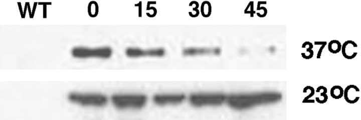
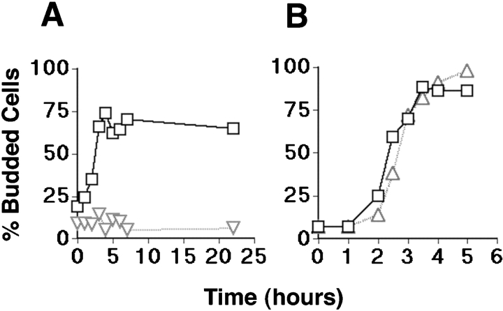
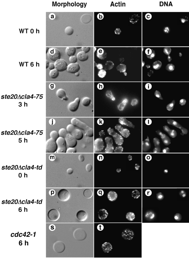
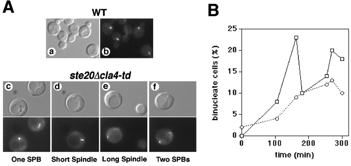
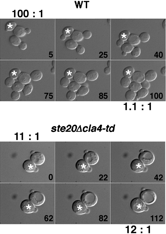
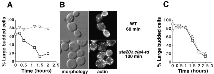
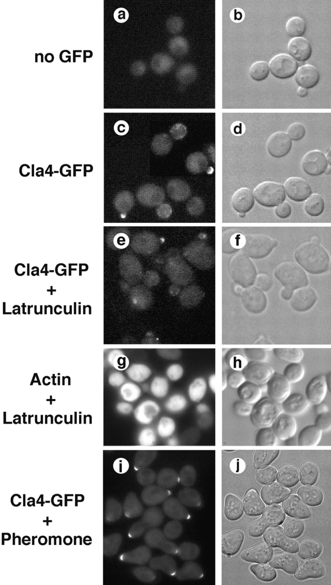
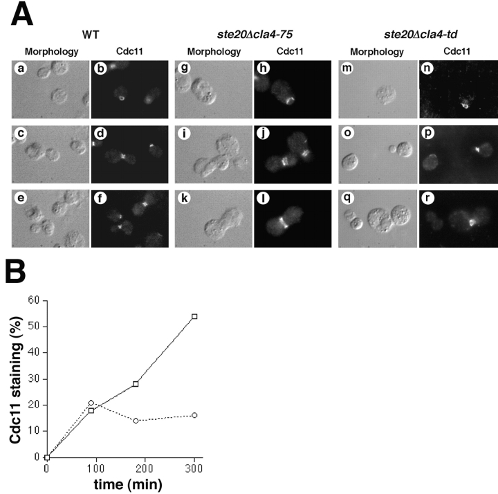
During the cell cycle of the yeast Saccharomyces cerevisiae, the actin cytoskeleton and cell surface growth are polarized, mediating bud emergence, bud growth, and cytokinesis. We have determined whether p21-activated kinase (PAK)-family kinases regulate cell and actin polarization at one or several points during the yeast cell cycle. Inactivation of the PAK homologues Ste20 and Cla4 at various points in the cell cycle resulted in loss of cell and actin cytoskeletal polarity, but not in depolymerization of F-actin. Loss of PAK function in G1 depolarized the cortical actin cytoskeleton and blocked bud emergence, but allowed isotropic growth and led to defects in septin assembly, indicating that PAKs are effectors of the Rho-guanosine triphosphatase Cdc42. PAK inactivation in S/G2 resulted in depolarized growth of the mother and bud and a loss of actin polarity. Loss of PAK function in mitosis caused a defect in cytokinesis and a failure to polarize the cortical actin cytoskeleton to the mother-bud neck. Cla4-green fluorescent protein localized to sites where the cortical actin cytoskeleton and cell surface growth are polarized, independently of an intact actin cytoskeleton. Thus, PAK family kinases are primary regulators of cell and actin cytoskeletal polarity throughout most or all of the yeast cell cycle. PAK-family kinases in higher organisms may have similar functions.


Figures

Figure 1
Figure 1
Temperature-dependent degradation of DHFR-Cla4. Cells expressing an HA epitope-tagged form of a DHFR-Cla4 fusion protein were grown at permissive temperature (23°C) and either maintained at permissive temperature or shifted to the nonpermissive temperature (37°C) for the indicated times. DHFR-HA-Cla4 was detected by immunoblotting.
Figure 2
Figure 2
Dependence of bud emergence on Ste20 and Cla4 function. Wild-type cells (W303), a ste20Δ cla4-75 mutant (KBY211), and a ste20Δ cla4-td mutant (KBY212) were synchronized in G1 at permissive temperature (23°C) by nutritional deprivation, shifted to nonpermissive temperature (37°C), and released from the nutritional block at nonpermissive temperature (see Materials and Methods). Bud emergence was scored microscopically and expressed as the fraction of the cell population that had resumed growth and budded. Cells with buds and wide necks were scored as budded cells. (A) Budding kinetics of wild-type cells (indicated by squares) and a ste20Δ cla4-td mutant (indicated by triangles), and (B) wild-type cells (indicated by squares) and a ste20Δ cla4-75 mutant (indicated by triangles) are shown.
Figure 3
Figure 3
Effects of Ste20 and Cla4 inactivation on actin cytoskeletal organization in G1. Cells were arrested in G1, shifted to the nonpermissive temperature, released from the nutritional block at the nonpermissive temperature, fixed at the indicated times, and stained with rhodamine-phalloidin and DAPI. Wild-type cells (a–f), a ste20Δ cla4-75 mutant (g–l), a ste20Δ cla4-td mutant (m–r), and a cdc42-1 mutant (s and t) were analyzed.
Figure 4
Figure 4
Effect of Ste20 and Cla4 inactivation on nuclear division. A shows spindle phenotypes in wild-type cells (a and b) and a ste20Δ cla4-td mutant (c–f) expressing GFP-tubulin (GFP-Tub1) after incubation at restrictive temperature for 6 h. Wild-type cells showed spindle phenotypes appropriate for various stages of the cell cycle. Unbudded ste20Δ cla4-td mutant cells displayed unduplicated spindle pole bodies (c) like unbudded wild-type cells, but more frequently displayed spindle phenotypes inappropriate for unbudded cells including short spindles (d), anaphase spindles (e), and postmitotic segregated spindle pole bodies (f), indicating that the nuclear division cycle continued in unbudded cells. B shows the kinetics of nuclear division after wild-type cells (indicated by squares) and a ste20Δ cla4-td mutant (indicated by circles) were released from a G1 block at nonpermissive temperature, as described in Fig. 2. Nuclear division was scored over time by staining cells with DAPI.
Figure 5
Figure 5
Loss of polarized bud growth in mutants lacking Ste20 and Cla4. Asynchronous wild-type (upper panels) or ste20Δ cla4-td (lower panels) cells were incubated 2 h at 37°C before mounting on an agarose pad containing synthetic media. The ratio of mother to bud volume for a typical mother-bud pair (labeled with an asterisk, calculated assuming a spherical shape) is shown. Wild-type cells (6/6 cells) grew in a polarized manner, since the bud grew relative to the mother, thereby decreasing the mother bud ratio, whereas the mother did not grow appreciably (7% in 2 h for the cell shown). However, in the ste20Δ cla4-td mutant, cell growth occurred isotropically (7/7 cells). Both the mother and bud grew (mother and bud grew 31% and 30%, respectively, in 110 min in the cells shown), thereby maintaining a constant mother to bud volume ratio.
Figure 6
Figure 6
Dependence of cell division on Ste20 and Cla4 function. Cells were arrested before mitosis by nocodazole treatment at permissive temperature (23°C), shifted to the nonpermissive temperature (37°C) for 1–2 h and released from the nocodazole block at the nonpermissive temperature. Cell division was scored microscopically by the disappearance of large budded cells over time. A shows results obtained using wild-type cells (indicated by squares) and a ste20Δ cla4-td mutant (indicated by triangles). B shows the morphology and actin distribution of the wild-type cells and the ste20Δ cla4-td mutant after release from the nocodazole block at the nonpermissive temperature. C shows results obtained using wild-type cells (indicated by squares) and a ste20Δ cla4-75 mutant (indicated by triangles).
Figure 8
Figure 8
Localization of Cla4-GFP to sites of polarized cell surface growth. Cla4-GFP expressed from the chromosomal CLA4 locus was localized by fluorescence microscopy. Cla4-GFP was detected at the apex of small buds (c), and in dots distributed over the cortex of larger buds (c); depolymerization of the actin cytoskeleton by treating cells with latrunculin (10 min) did not affect the localization of Cla4-GFP (e). Cla4-GFP localized to the tips of polarized cell-surface projections in cells treated 2 h with mating pheromone (α-factor) (i).
Figure 7
Figure 7
Effect of Ste20 and Cla4 inactivation on septin stability and assembly. A shows the effects on septin stability. Asynchronous cultures of wild-type cells (a–f) and a ste20Δ cla4-75 mutant (g–l) were incubated at the nonpermissive temperature for 6 h; the ste20Δ cla4-td mutant (m–r) was incubated 2 h at the nonpermissive temperature. Cells were fixed and stained with anti-Cdc11 polyclonal antibodies and an FITC-labeled secondary antibody. Cells at different stages of the cell cycle are shown. B shows the effects on septin assembly. The kinetics of septin assembly in wild-type cells and the ste20Δ cla4-td mutant were determined after release from a G1 block at nonpermissive temperature. The proportion of the cell population that stained with anti-Cdc11 polyclonal antibodies and an FITC-labeled secondary antibody was scored.

Similar articles

Cited by

References

    1. Abo A., Qu J., Cammarano M.S., Dan C., Fritsch A., Baud V., Belisle B., Minden A. PAK4, a novel effector for Cdc42Hs, is implicated in the reorganization of the actin cytoskeleton and in the formation of filopodia. EMBO (Eur. Mol. Biol. Organ.) J. 1998;17:6527–6540 . - PMC - PubMed
    1. Adams A.E., Johnson D.I., Longnecker R.M., Sloat B.F., Pringle J.R. CDC42 and CDC43, two additional genes involved in budding and the establishment of cell polarity in the yeast Saccharomyces cerevisiae . J. Cell Biol. 1990;111:131–142 . - PMC - PubMed
    1. Amatruda J.F., Gattermeir D.J., Karpova T.S., Cooper J.A. Effects of null mutations and overexpression of capping protein on morphogenesis, actin distribution, and polarized secretion in yeast. J. Cell Biol. 1992;119:1151–1162 . - PMC - PubMed
    1. Ayscough K.R., Stryker J., Pokala N., Sanders M., Crews P., Drubin D.G. High rates of actin filament turnover in budding yeast and roles for actin in establishment and maintenance of cell polarity revealed using the actin inhibitor latrunculin-A. J. Cell Biol. 1997;137:399–416 . - PMC - PubMed
    1. Barral Y., Parra M., Bidlingmaier S., Snyder M. Nim1-related kinases coordinate cell cycle progression with the organization of the peripheral cytoskeleton in yeast. Genes Dev. 1999;13:176–187 . - PMC - PubMed

Publication types

MeSH terms

Substances