. 1999 Nov 15;147(4):879-90.
doi: 10.1083/jcb.147.4.879.

Yes-associated protein 65 localizes p62(c-Yes) to the apical compartment of airway epithelia by association with EBP50

Affiliations

Yes-associated protein 65 localizes p62(c-Yes) to the apical compartment of airway epithelia by association with EBP50

P J Mohler et al. J Cell Biol. .

Abstract

We recently showed that the COOH terminus of the cystic fibrosis transmembrane conductance regulator associates with the submembranous scaffolding protein EBP50 (ERM-binding phosphoprotein 50 kD; also called Na(+)/H(+) exchanger regulatory factor). Since EBP50 associates with ezrin, this interaction links the cystic fibrosis transmembrane conductance regulator (CFTR) to the cortical actin cytoskeleton. EBP50 has two PDZ domains, and CFTR binds with high affinity to the first PDZ domain. Here, we report that Yes-associated protein 65 (YAP65) binds with high affinity to the second EBP50 PDZ domain. YAP65 is concentrated at the apical membrane in airway epithelia and interacts with EBP50 in cells. The COOH terminus of YAP65 is necessary and sufficient to mediate association with EBP50. The EBP50-YAP65 interaction is involved in the compartmentalization of YAP65 at the apical membrane since mutant YAP65 proteins lacking the EBP50 interaction motif are mislocalized when expressed in airway epithelial cells. In addition, we show that the nonreceptor tyrosine kinase c-Yes is contained within EBP50 protein complexes by association with YAP65. Subapical EBP50 protein complexes, containing the nonreceptor tyrosine kinase c-Yes, may regulate apical signal transduction pathways leading to changes in ion transport, cytoskeletal organization, or gene expression in epithelial cells.


Figures

Figure 1
Figure 1
Domain organization of EBP50 and YAP65. EBP50 contains two PDZ domains and a COOH-terminal ezrin-binding domain. Human YAP65 contains a WW domain (residues 171–208), an SH3-binding motif (residues 240–249), a predicted coiled coil (CC; residues 259–292), and a COOH-terminal PDZ interaction motif.
Figure 2
Figure 2
In vitro association of EBP50 and YAP65. (A) Lysates of 16HBE14o− cells (100 μg) were incubated with immobilized GST or GST-EBP50 (10 μg), washed, and analyzed by SDS-PAGE followed by immunoblotting with rabbit anti-YAP65 antisera. (B) Primary sequence of biotinylated peptides used in binding and competition experiments. (C) Biotinylated peptides (10 μg) were immobilized on streptavidin agarose and incubated with 16HBE14o− cell lysates (100 μg). Bound (B) and unbound (U) fractions were electrophoresed on 10% SDS-PAGE gels and analyzed by immunoblotting using rabbit anti-EBP50 antisera. (D) Radiolabeled EBP50 was generated by coupled in vitro transcription/translation in the presence of [35S]methionine and incubated with immobilized wild type (CFTRwt, YAP65wt), mutant COOH-terminal (CFTRmut, YAP65mut) or YAP65 WW domain peptides (10 μg). Bound and unbound fractions were analyzed by SDS-PAGE and phosphorimage analysis.
Figure 3
Figure 3
Expression and subcellular localization of YAP65 in cultured cells. (A) 30 μg of total cell lysate were analyzed by Western blot analysis using rabbit anti-YAP65 antisera. (B) 16HBE14o− cells were lysed in TEE and centrifuged to obtain soluble (S) and particulate (P) fractions. 20 μg of each fraction was electrophoresed on 10% SDS-PAGE gels and analyzed using YAP65 antisera. (C) 16HBE14o− cells, grown on transwell filters, were fixed in paraformaldehyde, blocked, and incubated with mouse anti-ezrin (1:300), rat anti-ZO-1 (1:200), rabbit anti-EBP50 (1:500), or rabbit anti-YAP65 (1:300). After washes, the cultures were incubated with appropriate Texas red– or FITC-conjugated secondary antisera and XZ sections were analyzed by confocal microscopy. Bar, 10 μm.
Figure 4
Figure 4
Localization of YAP65, ezrin, and EBP50 in primary well differentiated human nasal epithelia. (A) Well differentiated, pseudostratified primary cultures of human nasal epithelia were counterstained with hemotoxylin and eosin and analyzed by confocal microscopy. A higher magnification of the lumenal surface is shown on the right. (B) Primary human nasal epithelial cultures were incubated with antiserum directed against ezrin (1:300) and EBP50 (1:200). (C) Primary human nasal epithelial cultures were incubated with antiserum directed against YAP65 (1:200) and labeled with 70 nM FITC-conjugated phalloidin to visualize actin. (D) Primary human nasal epithelial cultures were incubated with antiserum directed against ezrin (1:300) and YAP65 (1:200). After washes, the cultures (B–D) were incubated with appropriate Texas red– or FITC-conjugated secondary antisera and mounted for confocal analysis. Bar, 10 μm.
Figure 5
Figure 5
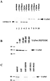
Identification of YAP65 binding site in EBP50. (A) GST-EBP50 (10 μg) was immobilized on beads and incubated with (+ peptide) or without (− peptide) 400 nM COOH-terminal peptides as indicated. Radiolabeled CFTR-CT or full-length YAP65 were added to binding reactions. Bound proteins were extensively washed in binding buffer, eluted, and analyzed by SDS-PAGE and applied to phosphorimage screens. (B) Immobilized GST-EBP50 (10 μg) was incubated with radiolabeled full-length YAP65 in the presence of increasing concentrations of YAP65wt peptide. Bound proteins were analyzed as described in A. (C) 10 μg of GST, GST-EBP50, GST-PDZ1, or GST-PDZ2 were immobilized on glutathione agarose beads and incubated with radiolabeled YAP65. After washing in buffers containing 1 M NaCl, bound proteins were eluted from the beads and analyzed as described in A.
Figure 6
Figure 6
In vivo association of YAP65 and EBP50. (A) Whole cell lysates from wild-type 16HBE14o− cells were prepared in RIPA buffer and ∼300 μg of total protein was immunoprecipitated using EBP50-specific antisera (Ab) or normal rabbit IgG. Bound proteins were extensively washed in RIPA buffer, eluted, electrophoresed on 10% SDS-PAGE and blotted with rabbit anti-YAP65 (1:1,000) or mouse anti-ezrin (1:1,000) antisera. Input: 10% or 20% of the material added into each binding reaction. (B) Lysates from 16HBE14o− cells stably expressing GFP-YAP65 or GFP-YAP65/−4 (200 μg) were immunoprecipitated using EBP50-specific antisera as described in A. Bound proteins were electrophoresed on 10% SDS-PAGE and blotted with mouse anti-GFP (1:1,000). Input: 20% of the material added into each binding reaction. B, protein A beads alone; IgG, normal rabbit IgG; and Ab, rabbit anti-EBP50. (C) 16HBE14o− cells stably expressing GFP-YAP65 or GFP-YAP65/−4 were grown to confluence on transwell filters, fixed in 4% paraformaldehyde, and analyzed by confocal microscopy. At least two independent clones were analyzed for each cell line. Bars, 10 μm.
Figure 6
Figure 6
In vivo association of YAP65 and EBP50. (A) Whole cell lysates from wild-type 16HBE14o− cells were prepared in RIPA buffer and ∼300 μg of total protein was immunoprecipitated using EBP50-specific antisera (Ab) or normal rabbit IgG. Bound proteins were extensively washed in RIPA buffer, eluted, electrophoresed on 10% SDS-PAGE and blotted with rabbit anti-YAP65 (1:1,000) or mouse anti-ezrin (1:1,000) antisera. Input: 10% or 20% of the material added into each binding reaction. (B) Lysates from 16HBE14o− cells stably expressing GFP-YAP65 or GFP-YAP65/−4 (200 μg) were immunoprecipitated using EBP50-specific antisera as described in A. Bound proteins were electrophoresed on 10% SDS-PAGE and blotted with mouse anti-GFP (1:1,000). Input: 20% of the material added into each binding reaction. B, protein A beads alone; IgG, normal rabbit IgG; and Ab, rabbit anti-EBP50. (C) 16HBE14o− cells stably expressing GFP-YAP65 or GFP-YAP65/−4 were grown to confluence on transwell filters, fixed in 4% paraformaldehyde, and analyzed by confocal microscopy. At least two independent clones were analyzed for each cell line. Bars, 10 μm.
Figure 7
Figure 7
Association of EBP50 with Src family kinase activity in 16HBE14o− cells. GST fusion proteins (10 μg), immobilized on glutathione agarose, were incubated with ∼200 μg of 16HBE14o− cell lysate. For competition experiments, immobilized GST-EBP50 was preincubated with 400 nM YAPwt peptide for 3 h before addition of lysates. Beads were washed, and kinase activity was measured by adding an exogenous Src family substrate, p34cdc2 and 1 μCi γ-[32P]ATP. Statistical analysis was performed using the t test (*P < 0.01; n = 3).
Figure 8
Figure 8
Association of YAP65 and c-Yes in 16HBE14o− cells. (A) 30 μg of total cell lysate from CalU3 (C), 16HBE14o− (H), or MDCK (M) cells was electrophoresed on 10% SDS-PAGE gels and analyzed by immunoblotting with c-Src (1:1,000−) or c-Yes (1:1,000) antisera as noted above the blot. (B and C) Immobilized GST fusion proteins (10 μg) were incubated with 16HBE14o− cell lysates (∼200 μg) diluted in binding buffer. After extensive washing in binding buffer, bound proteins were eluted from the beads, electrophoresed on 10% SDS-PAGE and analyzed by immunoblotting using c-Yes (1:1,000) or c-Src (1:1,000) antisera. Input (In): 20% (B) or 25% (C) of the material added into each binding reaction. (D) Lysates from wild-type 16HBE14o− cells (∼200 μg) were immunoprecipitated using EBP50-specific antisera (Ab), rabbit IgG, or protein A beads alone (B). Bound proteins were washed and electrophoresed on 10% SDS-PAGE and blotted with mouse anti–c-Yes antisera. Input (In): 20% of the material added into each reaction.
Figure 9
Figure 9
Localization of c-Yes in airway epithelial cells. (A) Well differentiated primary human nasal epithelia and (B)16HBE14o− cells grown to confluence on transwell filters were fixed and blocked as described in Materials and Methods followed by incubation in antisera specific for c-Yes (1:100), YAP65 (1:200), or EBP50 (1:200). After washes, the cultures were incubated with appropriate Texas red– or FITC-conjugated secondary antisera and analyzed on the confocal microscope. Bars, 10 μm.
Figure 10
Figure 10
Stable association of YAP65 and c-Yes in airway epithelial cells. 16HBE14o− cells stably expressing (A) GFP-YAP65 or (B) GFP-YAP65/−4 were grown to confluence on transwell filters, fixed in 4% paraformaldehyde, stained with mouse anti–c-Yes antisera (1:100) and analyzed by XZ scanning confocal microscopy. Bar, 10 μm.

Similar articles

Cited by

References

    1. Biemesderfer D., Rutherford P.A., Nagy T., Pizzonia J.H., Abu-Alfa A.K., Aronson P.S. Monoclonal antibodies for high-resolution localization of NHE3 in adult and neonatal rat kidney. Am. J. Physiol. 1997;273:289–299 . - PubMed
    1. Boyer B., Roche S., Denoyelle M., Thiery J.P. Src and Ras are involved in separate pathways in epithelial cell scattering. EMBO (Eur. Mol. Biol. Organ.) J. 1997;16:5904–5913 . - PMC - PubMed
    1. Brant S.R., Yun C.H., Donowitz M., Tse C.M. Cloning, tissue distribution, and functional analysis of the human Na+/H+ exchanger isoform, NHE3. Am. J. Physiol. 1995;269:198–206 . - PubMed
    1. Brenman J.E., Chao D.S., Gee S.H., McGee A.W., Craven S.E., Santillano D.R., Wu Z., Huang F., Xia H., Peters M.F., Froehner S.C., Bredt D.S. Interaction of nitric oxide synthase with the postsynaptic density protein PSD-95 and α1-syntrophin mediated by PDZ domains. Cell. 1996;84:757–767 . - PubMed
    1. Chen H.I., Einbond A., Kwak S.J., Linn H., Koepf E., Peterson S., Kelly J.W., Sudol M. Characterization of the WW domain of human Yes-associated protein and its polyproline-containing ligands. J. Biol. Chem. 1997;272:17070–17077 . - PubMed

Publication types

MeSH terms