. 1999 Nov 15;147(4):891-903.
doi: 10.1083/jcb.147.4.891.

Manner of interaction of heterogeneous claudin species within and between tight junction strands

Affiliations

Manner of interaction of heterogeneous claudin species within and between tight junction strands

M Furuse et al. J Cell Biol. .

Abstract

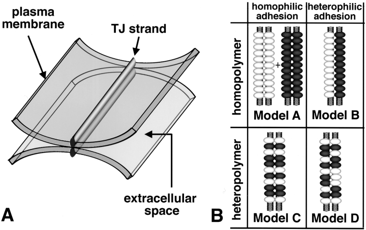
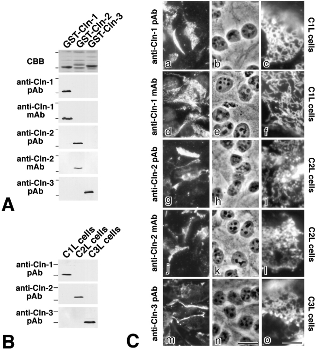
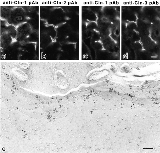
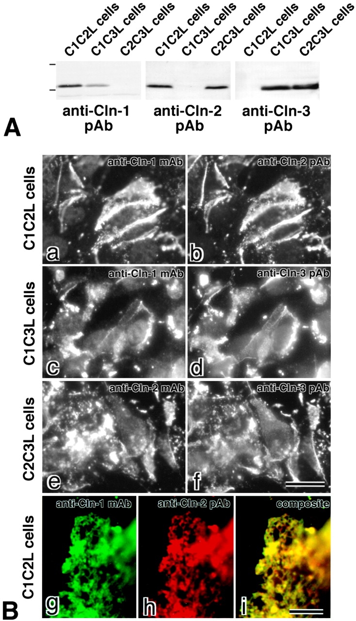
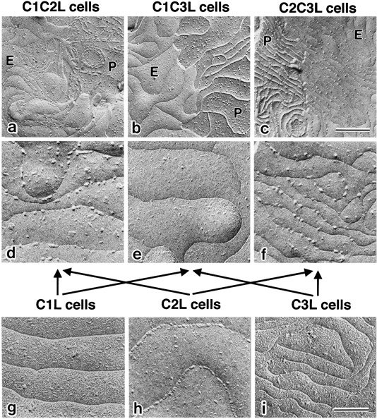
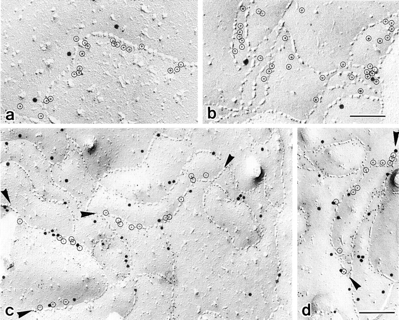
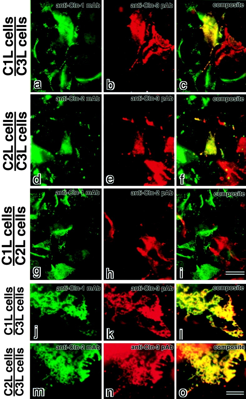
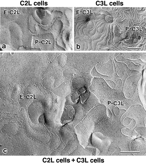
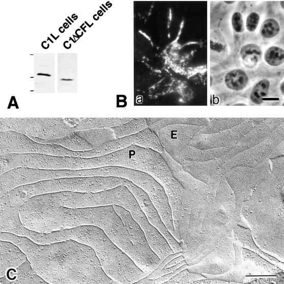
In tight junctions (TJs), TJ strands are associated laterally with those of adjacent cells to form paired strands to eliminate the extracellular space. Claudin-1 and -2, integral membrane proteins of TJs, reconstitute paired TJ strands when transfected into L fibroblasts. Claudins comprise a multigene family and more than two distinct claudins are coexpressed in single cells, raising the questions of whether heterogeneous claudins form heteromeric TJ strands and whether claudins interact between each of the paired strands in a heterophilic manner. To answer these questions, we cotransfected two of claudin-1, -2, and -3 into L cells, and detected their coconcentration at cell-cell borders as elaborate networks. Immunoreplica EM confirmed that distinct claudins were coincorporated into individual TJ strands. Next, two L transfectants singly expressing claudin-1, -2, or -3 were cocultured and we found that claudin-3 strands laterally associated with claudin-1 and -2 strands to form paired strands, whereas claudin-1 strands did not interact with claudin-2 strands. We concluded that distinct species of claudins can interact within and between TJ strands, except in some combinations. This mode of assembly of claudins could increase the diversity of the structure and functions of TJ strands.


Figures

Figure 1
Figure 1
TJ strands and claudins. A, Schematic drawing of the TJ. At TJs, two apposing membranes lie close together, and TJ strands run within the lipid bilayer of each membrane. TJ strands in apposing membranes laterally associate to form paired strands, where the extracellular space is completely eliminated. B, Possible models of the arrangements of two distinct species of claudins (white and black spheroids) in each paired TJ strand. When each TJ strand is composed of a single species of claudin (homopolymer), two types of paired strands are formed by homophilic lateral association (homophilic adhesion; Model A) or heterophilic lateral association (heterophilic adhesion; Model B) of each TJ strand. When each TJ strand is a heteropolymer of two distinct species of claudins (heteropolymer), paired strands are formed by homophilic interaction (Model C) or heterophilic adhesion (Model D) of each claudin molecule.
Figure 2
Figure 2
Anticlaudin antibodies and L transfectants expressing claudin-1, -2, or -3. A, GST fusion proteins with the COOH-terminal cytoplasmic domains of claudin-1 (GST-Cln-1), claudin-2 (GST-Cln-2), and claudin-3 (GST-Cln-3) were separated by SDS-PAGE (CBB), and immunoblotted with guinea pig anticlaudin-1 pAb (anti-Cln-1 pAb), rat anticlaudin-1 mAb (anti-Cln-1 mAb), rabbit anticlaudin-2 pAb (anti-Cln-2 pAb), rat anticlaudin-2 mAb (anti-Cln-2 mAb), and rabbit anticlaudin-3 pAb (anti-Cln-3 pAb). Each GST fusion protein was specifically detected by respective pAb or mAb. B, Total cell lysates of L transfectants expressing claudin-1 (C1L cells), claudin-2 (C2L cells), or claudin-3 (C3L cells) were separated by SDS-PAGE and immunoblotted with anticlaudin-1 pAb (anti-Cln-1 pAb), anticlaudin-2 pAb (anti-Cln-2 pAb), or anticlaudin-3 pAb (anti-Cln-3 pAb). The bands of respective claudins with expected molecular masses were specifically detected. Bars indicate molecular masses of 31 and 21 kD, respectively, from the top. C, C1L, C2L, and C3L cells were immunofluorescently stained with respective antibodies (a, d, g, j, and m). b, e, h, k, and n, Corresponding phase-contrast images. Expressed claudins were specifically found to be concentrated at cell–cell borders as planes. At higher magnification, claudins were seen to be distributed in an elaborate network pattern in these planes (c, f, i, l, and o). Bars: (a, b, d, e, g, h, j, k, m, and n) 10 μm; (c, f, i, l, and o) 3 μm.
Figure 3
Figure 3
Codistribution of claudin-1, -2, and -3 in TJs of the mouse liver. a–d, Frozen sections of the liver were double stained with guinea pig anticlaudin-1 pAb (a)/rabbit anticlaudin-2 pAb (b) or guinea pig anticlaudin-1 pAb (c)/rabbit anticlaudin-3 pAb (d). Claudin-1, -2, and -3 were precisely colocalized at the junctional complex regions, although the expression levels of claudin-1 and -2 appeared to vary significantly, depending on the position of hepatocytes in lobules (data not shown). e, Freeze-fracture replicas of mouse liver were double labeled with anticlaudin-1 pAb (15-nm gold) and anticlaudin-3 pAb (5-nm gold). 5-nm gold particles were encircled. Although the labeling ability of anticlaudin-1 pAb was very low, these images suggested that claudin-1 and -3 were copolymerized into individual TJ strands in the liver. Bars: (a–d) 6 μm; (e) 100 nm.
Figure 4
Figure 4
L transfectants coexpressing claudin-1 and -2 (C1C2L cells), claudin-1 and -3 (C1C3L cells), and claudin-2 and -3 (C2C3 cells). A, Total cell lysates of C1C2L, C1C3L, and C2C3L cells were immunoblotted with anticlaudin-1 pAb (anti-Cln-1 pAb), anticlaudin-2 pAb (anti-Cln-2 pAb), or anticlaudin-3 pAb (anti-Cln-3 pAb). Respective claudins with expected molecular masses were detected. Bars indicate molecular masses of 31 and 21 kD, respectively, from the top. B, Semiconfluent cultures of C1C2L, C1C3L, and C2C3L cells were double stained with anticlaudin-1 mAb (a)/anticlaudin-2 pAb (b), anticlaudin-1 mAb (c)/anticlaudin-3 pAb (d), and anticlaudin-2 mAb (e)/anticlaudin-3 pAb (f), respectively. Coexpressed claudins were precisely colocalized. At higher magnification of C1C2L cells, in the cell–cell contact planes, claudin-1 and -2 were precisely coconcentrated in an elaborate network (g–i), suggesting that claudin-1 and -2 were coincorporated into rTJ strands. Bars: (a–f) 10 μm; (g–i) 3 μm.
Figure 5
Figure 5
Freeze-fracture replica images of rTJ strands in C1C2L, C1C3L, and C2C3L cells. In the cell–cell contact planes of these cells, well-developed networks of rTJ strands were observed. As previously shown in C1FL and C2FL cells ( Furuse et al. 1998b), in glutaraldehyde-fixed C1L cells, claudin-1–induced strands were largely associated with the P-face as mostly continuous structures (data not shown) with vacant grooves at the E-face (g; P-face–associated TJs), whereas in C2L cells, claudin-2–induced strands were discontinuous at the P-face (data not shown) with complementary grooves at the E-face that were occupied by chains of particles (h; see Fig. 8 a; E-face–associated TJs). Similar to C1L cells, P-face–associated TJs with vacant grooves on the E-face (i) were induced in C3L cells (see Fig. 8 b). C1C3L cells bore typical P-face–associated TJs (b), whereas C1C2L and C2C3L cells showed an intermediate morphology of rTJ strands/grooves between P- and E-face–associated TJs (a and c); the grooves at the E-face of C1C3L cells were completely vacant (e), whereas those in C1C2L and C2C3L cells were characterized by evenly scattered particles (d and f). Bars: (a–c) 200 nm; (d–i) 100 nm.
Figure 6
Figure 6
Immunoreplica EM of C1C2L and C1C3L cells. a and b, Freeze-fracture replicas of C1C2L and C1C3L cells were double labeled with guinea pig anticlaudin-1 pAb (15-nm gold)/rabbit anticlaudin-2 pAb (5-nm gold; a) and guinea pig anticlaudin-1 pAb (15-nm gold)/rabbit anticlaudin-3 pAb (5-nm gold; b), respectively. 5-nm gold particles were encircled. Although the labeling ability of anticlaudin-1 pAb was fairly low, the 15- and 5-nm gold particles appeared to be admixed along individual rTJ strands. c and d, Freeze-fracture replicas of C1FC2L cells were double labeled with mouse anti-FLAG mAb (15-nm gold) and rabbit anticlaudin-2 pAb (5-nm gold). 5-nm gold particles on selected rTJ strands (between two arrowheads) were encircled. Numerous 15- and 5-nm gold particles distribute evenly and specifically along individual rTJ strands. Bars: (a and b) 100 nm; (c and d) 150 nm.
Figure 7
Figure 7
Coculture experiments of two of L transfectants expressing claudin-1 (C1L cells), -2 (C2L cells), or -3 (C3L cells). C1L/C3L (a–c, j–l), C2L/C3L (d–f, m–o), or C1L/C2L (g–i) cocultured cellular sheets were double stained with anticlaudin-1 mAb (green)/anticlaudin-3 pAb (red), anticlaudin-2 mAb (green)/anticlaudin-3 pAb (red), or anticlaudin-1 mAb (green)/anticlaudin-2 pAb (red), respectively. Merged images in the coculture of C1L/C3L (c) and C2L/C3L (f) identified three types of cell–cell contact planes in terms of immunofluorescence staining; green, red, and yellow. Yellow planes were formed between adjacent C1L/C3L cells or C2L/C3L cells. At higher magnification, distinct species of claudins were seen to be recruited to these planes, being precisely coconcentrated in an elaborate network pattern (j–l, m–o). In cocultures of C1L/C2L (i), however, such yellow planes were not observed. We observed 50 fields for C1L/C3L, C2L/C3L, and C1L/C2L coculture experiments and identified yellow planes in 50, 50, and 0 fields, respectively. Bars: (a–i) 10 μm; (j–o) 3 μm.
Figure 8
Figure 8
Freeze-fracture replica images of cell–cell contact planes between adjacent C2L and C3L cells. When the cell–cell contact planes in the C2L confluent culture were fractured, the fracture planes were characterized by discontinuous strands on the P-face (P-C2L; a) and grooves occupied with chains of particles on the E-face (E-C2L; a). In contrast, the fracture planes at the contact regions in the C3L confluent culture showed continuous strands on the P-face (P-C3L; b) and vacant grooves on the E-face (E-C3L; b). In the confluent C2L/C3L coculture, in addition to these C2L/C2L and C3L/C3L fracture planes, which would be derived from the contact regions between adjacent C2L and C3L cells, were occasionally identified (c). In these planes, as expected, combinations of E-C2L and P-C3L (c) or E-C3L and P-C2L (data not shown) were observed. When the fracture plane jumped from the E- to the P-face, the continuity of network pattern of grooves of E-C2L and strands of P-C3L were seen to be completely maintained, indicating that in these planes individual claudin-2 homopolymers in C2L cells always associated laterally with claudin-3 homopolymers in adjacent C3L cells. Bars: (a and b) 200 nm; (c) 200 nm.
Figure 9
Figure 9
Formation of rTJ strands from claudin-1 mutant lacking its COOH-terminal domain in L cells. A, Total cell lysates of L transfectants expressing claudin-1 (C1L cells) or FLAG-tagged claudin-1 mutant lacking its COOH-terminal domain (C1ΔCFL cells) were immunoblotted with anticlaudin-1 pAb and anti-FLAG mAb, respectively. Bars indicate molecular masses of 31, 21, and 14 kD, respectively, from the top. B, Semiconfluent cultures of C1ΔCFL cells were stained with anti-FLAG mAb (a). Expressed claudin-1 mutant was highly concentrated at cell–cell borders as planes. b, Phase-contrast image. C, Freeze-fracture replica images of induced rTJ strands in C1ΔCFL cells. FLAG-tagged claudin-1 mutant lacking its COOH-terminal domain can form a well-developed network of rTJ strands on the P-face (P) in L cells. E, E-face. Bars: (B) 10 μm; (C) 200 nm.

Similar articles

Cited by

References

    1. Anderson J.M., van Itallie C.M. Tight junctions and the molecular basis for regulation of paracellular permeability. Am. J. Physiol. 1995;269:G467–G475 . - PubMed
    1. Ando-Akatsuka Y., Saitou M., Hirase T., Kishi M., Sakakibara A., Itoh M., Yonemura S., Furuse M., Tsukita Sh. Interspecies diversity of the occludin sequencecDNA cloning of human, mouse, dog, and rat-kangaroo homologues. J. Cell Biol. 1996;133:43–47 . - PMC - PubMed
    1. Balda M.S., González-Mariscal L., Matter K., Cereijido M., Anderson J.M. Assembly of the tight junctionthe role of diacylglycerol. J. Cell Biol. 1993;123:293–302 . - PMC - PubMed
    1. Balda M.S., Whitney J.A., Flores C., González S., Cereijido M., Matter K. Functional dissociation of paracellular permeability and transepithelial electrical resistance and disruption of the apical–basolateral intramembrane diffusion barrier by expression of a mutant tight junction membrane protein. J. Cell Biol. 1996;134:1031–1049 . - PMC - PubMed
    1. Chen Y.-H., Merzdorf C., Paul D.L., Goodenough D.A. COOH terminus of occludin is required for tight junction barrier function in early Xenopus embryos. J. Cell Biol. 1997;138:891–899 . - PMC - PubMed

Publication types

MeSH terms