. 1999 Nov;104(10):1343-51.
doi: 10.1172/JCI8088.

A murine model of hereditary hemorrhagic telangiectasia

Affiliations

A murine model of hereditary hemorrhagic telangiectasia

A Bourdeau et al. J Clin Invest. 1999 Nov.

Abstract

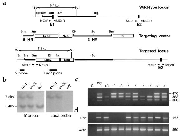
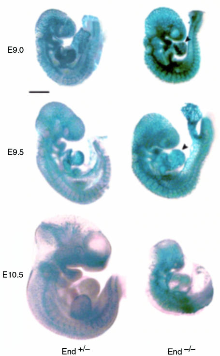
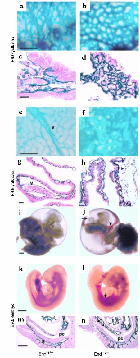
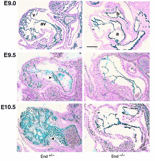
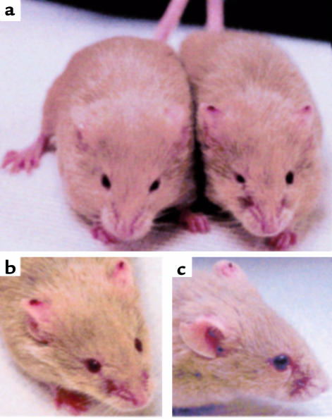
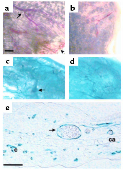
Endoglin (CD105), an accessory protein of the TGF-beta receptor superfamily, is highly expressed on endothelial cells. Hereditary hemorrhagic telangiectasia type 1 (HHT1) is associated with mutations in the Endoglin gene, leading to haploinsufficiency. To generate a disease model and ascertain the role of endoglin in development, we generated mice lacking 1 or both copies of the gene. Endoglin null embryos die at gestational day 10.0-10.5 due to defects in vessel and heart development. Vessel formation appears normal until hemorrhage occurs in yolk sacs and embryos. The primitive vascular plexus of the yolk sac fails to mature into defined vessels, and vascular channels dilate and rupture. Internal bleeding is seen in the peritoneal cavity, implying fragile vessels. Heart development is arrested at day 9.0, and the atrioventricular canal endocardium fails to undergo mesenchymal transformation and cushion-tissue formation. These data suggest that endoglin is critical for both angiogenesis and heart valve formation. Some heterozygotes, either with an inbred 129/Ola or mixed C57BL/6-129/Ola background, show signs of HHT, such as telangiectases or recurrent nosebleeds. In this murine model of HHT, it appears that epigenetic factors and modifier genes, some of which are present in 129/Ola, contribute to disease heterogeneity.


Figures

Figure 1
Figure 1
Generation of End–/– mice. (a) Targeting strategy. Homologous recombination deletes 609 bp of the End gene including exon 1 (E1; 66 bp), placing the LacZ gene under control of its promoter. The targeting construct contains a 1.4-kb SmaI fragment (5′ HR) and a 5.1-kb SmaI-BglII fragment (3′ HR) of the End gene flanking the LacZ-Neo cassette. Proper recombination events were screened by Southern blot analysis with 5′ external and LacZ internal probes. Bm, BamHI; Bg, BglII; EI, EcoRI; Sa, SacI; Sc, ScaI; Sm, SmaI; Xb, XbaI. (b) Identification of targeted ES cell lines. DNA from 2 geneticin- and ganciclovir-resistant targeted ES cells (4A-11, 4A-36) and a wild-type clone (WT) were digested with ScaI. The 5.4-kb WT and 7.3-kb recombinant alleles hybridizing with the 5′ external probe and the recombinant allele reacting with the LacZ probe are shown. (c) Genotyping of embryos by multiplex PCR. DNA from yolk sacs of an E9.5 litter derived from a C57BL/6 End+/– mating is shown; #21 represents the mother and C represents the PCR control reaction without DNA. Primers ME1F and ME1R amplify normal exon 1 (300 bp), ME2F and ME2R amplify normal exon 2 (383 bp), and ME1F and MEZR specifically amplify the recombinant product (476 bp). (d) Absence of endoglin mRNA in End–/– embryos. RNA was prepared from the same embryos as in c and analyzed for endoglin and β-actin mRNA by RT-PCR.
Figure 2
Figure 2
Arrest in development in End–/– embryos. β-gal expression driven by the End promoter was analyzed in whole End+/– and End–/– embryos. At E9.0, embryos are of similar size and their vasculature appears normal. The End–/– embryo shows an enlarged heart protruding forward (arrowhead). At E9.5, edematous pericardium (arrowhead) is observed in the End–/– embryo, which is growth-retarded compared with the End+/– embryo. At E10.5, the End –/– embryo is dead and is much smaller that the End+/– embryo. Bar: 500 μm.
Figure 3
Figure 3
Vascular defects in End–/– mice. Whole-mount embryos in their yolk sacs were stained for β-gal activity. (a, b, e, and f) Direct microscopic examination of the yolk sacs. (c, d, g, and h) Sagittal histological sections. At E9.0, the normal capillary plexus and initiation of branching are seen in the End+/– embryo (a and c). The End–/– yolk sac also has a highly vascularized plexus, but no vessel branching is observed (b and d). At E9.5, a vitelline vessel (v) is readily detectable in the End+/– yolk sac (e); this intact vessel is full of primitive red cells (g). The End–/– yolk sac shows a disorganized capillary plexus and no vitelline vessels (f); abnormally dilated blood islands are seen (h), which have ruptured, releasing primitive erythroblasts toward the amnion (arrowhead). (i and j) Unstained E9.5 embryos, with yolk sac and placenta still attached. Bleeding in the yolk sac cavity (arrowhead) and edematous pericardium (arrow) are observed in End–/– embryo (j), compared with a littermate control (i). An unstained E9.0 End+/– embryo (k), dissected away from the yolk sac, is compared with an End–/– littermate (l), which shows internal bleeding (arrow). Sagittal sections stained for β-gal demonstrate the presence of blood (arrow) in the peritoneal cavity of the End–/– embryo (n), which is absent from the End+/– embryo (m). a, dorsal aorta; pc, peritoneal cavity; u, umbilical vein. Bar: 100 μm (ah), 500 μm (i and j), 250 μm (kn).
Figure 4
Figure 4
Heart deficiency in End–/– mice. Transverse sections from whole-mount embryos stained for β-gal activity are shown. The E9.0 End+/– embryo shows staining associated with the endocardium in the ventricular compartment (v) and tightly delineated in the atrioventricular region (av). The endocardium of the End–/– embryo is rudimentary, with an abnormally large lumen in the common atrial chamber (a) and the bulbus cordis (b). At E9.5 increased trabeculation is observed in the ventricle of the End+/– embryo; a few β-gal–stained mesenchymal cells (arrowhead) are seen adjacent to the atrioventricular canal. The heart of the End–/– embryo is much larger than that of the control and is devoid of trabeculae; mesenchymal cells are absent. At E10.5 cushion-tissue mesenchyme (arrowhead) from the End+/– embryo stains strongly positive for β-gal; in contrast, the heart of the End–/– embryo shows edema and signs of necrosis. Bar: 100 μm.
Figure 5
Figure 5
End+/– 129/Ola mice have nosebleeds and telangiectases characteristic of HHT. (a) A 129/Ola End+/– male mouse with recurrent nosebleeds and telangiectases is shown on the right of a female littermate with no disease manifestation. (b) Telangiectases are present on the upper part of both ears. (c) A side view provides a closer look at the affected nasal area.
Figure 6
Figure 6
Microscopic examination of a telangiectasia from a 129/Ola End+/– mouse. Ear lobe biopsies from an End+/– mouse and normal littermate were left unstained (a and b) or were stained for β-gal activity (c and d). (a) A dilated vessel (arrow), engorged with blood, is seen in the affected End+/– ear lobe, together with a necrotic area (arrowhead) from a telangiectasia ruptured previously. (b) The corresponding area from a nonaffected control 129/Ola mouse shows much smaller vessels. (c) A small telangiectasia (arrow), close to the edge of the affected ear, appears as a dilated vessel distinct from the adjacent network of capillaries. (d) Normal capillary network from a nonaffected ear. (e) A cross-section of the resected ear segment from the affected 129/Ola End+/– mouse was stained for β-gal. An enlarged vessel filled with blood (arrow), corresponding to a telangiectasia, is seen. The endothelial cells of this vessel and of several normal small capillaries are stained; a layer of cartilage (ca) is seen across the section. Bar: 500 μm (ad) and 100 μm (e).

Comment in

Similar articles

Cited by

References

    1. Guttmacher AE, Marchuk DA, White RIJ. Hereditary hemorrhagic telangiectasia. N Engl J Med. 1995;333:918–924. - PubMed
    1. McAllister KA, et al. Endoglin, a TGF-β binding protein of endothelial cells is the gene for hereditary haemorrhagic telangiectasia type 1. Nat Genet. 1994;8:345–351. - PubMed
    1. Heutink P, et al. Linkage of hereditary haemorrhagic telangiectasia to chromosome 9q34 and evidence for locus heterogeneity. J Med Genet. 1994;31:933–936. - PMC - PubMed
    1. McAllister KA, et al. Genetic heterogeneity in hereditary haemorrhagic telangiectasia: possible correlation with clinical phenotype. J Med Genet. 1994;31:927–932. - PMC - PubMed
    1. Porteous MEM, et al. Genetic heterogeneity in hereditary haemorrhagic telangiectasia. J Med Genet. 1994;31:925–926. - PMC - PubMed

Publication types

MeSH terms