. 1999 Nov;104(10):1375-82.
doi: 10.1172/JCI6097.

Allergen-induced IL-9 directly stimulates mucin transcription in respiratory epithelial cells

Affiliations

Allergen-induced IL-9 directly stimulates mucin transcription in respiratory epithelial cells

M Longphre et al. J Clin Invest. 1999 Nov.

Abstract

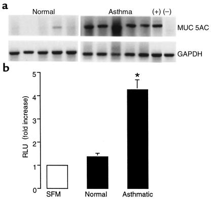
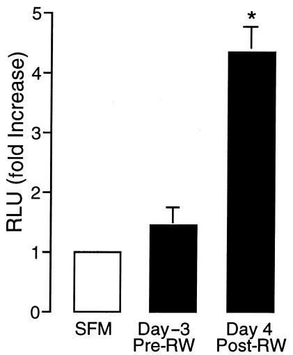
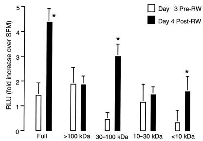
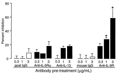
A hallmark of asthma is mucin overproduction, a condition that contributes to airway obstruction. The events responsible for mucin overproduction are not known but are thought to be associated with mediators of chronic inflammation. Others have shown that T-helper 2 (Th2) lymphocytes are required for mucous cell metaplasia, which then leads to mucin overproduction in animal models of allergy. We hypothesized that Th2 cell mediators are present in asthmatic airway fluid and directly stimulate mucin synthesis in airway epithelial cells. Results in cultured airway epithelial cells showed that samples of asthmatic fluid stimulated mucin (MUC5AC) synthesis severalfold more potently than non-asthmatic fluid. Consistent with this, lavage fluid from the airways of allergen-challenged dogs stimulated mucin synthesis severalfold more potently than that from non-allergen-challenged dogs. Fractionation of dog samples revealed 2 active fractions at <10 kDa and 30-100 kDa. Th2 cytokines in these molecular weight ranges are IL-9 (36 kDa), IL-5 (56 kDa), and IL-13 (10 kDa). Antibody blockade of ligand-receptor interaction for IL-9 (but not IL-5 or IL-13) inhibited mucin stimulation by dog airway fluid. Furthermore, recombinant IL-9, but not IL-5 or IL-13, stimulated mucin synthesis. These results indicate that IL-9 may account for as much as 50-60% of the mucin-stimulating activity of lung fluids in allergic airway disease.


Figures

Figure 1
Figure 1
Human tracheal aspirates from asthmatics induce MUC5AC in airway epithelial cells in culture. (a) NCI-H292 cells were treated for 8 hours with aspirates from 5 healthy control (Normal) and 5 asthmatic human subjects. Cells used as negative (–) controls were in SFM and cells used as positive (+) controls were exposed to vanadate. RT-PCR was performed using primers for MUC5AC and GAPDH. (b) NCI-H292 cells were transfected with the 3.7-kb MUC5AC luciferase construct and challenged with asthmatic or non-asthmatic tracheal aspirates for 16 hours. RLU, relative luciferase units. Bars represent means ± SEM (n = 5 patients/group in duplicate). *Significantly different from SFM control; P < 0.05.
Figure 2
Figure 2
Ragweed-challenged dog lavage stimulates MUC5AC reporter in airway epithelial cells in culture. NCI-H292 cells were transfected with the 3.7-kb MUC5AC luciferase construct and challenged with BAL for 16 hours. RW, ragweed. Bars represent means ± SEM (n = 4 dogs in triplicate). *Significantly different from SFM control; P < 0.05.
Figure 3
Figure 3
Fractionated dog lavage stimulates MUC5AC reporter in airway epithelial cells in culture. NCI-H292 cells were transfected with the 3.7-kb MUC5AC luciferase construct and challenged with BAL for 16 hours. Bars represent means (n = 4 dogs in duplicate).
Figure 4
Figure 4
Demonstration of receptor RNA for IL-9, IL-5, and IL-13 in NCI-H292 airway epithelial cell line (H292) and primary cultures of human primary bronchial epithelial cells (HBE) by RT-PCR of unique receptor α subunits.
Figure 5
Figure 5
Demonstration of cytokines in dog airway lavage and human airway aspirate. (a) ELISA showing IL-5, IL-9, and IL-13 in post-RW lavage compared with pre-RW lavage. (b) ELISA showing IL-5, IL-9, and IL-13 in normal and asthmatic aspirates. Bars represent means ± SEM (n = 4 in duplicate). *Significantly different from pre-RW (a) or normal (b) control.
Figure 6
Figure 6
The effect of cytokine antibody or cytokine receptor antibody pretreatment on MUC5AC luciferase activity elicited by day 4 post-ragweed dog lavage. NCI-H292 cells were transfected with the 3.7-kb MUC5AC luciferase construct and challenged with BAL for 16 hours. Bars represent means ± SEM (n = 3 wells in duplicate). *Significantly different from isotype control; P < 0.05).
Figure 7
Figure 7
The effect of recombinant human cytokines on MUC5AC. (a) NCI-H292 cells were transfected with the 3.7-kb MUC5AC luciferase construct and challenged with rhIL-9 (circles), rhIL-5 (squares), or rhIL-13 (triangles) for 14–16 hours. Bars represent means ± SEM (n = 3 wells in triplicate). (b) Steady-state MUC5AC and cyclophilin were measured by RPA in NCI-H292 cells treated with IL-9. (c) MUC5AC and GAPDH mRNA were also quantified by RT-PCR in human primary bronchial epithelial cells treated with rhIL-9.
Figure 8
Figure 8
IL-9R is found in the human airway. Bronchial biopsies were immunostained for the presence of IL-9R with DAB (brown) and counterstained with hematoxylin (purple). (a) A representative slide (×40 magnification) is shown; the receptor for IL-9 was found dispersed throughout the pseudostratified bronchial epithelium (e) and in the lamina propria associated with vascular endothelium (arrow). The basal lamina (BL) is also indicated for reference. (b) An Ig isotype control (anti-trinitrophenol) showed no DAB staining. (c) The IL-9 receptor was also shown by Western blot and immunostaining to be present on NCI-H292 cells in culture.
Figure 9
Figure 9
Tracheal instillation of rmIL-9 in C57BL6/J mice. The mean number (±SEM) of PAS-positive (PAS+) cells per millimeter basal lamina in the trachea and mainstem bronchi of mice instilled with 100 μL of either IL-9 (10 μg/mL in PBS; solid bars) or PBS (open bars). *Significantly different from PBS-instilled; P < 0.05.

References

    1. Fahy JV, Steiger DJ, Liu J, Basbaum CB, Finkbeiner WE, Boushey HA. Markers of mucus secretion and DNA levels in induced sputum from asthmatic and from health subjects. Am Rev Respir Dis. 1993;147:1132–1137. - PubMed
    1. Thurlbeck WM. Small airways disease. Hum Pathol. 1973;4:150–152. - PubMed
    1. Takizawa T, Thurlbeck W. Muscle and mucous gland size in the major bronchi of patients with chronic bronchitis, asthma and asthmatic bronchitis. Am Rev Respir Dis. 1971;104:331–336. - PubMed
    1. Reid L. Pathology of chronic bronchitis. Lancet. 1954;1:275–278. - PubMed
    1. Sperber K, Goswami SK, Gollub E, Mayer L, Marom Z. Mucus secretagogue production by a human macrophage hybridoma. J Allergy Clin Immunol. 1991;87:490–498. - PubMed

Publication types

MeSH terms