

A proinflammatory role for IL-18 in rheumatoid arthritis
- PMID: 10562301
- PMCID: PMC409841
- DOI: 10.1172/JCI7317
A proinflammatory role for IL-18 in rheumatoid arthritis
Abstract
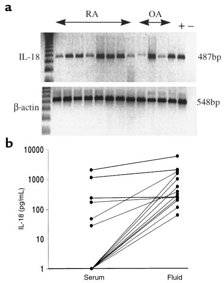
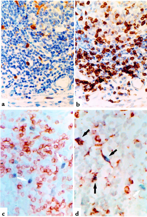
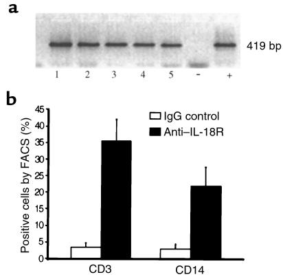
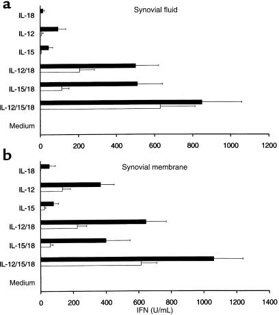
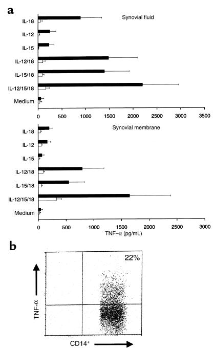
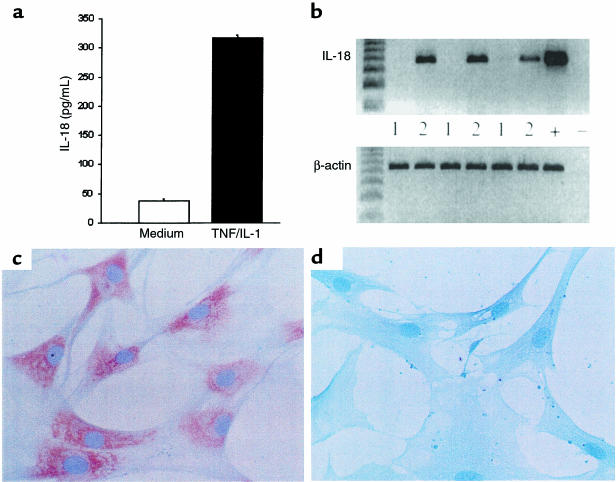
IL-18 is a novel cytokine with pleiotropic activities critical to the development of T-helper 1 (Th1) responses. We detected IL-18 mRNA and protein within rheumatoid arthritis (RA) synovial tissues in significantly higher levels than in osteoarthritis controls. Similarly, IL-18 receptor expression was detected on synovial lymphocytes and macrophages. Together with IL-12 or IL-15, IL-18 induced significant IFN-gamma production by synovial tissues in vitro. IL-18 independently promoted GM-CSF and nitric oxide production, and it induced significant TNF-alpha synthesis by CD14(+) macrophages in synovial cultures; the latter effect was potentiated by IL-12 or IL-15. TNF-alpha and IFN-gamma synthesis was suppressed by IL-10 and TGF-beta. IL-18 production in primary synovial cultures and purified synovial fibroblasts was, in turn, upregulated by TNF-alpha and IL-1beta, suggesting that monokine expression can feed back to promote Th1 cell development in synovial membrane. Finally, IL-18 administration to collagen/incomplete Freund's adjuvant-immunized DBA/1 mice facilitated the development of an erosive, inflammatory arthritis, suggesting that IL-18 can be proinflammatory in vivo. Together, these data indicate that synergistic combinations of IL-18, IL-12, and IL-15 may be of importance in sustaining both Th1 responses and monokine production in RA.
Figures
Comment in
-
Interleukin-18, rheumatoid arthritis, and tissue destruction.J Clin Invest. 1999 Nov;104(10):1337-9. doi: 10.1172/JCI8731. J Clin Invest. 1999. PMID: 10562294 Free PMC article. Review. No abstract available.
References
-
- Mauri C, Williams RO, Walmsley M, Feldmann M. Relationship between Th1/Th2 cytokine patterns and the arthritogenic response in collagen induced arthritis. Eur J Immunol. 1996;26:1511–1518. - PubMed
-
- Katz JD, Benoist C, Mathis D. T helper cell subsets in insulin dependent diabetes. Science. 1995;268:1185–1188. - PubMed
-
- Mosmann TR, Sad S. The expanding universe of T cell subsets: Th1, Th2 and more. Immunol Today. 1996;17:13–17. - PubMed
Publication types
MeSH terms
Substances
Grants and funding
LinkOut - more resources
Full Text Sources
Other Literature Sources
Medical
Research Materials
Miscellaneous
