. 1999 Nov;104(10):1449-57.
doi: 10.1172/JCI7936.

Naturally processed and presented epitopes of the islet cell autoantigen IA-2 eluted from HLA-DR4

Affiliations

Naturally processed and presented epitopes of the islet cell autoantigen IA-2 eluted from HLA-DR4

M Peakman et al. J Clin Invest. 1999 Nov.

Abstract

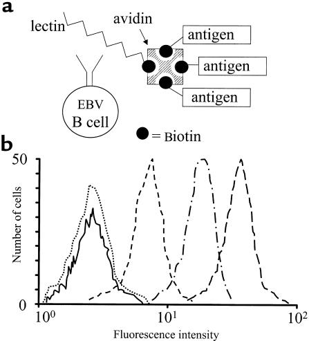
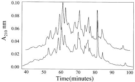
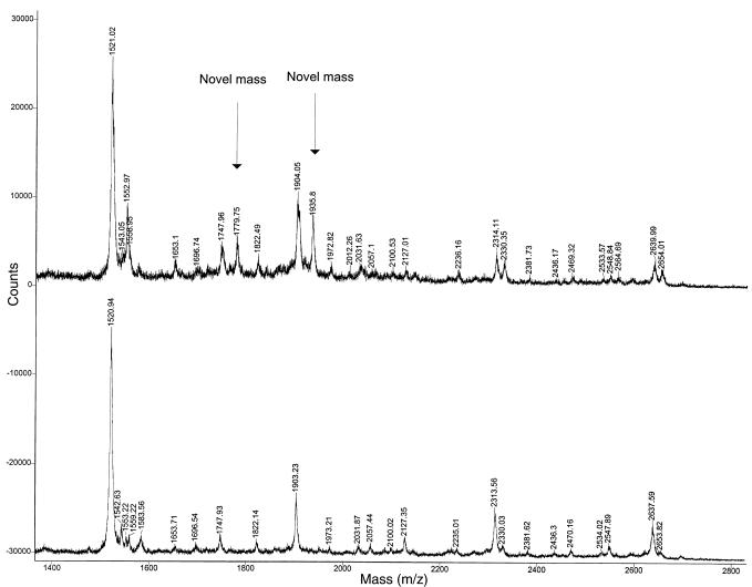
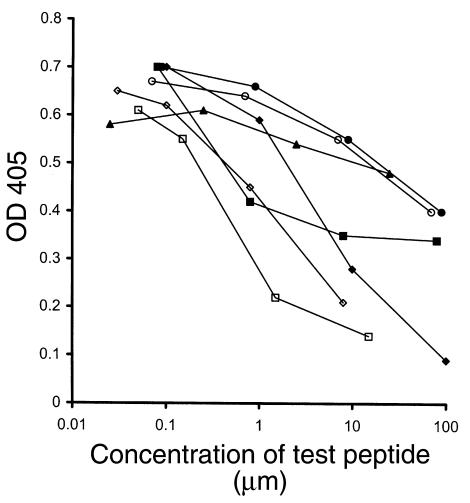
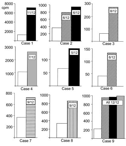
During immune responses, antigen-presenting cells (APCs) process antigens and present peptide epitopes complexed with human leukocyte antigen (HLA) molecules. CD4 cells recognize these naturally processed and presented epitopes (NPPEs) bound to HLA class II molecules. Epitope identification is important for developing diagnostic and therapeutic tools for immune-mediated diseases and providing insight into their etiology, but current approaches overlook effects of natural processing on epitope selection. We have developed a technique to identify NPPEs using mass spectrometry (MS) after antigen is targeted onto APCs using a lectin-based antigen delivery system (ADS). We applied the technique to identify NPPEs of the intracellular domain of the type 1 diabetes mellitus-associated (type 1 DM-associated) autoantigen insulinoma-associated-2 (IA-2ic), presented by HLA-DR4 (0401). IA-2ic-derived NPPEs eluted from HLA-DR4 constitute 6 sets of peptides nested around distinct core regions. Synthetic peptides based on these regions bind HLA-DR4 and elicit primary T-cell proliferation frequently in HLA-DR4-positive type 1 DM patients, but rarely in non-HLA-DR4 patients, and in none of the HLA-DR4 nondiabetic controls we tested. This flexible, direct approach identifies an HLA allele-specific map of NPPEs for any antigen, presented by any HLA class II molecule. This method should enable a greater understanding of epitope selection and lead to the generation of sensitive and specific reagents for detecting autoreactive T cells.


Figures

Figure 1
Figure 1
ADS delivers antigen to the EBV B–cell surface at high concentration. (a) The b-PWM has preferential affinity for disulfide-bonded Ig-like molecules such as surface Ig on the EBV B cell. The b-PWM is then bound to avidin, which can bind up to 3 molecules of biotinylated antigen. (b) The PWM-based ADS is completely internalized in 6 hours. Priess EBV B cells were incubated sequentially on ice with each component of the ADS (b-PWM, avidin-D, biotinylated antigen [TT]) and then incubated in culture medium at 37°C for 0, 1, 3, or 6 hours and removed for analysis of surface TT by flow cytometry, using rabbit anti-TT antiserum and FITC goat anti-rabbit Ig. Compared with background (solid line), surface expression was high at 0 hours (long dashes) but had diminished by 1 (dashes broken by dots) and 3 hours (short dashes). ADS-delivered antigen was completely internalized by 6 hours (dotted line).
Figure 2
Figure 2
RP-HPLC analysis of peptides eluted from HLA-DR4 purified from Priess cells pulsed with ADS plus IA-2ic (upper trace) or ADS alone (lower trace). The chromatograms are largely identical.
Figure 3
Figure 3
Subtractive approach to identification of IA-2ic–derived, naturally processed and presented peptides by mass spectrometric analysis of representative RP-HPLC fraction of IA-2ic and control-pulsed EBV B cells. Peptide masses in the upper trace have been eluted from HLA-DR4 from Priess EBV B cells pulsed with IA-2ic using the ADS; in the lower trace, B cells were pulsed with the ADS components but no IA-2ic. IA-2ic–derived peptides are marked in the upper trace as novel masses (see Table 1).
Figure 4
Figure 4
Binding of IA-2ic peptides to HLA-DR4 (0401). Peptides were tested at a range of concentrations for the ability to compete with a fixed concentration (2.5 μM) of biotinylated invariant chain (Ii) peptide. Filled diamonds represent the binding of nonbiotinylated Ii peptide competing with its biotinylated form. Test peptides of IA-2ic representing residues 654-674 (open squares), 709-732 (open diamonds), and 797-817 (filled squares) are good HLA-DR4 binders, having higher affinity than Ii. Test peptides of IA-2ic representing residues 753-771 (open circles), 854-872 (filled circles), and 955-975 (filled triangles) bind HLA-DR4, but with lower affinity than Ii.
Figure 5
Figure 5
T-cell responses to IA-2ic synthetic peptides with sequences based on NPPEs identified by elution from HLA-DR4. Vertical axes show T-cell proliferation in cpm. Case numbers 1–9 refer to the HLA-DR4 type 1 DM patients 1–9 described in Table 1 who had positive responses to IA-2ic peptides. Bars represent mean cpm from 12 replicate wells, and notations on bars indicate number of test wells positive (> mean + 2 SD of background wells). Background, open bars; peptide 654-674, hatched bars; peptide 709-732, vertically striped bar; peptide 797-817, filled bars; peptide 854-872, gray bars; and peptide 955-975, horizontally striped bars.

Comment in

Similar articles

Cited by

References

    1. Germain RN. MHC-dependent antigen processing and peptide presentation: providing ligands for T lymphocyte activation. Cell. 1994;76:287–299. - PubMed
    1. Zerva L, et al. Arginine at positions 13 or 70-71 in pocket 4 of HLA-DRB1 alleles is associated with susceptibility to tuberculoid leprosy. J Exp Med. 1996;183:829–836. - PMC - PubMed
    1. Cramp ME, et al. Association between HLA class II genotype and spontaneous clearance of hepatitis C viraemia. J Hepatol. 1998;29:207–213. - PubMed
    1. Pardoll DM, Topalian SL. The role of CD4+ T cell responses in antitumor immunity. Curr Opin Immunol. 1998;10:588–594. - PubMed
    1. Czarnecki D, Nicholson I, Tait B, Nash C. HLA DR4 is associated with the development of multiple basal cell carcinomas and malignant melanoma. Dermatology. 1993;187:16–18. - PubMed

MeSH terms