. 1999 Nov;104(10):1469-80.
doi: 10.1172/JCI7251.

Regulation of smooth muscle cell migration and integrin expression by the Gax transcription factor

Affiliations

Regulation of smooth muscle cell migration and integrin expression by the Gax transcription factor

B Witzenbichler et al. J Clin Invest. 1999 Nov.

Abstract

Homeobox transcription factors specify body plan by regulating differentiation, proliferation, and migration at a cellular level. The homeobox transcription factor Gax is expressed in quiescent vascular smooth muscle cells (VSMCs), and its expression is downregulated by vascular injury or other conditions that lead to VSMC proliferation. Previous investigations demonstrate that Gax may regulate VSMC proliferation by upregulating the cyclin-dependent kinase (cdk) inhibitor p21. Here we examined whether Gax influences VSMC migration, a key feature in the development of stenotic lesions after balloon injury. Transduction of a Gax cDNA inhibited the migratory response of VSMCs toward PDGF-BB, basic fibroblast growth factor, or hepatocyte growth factor/scatter factor. Gax expression also inhibited migration of NIH.3T3 fibroblasts and embryonic fibroblasts lacking p53. Gax was unable to inhibit the migration of fibroblasts lacking p21, but this effect could be restored in these cells by providing exogenous p21 or by overexpressing another cdk inhibitor, p16. Flow cytometric analysis implicated a Gax-mediated downregulation of alpha(v)beta(3) and alpha(v)beta(5) integrin expression in VSMCs as a potential cause for reduced cell motility. Gax specifically downregulated beta(3) and beta(5) in VSMCs in culture and after acute vascular injury in vivo. Repression of integrin expression was also found in NIH 3T3 cells and p53 knockout fibroblasts, but not in p21-knockout fibroblasts, unless these cells express exogenous p21 or p16. These data suggest that cycle progression, integrin expression, and cell migration can be regulated in VSMCs by the homeobox gene product Gax.


Figures

Figure 1
Figure 1
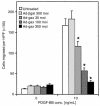
Effect of gax transduction on VSMC migration. Quiescent VSMCs were transduced with Ad-βgal or Ad-gax at an moi of 300 for 24 hours. The migration assay was performed 12 hours later in a chemotaxis chamber for 5 hours with a polycarbonate membrane coated with type I collagen and vitronectin and designated concentrations of PDGF-BB in the lower well. (a) Representative membranes using 10 ng/mL PDGF-BB as chemoattractant and stained with a Giemsa-solution. Untreated (left), Ad-βgal–transduced (middle), and Ad-gax–transduced (right) cells. (b) Quantitative analysis of the chemotactic response of VSMCs toward PDGF-BB at different concentrations. Results represent the average of 3 experiments. Each condition was performed in quadruplicate, and 3 high-power fields were counted per well. The bars show the mean ± SEM. *P < 0.01 versus Ad-βgal–transduced cells. There was no statistically significant difference between untreated and Ad-βgal–transduced cells. HPF = high power field.
Figure 2
Figure 2
Effect of gax gene dosage on VSMC migration using increasing moi’s. Migratory response of untreated (no virus applied), Ad-βgal–transduced (300 moi), or Ad-gax–transduced (30, 100, and 300 moi) rat VSMCs toward PDGF-BB (10 ng/mL). The experiment was done as described in Figure 1. *P < 0.01 versus Ad-βgal–transduced cells. There was no statistically significant difference between untreated and Ad-βgal–transduced cells. Bars show mean ± SEM.
Figure 3
Figure 3
Transduction of gax does not alter PDGF receptor expression or affinity for ligand. Specific binding curve of [125I]PDGF-BB to rat VSMCs. Subconfluent rat VSMCs were transduced with Ad-βgal or Ad-gax at an moi of 300 for 24 hours. Twelve hours after removal of virus, cell were incubated in binding buffer containing 25–800 pM [125I]PDGF-BB for 2 hours on ice. Triplicate cell lysates were counted using a gamma counter. Nonspecific binding, determined by competition with a 200-fold molar excess of non-radiolabeled PDGF-BB, was subtracted. The graph insert shows the Scatchard plot of both binding curves, indicating a single high-affinity PDGF-BB binding site. No statistically significant differences were obtained for either KD or total receptor number/well (Bmax) between Ad-gax– and Ad-βgal–transduced VSMCs.
Figure 4
Figure 4
Transduction of gax inhibits VSMC migration to multiple growth factors. Effect of adenoviral overexpression of Gax on chemotaxis toward different VSMC growth factors. The migration experiment was performed using PDGF-BB, bFGF, and HGF/SF. The concentration used for each growth factor was previously determined to provide the optimal migratory response. *P < 0.01 versus Ad-βgal–transduced cells. Bars show mean ± SEM.
Figure 5
Figure 5
Transduction of cell-cycle regulatory genes p21 and p16 does not inhibit VSMC migration. Effect of adenoviral overexpression of p16 (Ad-p16) or p21CIP1 (Ad-p21) on chemotaxis of rat VSMCs toward PDGF-BB. Subconfluent rat VSMCs were transduced with Ad-βgal, Ad-p16, or Ad-p21 at an moi of 300 for 24 hours. The migration experiment was performed 12 hours after removal of virus, using PDGF-BB as chemoattractant. Bars show mean ± SEM. The differences between uninfected and Ad-βgal–, Ad-p16–, and Ad-p21–transduced cells are not statistically significant.
Figure 6
Figure 6
The cdk inhibitor p21 is essential for Gax-mediated inhibition of migration. Effect of adenoviral overexpression of Gax at different moi’s (30, 100, 300) compared with Ad-βgal (moi 300) on the migratory response toward PDGF-BB (10 ng/mL) of wild-type 3T3 cells, p53–/–MEFs, and p21–/– MEFs, derived from homozygous disruption of the gene encoding p53 and p21, respectively. The experiment was done as described in Figure 1. *P < 0.01 versus Ad-βgal–transduced cells. Bars show mean ± SEM. Migration of untreated cells did not differ from that of Ad-βgal–infected cells (not shown).
Figure 7
Figure 7
The antimigratory effect of Gax in p21–/– cells can be rescued by reconstitution with p21 or by overexpression of p16. Effect of adenoviral overexpression of Gax, p21, or a combination of Gax and p21 or Gax and p16 at different moi’s (30, 100, or 300 each) is compared with the effect of Ad-βgal (moi 300) on the migratory response of p21–/– MEFs toward 10 ng/mL PDGF-BB. The bars indicate the percentage of cells migrated compared with their untreated controls. The experiment was done as described in Figure 1. *P < 0.01 versus Ad-βgal–transduced cells. Bars show mean ± SEM.
Figure 8
Figure 8
Gax downregulates αvβ3 and αvβ5 expression on VSMCs. (a) Comparison of integrin signal intensities on rat VSMCs and HUVECs by flow cytometric analysis. (b) Flow cytometric analysis of integrin expression in rat VSMCs after transduction with Ad-βgal (upper row) or Ad-gax (lower row). Quiescent cells were transduced at an moi of 300 for 24 hours, and fresh medium was added for an additional 12 hours. VSMCs were harvested with EDTA, incubated with anti-integrin antibodies for 45 minutes followed by incubation with FITC-conjugated anti-mouse IgG, and analyzed by flow cytometry. The histograms show fluorescence intensity (x axis) plotted against number of events (y axis). The fluorescence of the corresponding isotype-matched control IgG appears on the left of each histogram.
Figure 9
Figure 9
Gax specifically downregulates expression of β3 and β5 integrin subunits in rat and human VSMCs. Cultured VSMCs (rat: left panels; human: right panels) were infected with adenovirus at an moi of 300 (Ad-gax: Gax; Ad-βgal: β-gal) or uninfected (mock) in low-serum media (0.5% FBS) for 12 hours. After infection, cell were incubated with 10% FBS–containing media for 24 hours as described in Methods. Whole-cell extracts (30 μg protein) were prepared from the cultures and subjected to SDS-PAGE on 7.5% polyacrylamide gels. Immunoblot analysis was performed for each indicated integrin subtype (β3, β5, β1, α1) or tubulin as a control, and immunoreactive bands were viewed by chemiluminescence.
Figure 10
Figure 10
Transduction of the gax gene inhibits injury-induced integrin expression and intimal hyperplasia in rat carotid arteries. (a) Upregulation of β3 integrin and β5 integrin is inhibited by adenovirus-mediated gax gene transfer. Arteries were harvested 3 days after balloon injury and incubation with the indicated adenoviral construct (βgal or Gax) or saline. Uninjured, contralateral arteries served as a control. β3 and β5 integrin antibody immunoreactivity was detected in frozen sections using SuperSensitive Immuno Detection System (red). Each specimen was counterstained with hematoxylin (blue). (b) Immunoblot analysis was performed using extracts (30 μg) prepared from rat carotid arteries treated as described in the text. Immunoreactivity to integrin β3, β5, and tubulin was detected by chemiluminescence. (c) Transduction of the gax gene inhabits neointima formation at 2 weeks after injury and gene transfer. The ratio of intimal (I) to medial (M) area is shown.
Figure 11
Figure 11
The cdk inhibitor p21 is essential for Gax-mediated downregulation of integrin expression, and reconstitution of p21–/– cells with p21, or overexpression of p16, will restore the ability of Gax to suppress integrin expression. (a) Flow cytometric analysis of αvβ3 integrin expression in p21–/– MEFs after transduction with Ad-βgal, Ad-p21, Ad-gax or a combination of Ad-gax and Ad-p21 at an moi of 300. The experiment was done as described in Figure 8. (b) Integrin β1, αvβ3, and αvβ5 expression in p21–/– cells after transduction with either Ad-βgal, Ad-p21, Ad-p16 or Ad-gax alone, or Ad-gax in combination with Ad-p21 or Ad-p16. Cells were transduced at an moi of 300 for 24 hours followed by addition of fresh medium for further 12 hours. Bars show mean ± SEM from 3 experiments. *P < 0.01 versus Ad-βgal–transduced cells.

Similar articles

Cited by

References

    1. Niehrs C, Keller R, Cho KWY, De Robertis EM. The homeobox gene goosecoid controls cell migration in Xenopus embryos. Cell. 1993;72:491–503. - PubMed
    1. Salser SJ, Kenyon C. Activation of a C. elegans Antennapedia homologue in migrating cells controls their direction of migration. Nature. 1992;355:255–258. - PubMed
    1. Gorski DH, Patel CV, Walsh K. Homeobox transcription factor regulation in the cardiovascular system. Trends Cardiovasc Med. 1993;3:184–190. - PubMed
    1. Olson EN, Rosenthal N. Homeobox genes and muscle patterning. Cell. 1994;79:9–12. - PubMed
    1. Scott MP, Tamkun JW, Hartzell GW. The structure and function of the homeodomain. Biochim Biophys Acta. 1989;989:25–48. - PubMed

Publication types

MeSH terms