. 1999 Nov 15;190(10):1405-16.
doi: 10.1084/jem.190.10.1405.

Restricted immunoglobulin variable region (Ig V) gene expression accompanies secondary rearrangements of light chain Ig V genes in mouse plasmacytomas

Affiliations

Restricted immunoglobulin variable region (Ig V) gene expression accompanies secondary rearrangements of light chain Ig V genes in mouse plasmacytomas

L Diaw et al. J Exp Med. .

Abstract

The many binding studies of monoclonal immunoglobulin (Ig) produced by plasmacytomas have found no universally common binding properties, but instead, groups of plasmacytomas with specific antigen-binding activities to haptens such as phosphorylcholine, dextrans, fructofuranans, or dinitrophenyl. Subsequently, it was found that plasmacytomas with similar binding chain specificities not only expressed the same idiotype, but rearranged the same light (V(L)) and heavy (V(H)) variable region genes to express a characteristic monoclonal antibody. In this study, we have examined by enzyme-linked immunosorbent assay five antibodies secreted by silicone-induced mouse plasmacytomas using a broader panel of antigens including actin, myosin, tubulin, single-stranded DNA, and double-stranded DNA. We have determined the Ig heavy and light chain V gene usage in these same plasmacytomas at the DNA and RNA level. Our studies reveal: (a) antibodies secreted by plasmacytomas bind to different antigens in a manner similar to that observed for natural autoantibodies; (b) the expressed Ig heavy genes are restricted in V gene usage to the V(H)-J558 family; and (c) secondary rearrangements occur at the light chain level with at least three plasmacytomas expressing both kappa and lambda light chain genes. These results suggest that plasmacytomas use a restricted population of B cells that may still be undergoing rearrangement, thereby bypassing the allelic exclusion normally associated with expression of antibody genes.


Figures
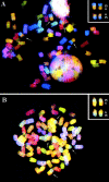
Figure 1
Figure 1
SKY™ images of SIPC tumors with variant T(6;15) chromosomal translocations, (A) SIPC3308 and (B) SIPC3282. The arrows mark chromosomal translocations that have occurred between CHR6 (yellow) and CHR15 (blue). There are two copies of each T(6;15) and the reciprocal T(15;6).
Figure 3
Figure 3
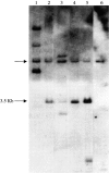
JH rearrangements in SIPC tumors. Genomic DNAs from SIPC tumors and BALB/c liver were digested with EcoR1 and hybridized with the JH probe: lane 1, SIPC3385; lane 2, SIPC3308; lane 3, SIPC3301; lane 4, SIPC3336; lane 5, SIPC3282; and lane 6, BALB/c. An arrow (left) depicts the germline (nonrearranged) JH fragment, whereas the 3.5-kb EcoR1 rearrangement seen in several tumors is also highlighted.
Figure 2
Figure 2
Southern hybridization assay for c-Myc and Pvt 1 rearrangements in SIPC tumors. Shown top (c-Myc, EcoR1) and bottom (Pvt 1, BamH1) are the following tumors: lane 1, SIPC3385; lane 2, SIPC3308; lane 3, SIPC3301; lane 4, SIPC3336; lane 5, SIPC3282; and lane 6, BALB/c. Arrows depict the germline (nonrearranged fragments). Two samples (SIPC3385 and SIPC3301) contain rearranged bands with c-Myc, whereas no rearrangements are found with Pvt 1.
Figure 4
Figure 4
Nucleotide sequences of VH genes from SIPC tumors. The sequences obtained with the five SIPC tumors have been compared with the most homologous germline genes which are members of the VH-J558 family. Top: SIPC3336, SIPC3308, and SIPC3282 express VH13-3; middle: SIPC3301 expresses 26.4.1-α; bottom: SIPC3385 expresses 4ma. Dashes indicate sequence identities. Germline amino acids in bold indicate that they are replaced in SIPCs. These sequence data are available from EMBL/GenBank/DDBJ under accession nos. AF154880–AF154882, AF154909, and AF154910.
Figure 5
Figure 5
IgL rearrangements of SIPC tumors. Genomic DNAs from SIPC tumors and BALB/c liver were digested with EcoR1 and hybridized with the IVS probe: lane 1, SIPC3385; lane 2, SIPC3308; lane 3, SIPC3301; lane 4, SIPC3336; lane 5, SIPC3282; and lane 6, BALB/c. An arrow (left) depicts the germline (nonrearranged) Igκ fragment, whereas the 18 and 9.5-kb EcoR1 rearrangements seen in SIPC3308, SIPC3336, and SIPC3282 are also highlighted (right).
Figure 6
Figure 6
Nucleotide sequence of the Vκ24C-Jκ4 gene from the SIPC tumors. Sequences obtained from SIPC3282, SIPC3336, and SIPC3308 (bottom line) have been compared to that of the germline Vκ24C (reference 15). Dashes indicate sequence identities. Substitutions at the amino acid level are indicated in bold. These sequence data are available from EMBL/GenBank/DDBJ under accession no. AF154911.
Figure 7
Figure 7
Nucleotide sequences of Vκ1 genes expressed in SIPC tumors. Sequences were obtained from individual clones (designated A–F) of SIPC3282, SIPC3301, SIPC3336, SIPC3308, and SIPC3385. Although multiple subclones with identical sequences were obtained, only representative sequences that differ are shown. The sequences are compared with the most homologous germline gene K5.1 (Vκ1A, top) or K1A5 (Vκ1C, bottom). Dashes indicate sequence identities, and amino acid substitutions are indicated in bold. These sequence data are available from EMBL/GenBank/DDBJ under accession nos. AF154883–AF154898.
Figure 8
Figure 8
Nucleotide sequences of Vλ1 genes expressed in SIPC tumors. Nucleotide sequences of subclones (designated A–F) of Vλ genes from SIPC3282, SIPC3336, and SIPC3301 are compared with the most homologous germline gene. Dashes indicate sequence identities, and amino acid substitutions are indicated in bold. These sequence data are available from EMBL/GenBank/DDBJ under accession nos. AF154899–AF154908.
Figure 9
Figure 9
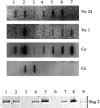
Transcript levels of Igκ, Igλ, and RAG-2 in SIPC tumors. (Top) Equal amounts of RNA from mouse liver (lane 1), spleen (lane 2), SIPC3301 (lane 3), SIPC3308 (lane 4), SIPC3282 (lane 5), SIPC3336 (lane 6), and SIPC3385 (lane 7) were loaded into slots and hybridized to nick-translated probes from Vκ24 (row 1), Vκ1 (row 2), Cκ (row 3), or Vλ (row 4). (Bottom) RNA from mouse spleen (lane 1), thymus (lane 2), SIPC3282 (lane 3), SIPC3301 (lane 4), SIPC3308 (lane 5), SIPC3336 (lane 6), SIPC3385 (lane 7), ABPC18 (lane 8), and MOPC104E (lane 9) was reverse transcribed (RT) and PCR amplified using primers specific for RAG-2.

Similar articles

Cited by

References

    1. Potter M., Wiener F. Plasmacytomagenesis in micemodel of neoplastic development dependent upon chromosomal translocations. Carcinogenesis. 1992;13:1681–1697. - PubMed
    1. Potter M. Antigen-binding myeloma proteins of mice. Adv. Immunol. 1977;25:141–211. - PubMed
    1. Guilbert B., Dighiero G., Avrameas S. Naturally occurring antibodies against nine common antigens in human sera. I. Detection, isolation and characterization. J. Immunol. 1982;128:2779–2787. - PubMed
    1. Dighiero G., Lymberi P., Mazie J.C., Rouyre S., Butler B.G., Whalen R.G., Avrameas S. Murine hybridomas secreting natural monoclonal antibodies reacting with self antigens. J. Immunol. 1983;131:2267–2272. - PubMed
    1. Diaw L., Magnac C., Pritsch O., Buckle M., Alzari P.M., Dighiero G. Structural and affinity studies of IgM polyreactive natural autoantibodies. J. Immunol. 1997;158:968–976. - PubMed

Publication types

Associated data