. 1999 Nov 15;190(10):1417-26.
doi: 10.1084/jem.190.10.1417.

In breast carcinoma tissue, immature dendritic cells reside within the tumor, whereas mature dendritic cells are located in peritumoral areas

Affiliations

In breast carcinoma tissue, immature dendritic cells reside within the tumor, whereas mature dendritic cells are located in peritumoral areas

D Bell et al. J Exp Med. .

Abstract

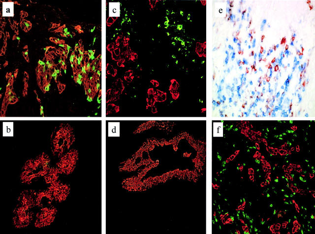
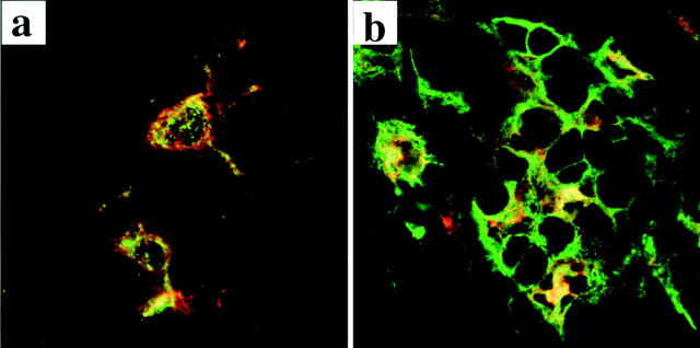
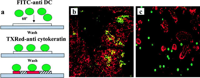
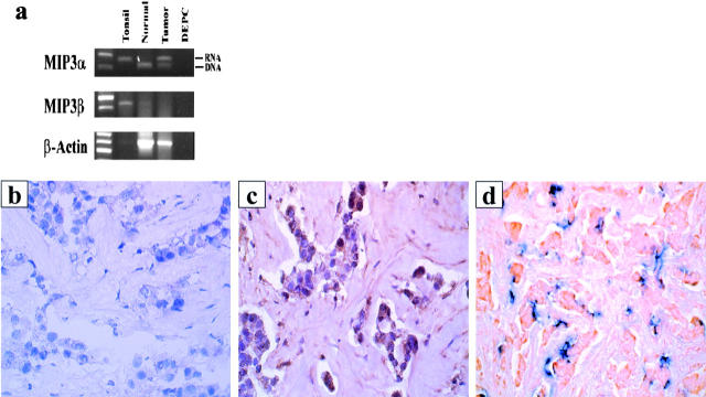
We have analyzed the presence of immature and mature dendritic cells (DCs) within adenocarcinoma of the breast using immunohistochemistry. Immature DCs were defined by expression of CD1a-, Langerin-, and intracellular major histocompatibility complex class II-rich vesicles. Mature DCs were defined by expression of CD83 and DC-Lamp. Breast carcinoma cells were defined by morphology and/or cytokeratin expression. We demonstrate two levels of heterogeneity of DCs infiltrating breast carcinoma tissue: (a) immature CD1a(+) DCs, mostly of the Langerhans cell type (Langerin(+)), were retained within the tumor bed in 32/32 samples and (b) mature DCs, CD83(+)DC-Lamp(+), present in 20/32 samples, are confined to peritumoral areas. The high numbers of immature DCs found in the tumor may be best explained by high levels of macrophage inflammatory protein 3alpha expression by virtually all tumor cells. Confirming the immature/mature DC compartmentalization pattern, in vitro-generated immature DCs adhere to the tumor cells, whereas mature DCs adhere selectively to peritumoral areas. In some cases, T cells are clustering around the mature DCs in peritumoral areas, thus resembling the DC-T cell clusters of secondary lymphoid organs, which are characteristic of ongoing immune reactions.


Figures

Figure 1
Figure 1
In breast carcinoma tissue, immature DCs are intratumoral, whereas mature DCs and T cells are peritumoral. Immunofluorescence and immunohistochemistry of frozen sections of breast carcinoma tissue. Breast carcinoma is defined by indirect staining with anticytokeratin antibody (red) or by morphology. Immature DCs were identified by labeling with CD1a mAb, mature DCs were identified by labeling with CD83 or DC-Lamp mAb, and T cells were labeled using CD3 or CD4 mAb. (a) Immature, CD1a+ DCs (green) are retained within the tumor bed (40×). Representative of 32 samples (patient 17). (b) In the normal mammary epithelium, CD1a staining is seldom detected. (c) Mature, DC-Lamp–labeled DCs (green) are selectively localized in peritumoral areas (20×). Pattern representative of 25 samples (patient 11). (d) No mature DCs, as judged by lack of CD83 staining, are present in the normal breast tissue. (e) CD4+ T cells (blue) cluster around CD83+ mature DCs (red; 40×) (patient 6). DC–T cell clusters were seen in three out of five cases. (f) CD3+ T cells (green) are spread within the peritumoral area. Representative of 19 cases (patient 11);in a majority of these cases, T cells were CD8+.
Figure 2
Figure 2
In breast carcinoma tissue, immature DCs express intracellular MHC class II vesicles, whereas mature DCs express surface MHC class II molecules. Immunofluorescence staining on frozen sections evaluated by confocal microscopy as described in the Fig. 1 legend. (a) MHC class II molecules, stained with directly coupled L243 mAb (green), mostly reside in intracellular compartments in DCs identified by labeling with CD1a mAb (red) within the tumor bed. (b) Mature DCs, identified with lysosome resident marker DC-Lamp (red), express high numbers of cell surface MHC class II molecules (green). This pattern has been observed in three out of three analyzed samples.
Figure 3
Figure 3
Immature DCs selectively adhere to tumor cells, whereas mature DCs adhere to tumor parenchyma. (a) Schematic presentation of DC binding experiments using a modified version of the Woodruff-Stamper assay. DCs generated in vitro from either CD34+ HPCs or blood monocytes were FITC labeled and overlaid (105 cells) on frozen sections of breast carcinoma tissue. After 60 min at 20°C and stringent washing, the sections were labeled with anticytokeratin antibody (Texas [TX]Red). (b) Immature CD34+, HPC–derived, sorted CD1a+ DCs adhere selectively to cytokeratin-labeled breast cancer cells (20×; patient 17). (c) Mature DCs, generated from blood monocytes and activated by CD40 ligation, adhere selectively to tumor stroma (20×; patient 11). Representative of six experiments using breast carcinoma tissue from six patients.
Figure 4
Figure 4
Breast carcinoma cells express MIP3α mRNA and protein. (a) Expression of MIP3α and MIP3β was determined by RT-PCR using total RNA extracted from fresh breast carcinoma tissue. Immunohistochemistry staining with a control (b) or anti-MIP3α (c) antibody reveals specific MIP3α staining of breast carcinoma cells (20×; patient 28). Representative pattern of MIP3α expression from seven samples. (d) CD1a-labeled immature DCs (blue) colocalize with MIP3α-expressing breast cancer cells (red) (patient 28).

Similar articles

Cited by

References

    1. Cella M., Sallusto F., Lanzavecchia A. Origin, maturation and antigen presenting function of dendritic cells. Curr. Opin. Immunol. 1997;9:10–16. - PubMed
    1. Hart D.N. Dendritic cellsunique leukocyte populations which control the primary immune response. Blood. 1997;90:3245–3287. - PubMed
    1. Banchereau J., Steinman R.M. Dendritic cells and the control of immunity. Nature. 1998;392:245–252. - PubMed
    1. Bell D., Young J.W., Banchereau J. Dendritic cells. Adv. Immunol. 1999;72:255–324. - PubMed
    1. Toes R.E.M., Ossendorp F., Offringa R., Melief C.J.M. CD4 T cells and their role in antitumor immune responses. J. Exp. Med. 1999;189:753–756. - PMC - PubMed

Publication types