

Type I collagen synthesis and degradation in peritendinous tissue after exercise determined by microdialysis in humans
- PMID: 10562353
- PMCID: PMC2269635
- DOI: 10.1111/j.1469-7793.1999.00299.x
Type I collagen synthesis and degradation in peritendinous tissue after exercise determined by microdialysis in humans
Abstract
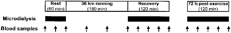
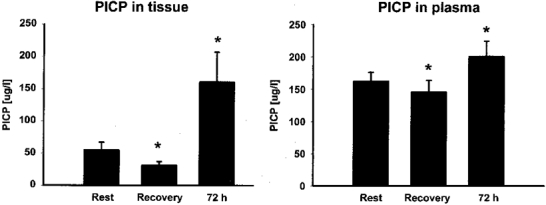
1. Physical activity is known to increase type I collagen synthesis measured as the concentration of biomarkers in plasma. By the use of microdialysis catheters with a very high molecular mass cut-off value (3000 kDa) we aimed to determine local type I collagen synthesis and degradation in the peritendinous region by measuring interstitial concentrations of a collagen propeptide (PICP; 100 kDa) and a collagen degradation product (ICTP; 9 kDa) as well as an inflammatory mediator (PGE2). 2. Seven trained human runners were studied before and after (2 and 72 h) 3 h of running (36 km). Two microdialysis catheters were placed in the peritendinous space ventral to the Achilles' tendon under ultrasound guidance and perfused with a Ringer-acetate solution containing 3H-labelled human type IV collagen and [15-3H(N)]PGE2 for in vivo recovery determination. Relative recovery was 37-59 % (range of the s.e.m. values) for both radioactively labelled substances. 3. PICP concentration decreased in both interstitial peritendinous tissue and arterial blood immediately after exercise, but rose 3-fold from basal 72 h after exercise in the peritendinous tissue (55 +/- 10 microg l-1, mean +/- s.e.m. (rest) to 165 +/- 40 microg l-1 (72 h), P < 0.05) and by 25 % in circulating blood (160 +/- 10 microg l-1 (rest) to 200 +/- 12 microg l-1 (72 h), P < 0.05). ICTP concentration did not change in blood, but decreased transiently in tendon-related tissue during early recovery after exercise only. PGE2 concentration increased in blood during running, and returned to baseline in the recovery period, whereas interstitial PGE2 concentration was elevated in the early recovery phase. 4. The findings of the present study indicate that acute exercise induces increased formation of type I collagen in peritendinous tissue as determined with microdialysis and using dialysate fibre with a very high molecular mass cut-off. This suggests an adaptation to acute physical loading also in non-bone-related collagen in humans.
Figures


Comment in
-
Teasing out the truth about collagen.J Physiol. 1999 Nov 15;521 Pt 1(Pt 1):1. doi: 10.1111/j.1469-7793.1999.00001.x. J Physiol. 1999. PMID: 10562329 Free PMC article.
References
-
- Almekinders LC, Banes AJ, Ballenger CA. Effects of repetitive motion on human fibroblasts. Medicine and Science in Sports and Exercise. 1993;25:603–607. - PubMed
-
- Almekinders LC, Banes AJ, Bracey LW. An in vitro investigation into the effects of repetitive motion and nonsteroidal antiinflammatory medication on human tendon fibroblasts. American Journal of Sports Medicine. 1995;23:119–123. - PubMed
-
- Ashizawa N, Ouchi G, Fujimura R, Yoshida Y, Tokuyama K, Suzuki M. Effects of a single bout of resistance exercise on calcium and bone metabolism in untrained young males. Calcified Tissue International. 1998;62:104–108. - PubMed
-
- Banes AJ, Tsuzaki M, Hu P, Brigman B, Brown T, Almekinders L, Lawrence WT, Fischer T. PDGF-BB, IGF-I and mechanical load stimulate DNA synthesis in avian tendon fibroblasts in vitro. Journal of Biomechanics. 1995;28:1505–1513. - PubMed
-
- Brahm H, Piehl-Aulin K, Ljunghall S. Biochemical markers of bone metabolism during distance running in healthy, regularly exercising men and women. Scandinavian Journal of Medicine and Science in Sports. 1996;6:26–30. - PubMed
Publication types
MeSH terms
Substances
LinkOut - more resources
Full Text Sources
Other Literature Sources
Medical
Research Materials