

Clinicopathological differences between colonic and rectal carcinomas: are they based on the same mechanism of carcinogenesis?
- PMID: 10562578
- PMCID: PMC1727756
- DOI: 10.1136/gut.45.6.818
Clinicopathological differences between colonic and rectal carcinomas: are they based on the same mechanism of carcinogenesis?
Abstract
Background: There is a difference in the location of colorectal mucosal lesions and invasive cancers.
Aims: To ascertain whether the location of colorectal neoplasms reflects the carcinogenesis pathway.
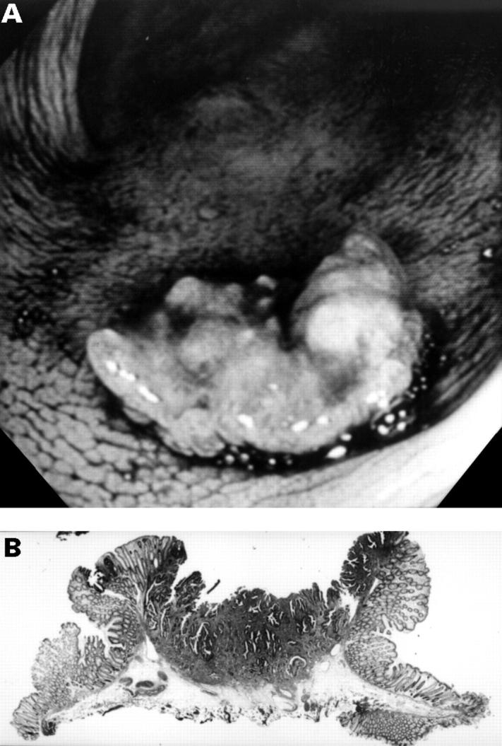
Methods: The subject material consisted of 4147 neoplastic lesions that had been resected endoscopically or surgically from 5025 patients. Mucosal lesions and submucosal cancers were classified into depressed and non-depressed types endoscopically or histologically. The relations between macroscopic type, size, histology, and location were investigated.
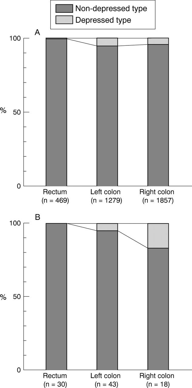
Results: (a) Non-depressed type. A total of 1774 of 3454 (51%) mucosal lesions were located in the right colon, 1212 (35%) in the left colon, and 468 (14%) in the rectum. The incidence of mucosal lesions larger than 10 mm was 10% (185/1774) in the right colon, 21% (254/1212) in the left colon, and 27% (127/468) in the rectum. The incidence of mucosal lesions with villous components was 2% (32/1774) in the right colon, 5% (63/1212) in the left colon, and 13% (62/468) in the rectum. The ratio of submucosal cancers to mucosal lesions was significantly higher in the rectum (0.064, 30/469) than in the left (0.034, 43/1279) or right (0.010, 18/1857) colon. (b) Depressed type. The incidences of depressed type mucosal lesions and submucosal cancers were 5% (83/1857) and 17% (3/18) in the right colon, 5% (67/1279) and 5% (2/43) in the left colon, and 0.2% (1/469) and 0% (0/30) in the rectum, respectively.
Conclusion: There may be some mechanisms that promote the progression of mucosal lesions to invasive cancers in the left colon and rectum, whereas a de novo pathway from depressed type lesions may be implicated in some cancers of the right colon.
Figures

Comment in
-
Colon cancer: the shape of things to come.Gut. 1999 Dec;45(6):794-5. doi: 10.1136/gut.45.6.794. Gut. 1999. PMID: 10562572 Free PMC article. No abstract available.
References
Publication types
MeSH terms
LinkOut - more resources
Full Text Sources