. 1999 Dec;45(6):864-73.
doi: 10.1136/gut.45.6.864.

Role of inducible nitric oxide synthase in trinitrobenzene sulphonic acid induced colitis in mice

Affiliations

Role of inducible nitric oxide synthase in trinitrobenzene sulphonic acid induced colitis in mice

D M McCafferty et al. Gut. 1999 Dec.

Abstract

Background: Studies using inhibitors of nitric oxide synthase (NOS) to date are inconclusive regarding the role of inducible NOS (iNOS) in intestinal inflammation.

Aims: (1) To examine the role of iNOS in the development of chronic intestinal inflammation; (2) to identify the cellular source(s) of iNOS.

Methods: Colitis was induced by an intrarectal instillation of trinitrobenzene sulphonic acid (TNBS, 60 mg/ml, 30% ethanol), in wild type (control) or iNOS deficient mice. Mice were studied over 14 days; the colons were scored for injury and granulocyte infiltration was quantified. Blood to lumen leakage of (51)Cr-EDTA was measured as a quantitative index of mucosal damage.

Results: At 24 and 72 hours, iNOS deficient mice had significantly increased macroscopic inflammation compared with wild type mice. Granulocyte infiltration increased significantly at 24 hours and remained elevated in iNOS deficient mice at 72 hours, but significantly decreased in controls. However, by seven days post-TNBS macroscopic damage, microscopic histology, granulocyte infiltration, and mucosal permeability did not differ between wild type and iNOS deficient mice. A four- to fivefold increase in iNOS mRNA was observed in wild type mice at 72 hours and seven days post-TNBS and was absent in iNOS deficient mice. Immunohistochemistry techniques showed that iNOS expression was predominantly localised in neutrophils, with some staining also in macrophages.

Conclusions: These results suggest that leucocyte derived iNOS ameliorates the early phase, but does not impact on the chronic phase of TNBS induced colitis despite the presence of iNOS.


Figures

Figure 1
Figure 1
(A) A representative 2% agarose gel of RT-PCR products. Base pair markers (M) denoting DNA size are shown in the far left lane. Glyceraldehyde-3-phosphate dehydrogenase (GAPDH; internal marker) and inducible nitric oxide synthase (iNOS) RT-PCR products are shown for non-inflamed (without trinitrobenzene sulphonic acid (−TNBS)) or inflamed (+TNBS) wild type (iNOS+/+) and iNOS deficient (iNOS−/−) tissue. (B) Mean data (n=4) after semiquantitative analysis of the iNOS band intensity expressed as a ratio of the GAPDH band for the various groups. The intensity of iNOS band in non-inflamed mice was equated to one and the other bands expressed as fold increase over this level. *Significantly greater than wild type non-inflamed group.
Figure 2
Figure 2
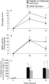
(A) Macroscopic damage score, (B) myeloperoxidase (MPO) activity, and (C) blood to lumen leakage of 51Cr-EDTA leakage as an indication of mucosal permeability in colonic tissue. n⩾6 in all groups. *Significant increase from healthy mice; δ significant decrease from 24 hour time point; τ significant increase from wild type mice. iNOS, inducible nitric oxide synthase.
Figure 3
Figure 3
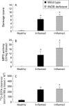
(A) Macroscopic damage score, (B) myeloperoxidase activity (MPO), and (C) blood to lumen leakage of 51Cr-EDTA leakage as an indication of mucosal permeability in colonic tissue from healthy or inflamed wild type or inducible nitric oxide synthase (iNOS) deficient mice. n⩾6 in all groups. *Significant increase from healthy mice. There were no significant differences between iNOS deficient and wild type animals after seven days of inflammation.
Figure 4
Figure 4
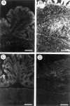
Fluorescence micrographs of inducible nitric oxide (iNOS) immunoreactivity (A and C) in healthy wild type animals. The same sections were double labelled to examine the cellular source of iNOS (B and D). Little or no iNOS was present in normal animals in macrophages (B). However, occasional small clusters of iNOS immunoreactive neutrophils (D) were observed in otherwise healthy tissue. Scale bar: 100 µm.
Figure 5
Figure 5
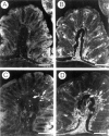
Fluorescence micrographs of inducible nitric oxide (iNOS) immunoreactivity in representative sections from a healthy wild type mouse (A), healthy iNOS deficient mouse (C), inflamed wild type mouse (B), and inflamed iNOS deficient mouse (D). There was a substantial increase in iNOS immunoreactivity in inflamed mouse colon seven days after treatment with trinitrobenzene sulphonic acid in wild type mice (B). No iNOS immunoreactivity was observed in the iNOS deficient mice (C and D). Scale bar: 100 µm.
Figure 6
Figure 6
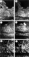
Fluorescence micrographs of inducible nitric oxide (iNOS) immunoreactivity (A, C, and E) in wild type animals seven days after treatment with trinitrobenzene sulphonic acid. The same sections were double labelled to examine the cellular source of iNOS (B, D, and F). Though there was an overlapping distribution, there were relatively few macrophages that expressed iNOS immunoreactivity (B). In contrast, iNOS immunoreactivity was extensively colocalised in neutrophils (D and F) in inflamed tissues. Scale bar: 100 µm (A-D), 50 µm (E and F).

References

    1. Am J Physiol. 1994 Feb;266(2 Pt 1):G222-9 - PubMed
    1. Proc Natl Acad Sci U S A. 1993 Nov 1;90(21):9813-7 - PubMed
    1. Am J Physiol. 1994 Sep;267(3 Pt 2):H931-7 - PubMed
    1. Cell. 1994 Sep 23;78(6):915-8 - PubMed
    1. J Pharmacol Exp Ther. 1994 Nov;271(2):1114-21 - PubMed

Publication types