. 1999 Nov;10(11):3835-48.
doi: 10.1091/mbc.10.11.3835.

Uptake and intracellular transport of acidic fibroblast growth factor: evidence for free and cytoskeleton-anchored fibroblast growth factor receptors

Affiliations
Free PMC article

Uptake and intracellular transport of acidic fibroblast growth factor: evidence for free and cytoskeleton-anchored fibroblast growth factor receptors

L Citores et al. Mol Biol Cell. 1999 Nov.
Free PMC article

Abstract

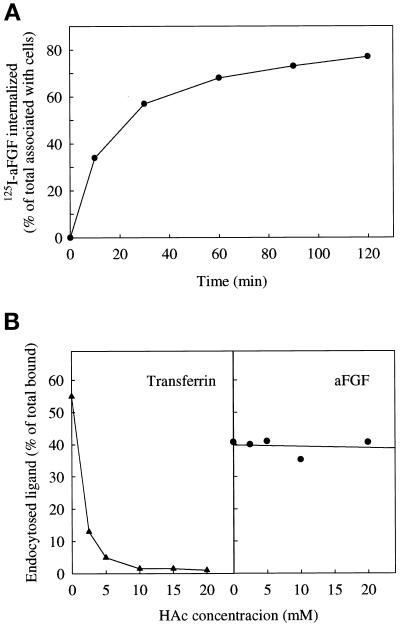
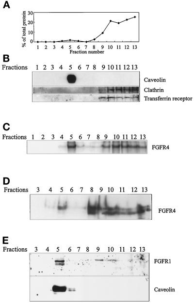
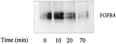
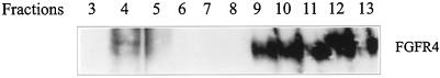
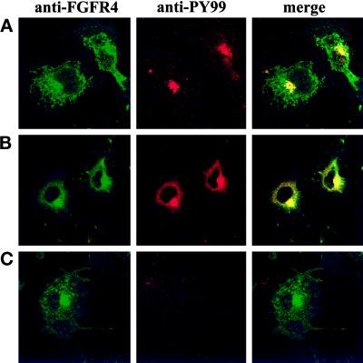
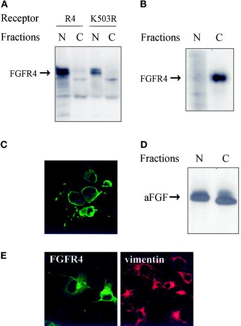
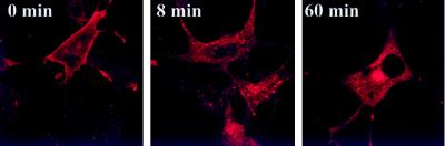
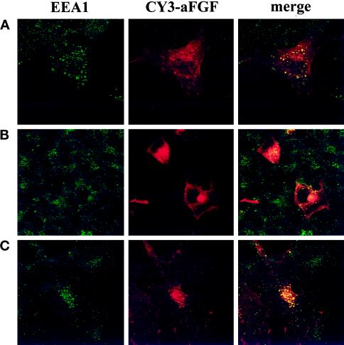
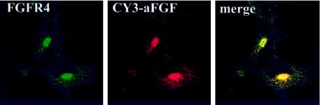
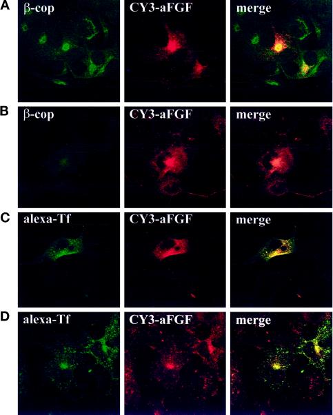
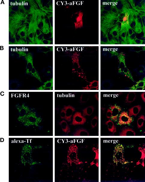
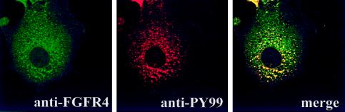
Endocytic uptake and intracellular transport of acidic FGF was studied in cells transfected with FGF receptor 4 (FGFR4). Acidification of the cytosol to block endocytic uptake from coated pits did not inhibit endocytosis of the growth factor in COS cells transfected with FGFR4, indicating that it is to a large extent taken up by an alternative endocytic pathway. Fractionation of the cells demonstrated that part of the growth factor receptor was present in a low-density, caveolin-containing fraction, but we were unable to demonstrate binding to caveolin in immunoprecipitation studies. Upon treatment of the cells with acidic FGF, the activated receptor, together with the growth factor, moved to a juxtanuclear compartment, which was identified as the recycling endosome compartment. When the cells were lysed with Triton X-100, 3-([3-chloramidopropyl]dimethylammonio)-2-hydroxy-1-propanesulfona te, or 2-octyl glucoside, almost all surface-exposed and endocytosed FGFR4 was solubilized, but only a minor fraction of the total FGFR4 in the cells was found in the soluble fraction. The data indicate that the major part of FGFR4 is anchored to detergent-insoluble structures, presumably cytoskeletal elements associated with the recycling endosome compartment.


Figures

Figure 1
Figure 1
Internalization of aFGF in cells transfected with FGFR4. (A) COS cells transiently transfected with FGFR4 were incubated at 37°C for the indicated periods of time in HEPES containing 10 U/ml heparin and 50 ng/ml 125I-aFGF. Then the cells were washed with PBS and treated for 6 min with 20 mM Na-acetate, pH 4, containing 2 M NaCl to remove surface-bound aFGF. The removed material was collected. The cells were subsequently dissolved, and the radioactivity in the medium and in the cells was measured. The data are expressed as the internalized radioactivity as a percentage of the total radioactivity associated with the cells. (B) Uptake of 125I-transferrin (▴) and 125I-aFGF (●) by COS cells transfected with FGFR4. The cells were incubated for 5 min at 37°C in HEPES medium, pH 5.5, with increasing concentrations of acetic acid. 125I-Transferrin or 125I-aFGF was then added as described above, and after 5 and 15 min of incubation, respectively, the amount of endocytosed proteins was measured as in A.
Figure 2
Figure 2
FGFRs partly comigrate with caveolin. (A) COS cells were homogenized in sodium carbonate, sonicated, and fractionated by floating in a sucrose density gradient. The protein content of each fraction is expressed as a percentage of the total amount of protein in the gradient. (B) Aliquots of the fractions in A were analyzed by SDS-PAGE and Western blotting with anti-caveolin, anti-clathrin, and anti-transferrin receptor antibodies. (C) COS cells transfected with FGFR4 were incubated with 125I-aFGF for 3 h at 4°C and treated for 20 min at 4°C with 0.3 mM disuccinimidyl suberate to cross-link the bound growth factor to the receptors. Then the cells were homogenized, sonicated, and fractionated as described above. The fractions were analyzed by SDS-PAGE and autoradiography. COS cells transfected with FGFR4 (D) and CPAE cells (E) were treated and fractionated as described above. The fractions were analyzed by Western blotting with anti-FGFR4 (D) or anti-FGFR1 and anti-caveolin (E).
Figure 3
Figure 3
Kinetics of aFGF-induced disappearance of FGFR4 from the caveolin-rich fraction. COS cells transfected with FGFR4 were grown for 12 h in the absence of serum and then incubated in the presence of 50 ng/ml aFGF for the indicated periods of time. Caveolin-rich fractions were prepared as described in Figure 2. Fractions 4 and 5 were pooled, dialyzed against distilled water, lyophilized, separated by SDS-PAGE, and visualized by immunoblotting with anti-FGFR4 antibodies.
Figure 4
Figure 4
Stimulation of tyrosine kinase activity in caveolin-rich fractions of FGFR4-transfected COS cells. The cells were grown in the absence of serum for 12 h and then incubated in the presence of 50 ng/ml aFGF for 10 min. The cells were fractionated as in Figure 2B, and the fractions were analyzed by SDS-PAGE and immunoblotted with anti-phosphotyrosine antibodies.
Figure 5
Figure 5
Localization of activated FGFR4 in cells. COS cells transfected with FGFR4 (A and B) or the FGFR4-K503R mutant (C) were starved of serum for 5 h at 37°C and then kept for 2 h at 4°C in the absence (A) or in the presence (B and C) of aFGF. The cells were then either fixed (A) or incubated at 37°C for 15 min before fixation (B and C). Double staining was performed with anti-FGFR4 and anti-phosphotyrosine antibodies.
Figure 6
Figure 6
Characterization of the expressed receptor. (A) Western blot analysis of FGFRs in subcellular fractions of COS cells. Transfected COS cells were lysed and fractionated into a nuclear (N) and a cytoplasmic (C) fraction. The fractions were precipitated with trichloroacetic acid and analyzed by SDS-PAGE. Blots were probed with anti-FGFR4 antibodies. (B) Localization of surface-labeled FGFR4. COS cells transfected with FGFR4 were surface labeled by incubating them with Na125I, lactoperoxidase, and H2O2 for 5 min, and then DTT and tyrosine were added to stop the reaction. The cells were subsequently lysed and fractionated into a nuclear (N) and a cytoplasmic (C) fraction. The fractions were sonicated and centrifuged, and the receptors in the supernatants were immunoprecipitated with anti-FGFR4 antibodies. The immunoprecipitates were analyzed by SDS-PAGE and autoradiography. (C) Localization of detergent-insoluble FGFR4. Transfected COS cells were grown on coverslips and extracted in buffer containing 1% Triton X-100 before fixation with 3% paraformaldehyde. Fixed cells were incubated with antibody to FGFR4 followed by FITC-conjugated secondary antibody. (D) Nuclear and cytosolic fractions contain functional FGFRs. COS cells transfected with FGFR4 were lysed and fractionated into a nuclear (N) and a cytoplasmic (C) fraction. Both fractions were carefully sonicated for 5 s, and insoluble material was removed by centrifugation. Then the supernatants were incubated with [35S]methionine-labeled aFGF and 10 U/ml heparin for 3 h at 4°C, immunoprecipitated with anti-FGFR4 antibodies, and analyzed by SDS-PAGE and autoradiography. (E) Effect of transfection on the distribution of vimentin. Transiently transfected COS cells were fixed and double stained with anti-FGFR4 and anti-vimentin antibodies followed by FITC- and CY3-conjugated secondary antibodies, respectively.
Figure 7
Figure 7
Transport of fluorescent aFGF in FGFR4-transfected COS cells. The cells were incubated at 4°C for 2 h with CY3-aFGF in the presence of heparin to allow binding of the growth factor and then either fixed immediately (0 min) or incubated at 37°C for 8 or 60 min before fixation.
Figure 8
Figure 8
Colocalization of aFGF with EEA1, a marker for early endosomes. FGFR4-transfected COS cells were incubated with CY3-aFGF for 8 min (A) or 2 h (B and C) at 37°C (A and B) or 16°C (C) and then fixed and stained with anti-EEA1 antibodies followed by FITC-conjugated secondary antibody.
Figure 9
Figure 9
Colocalization of FGFR4 and endocytosed aFGF. COS cells transfected with FGFR4 were incubated with CY3-aFGF for 2 h at 37°C, and then the cells were fixed and stained with anti-FGFR4.
Figure 10
Figure 10
Colocalization of aFGF with markers for different intracellular organelles. (A and B) FGFR4-transfected COS cells were either untreated (A) or pretreated for 30 min with brefeldin A (B), incubated for 2 h at 37°C with CY3-aFGF, and then fixed and stained for the Golgi marker β-cop. (C and D) COS cells were cotransfected with the human transferrin receptor and FGFR4. The cells were then either left untreated (C) or pretreated for 30 min with 2 μg/ml brefeldin A (D) and incubated with CY3-aFGF for 2 h at 37°C. Subsequently, alexa-transferrin was bound to the cells at 4°C, and then the cells were washed and incubated for 20 min at 37°C and fixed.
Figure 11
Figure 11
Effect of nocodazole on the intracellular localization of aFGF and FGFR4. COS cells transfected with FGFR4 (A–C) or cotransfected with FGFR4 and human transferrin receptor (D) were either left untreated (A) or pretreated for 30 min with 33 μM nocodazole (B–D). In some cases (A and B), the cells were then incubated with CY3-aFGF for 2.5 h at 37°C, fixed, and stained for tubulin. In C, the cells were stained with anti-FGFR4 and anti-tubulin. In D, the cells were treated with CY3-aFGF at 37°C for 2 h, alexa-transferrin was bound at 4°C, and then the cells were incubated for 20 min at 37°C.
Figure 12
Figure 12
Effect of nocodazole on the localization of tyrosine-phosphorylated proteins in aFGF-treated cells transfected with FGFR4. COS cells transfected with FGFR4 were pretreated for 30 min with 33 μM nocodazole and then incubated for 2 h in the presence of 50 ng/ml aFGF. Then the cells were fixed and stained with anti-FGFR4 and anti-PY99 antibodies.
Figure 13
Figure 13
Triton X-100 solubility of internalized aFGF. COS cells transfected with FGFR4 were incubated with [35S]methionine-labeled aFGF for 10 h at 37°C and then washed with PBS and acid-salt buffer to remove surface-bound aFGF. The cells were lysed and fractionated into a nuclear (N) and a cytoplasmic (C) fraction, and then both fractions were sonicated. After removal of insoluble material by centrifugation, FGFR4 with bound labeled aFGF was immunoprecipitated with anti-FGFR4 antibodies and analyzed by SDS-PAGE and fluorography.

Similar articles

Cited by

References

    1. Baldin V, Roman AM, Bosc-Bierne I, Amalric F, Bouche G. Translocation of bFGF to the nucleus is G1 phase cell cycle specific in bovine aortic endothelial cells. EMBO J. 1990;9:1511–1517. - PMC - PubMed
    1. Basilico C, Moscatelli D. The FGF family of growth factors and oncogenes. Adv Cancer Res. 1992;59:115–165. - PubMed
    1. Beck KA, Buchanan JA, Malhotra V, Nelson WJ. Golgi spectrin: identification of an erythroid beta-spectrin homolog associated with the Golgi complex. J Cell Biol. 1994;127:707–723. - PMC - PubMed
    1. Beck KA, Buchanan JA, Nelson WJ. Golgi membrane skeleton: identification, localization and oligomerization of a 195 kDa ankyrin isoform associated with the Golgi complex. J Cell Sci. 1997;110:1239–1249. - PubMed
    1. Bennett V. Ankyrins: adaptors between diverse plasma membrane proteins and the cytoplasm. J Biol Chem. 1992;267:8703–8706. - PubMed

Publication types

MeSH terms

LinkOut - more resources