. 1999 Nov;10(11):3909-26.
doi: 10.1091/mbc.10.11.3909.

The interaction and colocalization of Sam68 with the splicing-associated factor YT521-B in nuclear dots is regulated by the Src family kinase p59(fyn)

Affiliations
Free PMC article

The interaction and colocalization of Sam68 with the splicing-associated factor YT521-B in nuclear dots is regulated by the Src family kinase p59(fyn)

A M Hartmann et al. Mol Biol Cell. 1999 Nov.
Free PMC article

Abstract

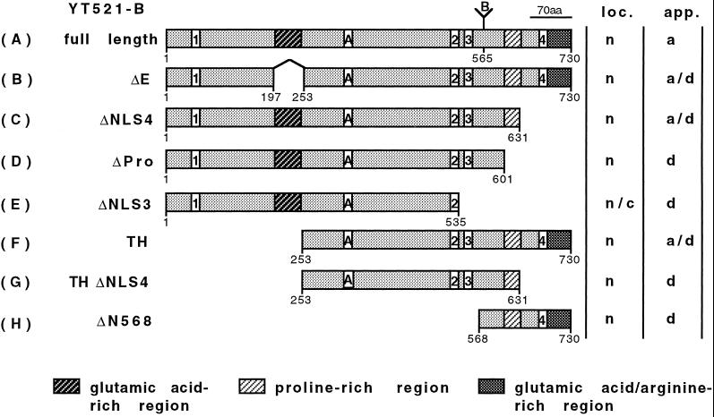
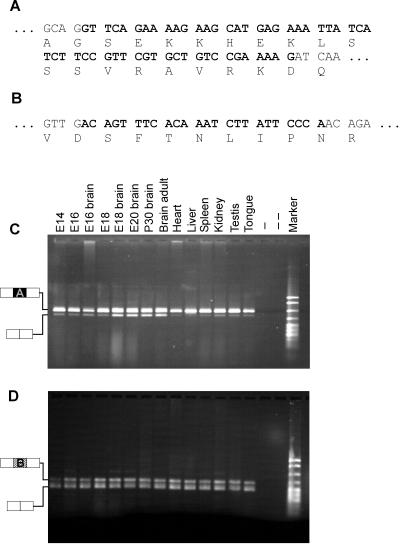
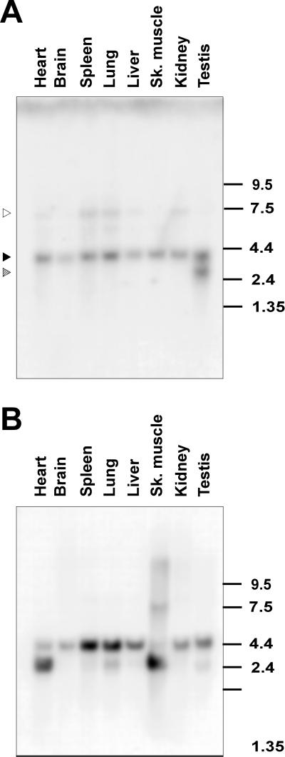
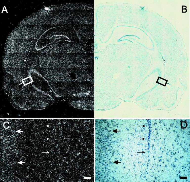
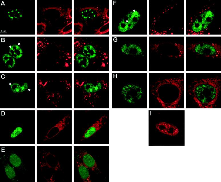
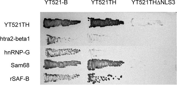
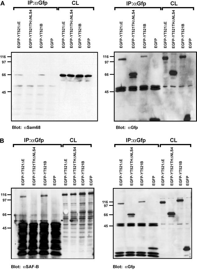
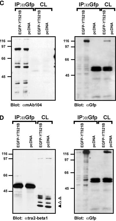
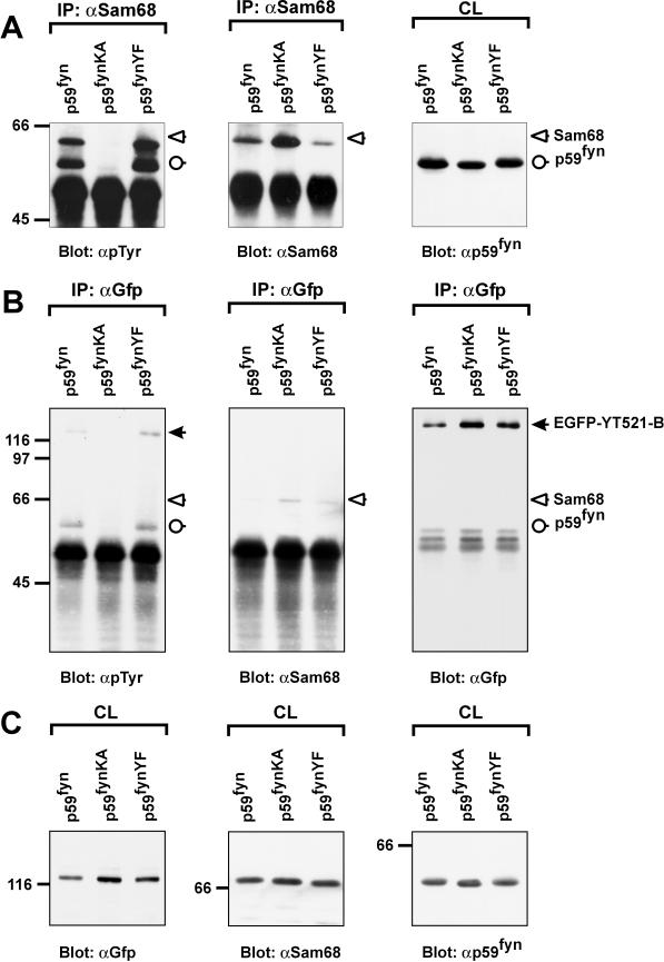
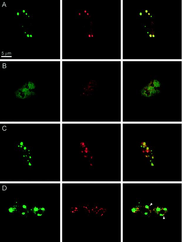
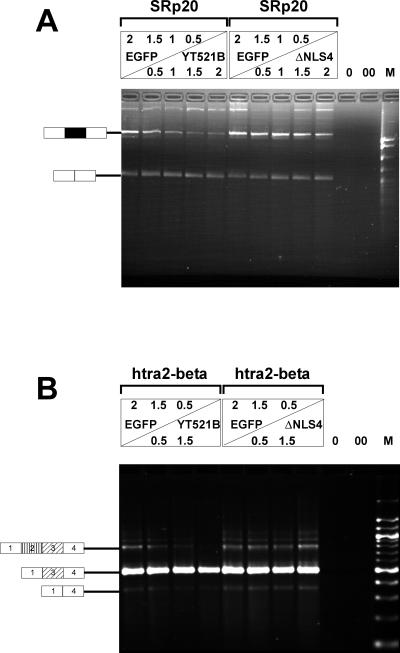
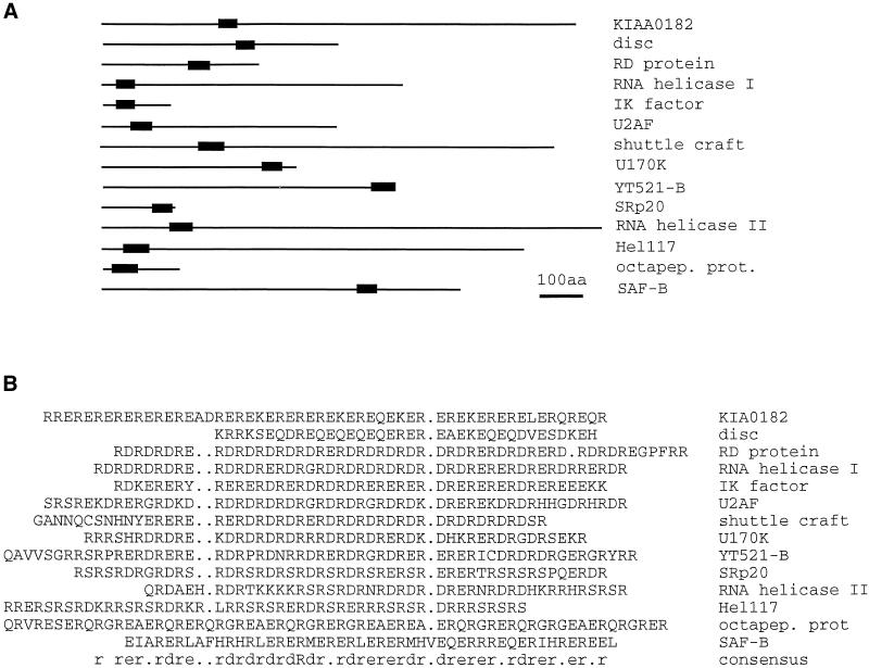
Alternative pre-mRNA splicing patterns can change an extracellular stimulus, but the signaling pathways leading to these changes are still poorly characterized. Here, we describe a tyrosine-phosphorylated nuclear protein, YT521-B, and show that it interacts with the nuclear transcriptosomal component scaffold attachment factor B, and the 68-kDa Src substrate associated during mitosis, Sam68. Northern blot analysis demonstrated ubiquitous expression, but detailed RNA in situ analysis revealed cell type specificity in the brain. YT521-B protein is localized in the nucleoplasm and concentrated in 5-20 large nuclear dots. Deletion analysis demonstrated that the formation of these dots depends on the presence of the amino-terminal glutamic acid-rich domain and the carboxyl-terminal glutamic acid/arginine-rich region. We show that the latter comprises an important protein-protein interaction domain. The Src family kinase p59(fyn)-mediated tyrosine phosphorylation of Sam68 negatively regulates its association with YT521-B, and overexpression of p59(fyn) dissolves nuclear dots containing YT521-B. In vivo splicing assays demonstrated that YT521-B modulates alternative splice site selection in a concentration-dependent manner. Together, our data indicate that YT521-B and Sam68 may be part of a signal transduction pathway that influences splice site selection.


Figures

Figure 1
Figure 1
Domain structure of YT521-B and its deletion constructs. The domain structure of the full-length YT521-B clone is shown on top (A). The amino-terminal glutamic acid-rich region, the carboxy-terminal proline-rich region, and the glutamic acid/arginine-rich region are indicated by shaded boxes. Boxes with numbers indicate the four nuclear localization signals. (A and B) Insertions most likely generated by alternative splicing. Numbers underneath the boxes indicate the locations of the amino acids. The deletion clones are shown schematically and named on the left (B–H). The intracellular localization (loc.; n, nuclear; c, cytosol) and staining appearance (app.; a, aggregates; d, diffuse) of the respective EGFP-tagged proteins is indicated on the right.
Figure 2
Figure 2
Alternative variants and their tissue distribution. The sequences of insertions A (A) and B (B) are shown in bold, and corresponding amino acids are indicated. RT-PCR was performed using RNA isolated from the tissues indicated to analyze expression of insertion A (C) and B (D). −, absence of reverse transcriptase; − −, absence of template. The structure of the PCR products is indicated on the left of C and D. Marker: pBr322 DNA MspI digest.
Figure 3
Figure 3
Northern blot analysis of YT521-B expression. (A) Northern blot analysis of YT521-B transcripts in various rat tissues. The location of the 4.0-kb band corresponding to YT521-B is indicated by a solid arrow. The locations of the 3- and 7.5-kb bands corresponding to minor transcript forms are indicated with striped and open arrows, respectively. The numbers on the right indicate the sizes in kilobases. (B) The same filter was rehybridized with an actin probe demonstrating loading in each lane.
Figure 4
Figure 4
In situ hybridization of YT521-B in the rat brain. (A) Dark-field picture of a coronal section of a brain hemisphere. (B) Same field shown in bright field. In C (darkfield) and D (brightfield), a higher magnification of the box indicated in A and B is shown. Large arrows point to YT521-B-positive cells; small arrows point to YT521-B-negative cells. Bars in C and D, 50 μm.
Figure 5
Figure 5
Intracellular localization of YT521-B and its deletion variants. BHK cells were transiently transfected with EGFP-YT521-B as well as various deletion constructs and analyzed by confocal microscopy. The deletion constructs (B–H) are schematically shown in Figure 1 in the same order. The staining in green (left column) corresponds to the EGFP-tagged YT521-B variants; the staining in red (middle column) shows membranes counterstained with (4-(4-(dihexadecylamino)styryl)-N-methylquinolinium iodide, and the right column shows the overlay of the green and red stainings (A–H). Arrows point to structures resembling dissolved dots with fuzzy edges. Bar, 5 μm. (I) Staining pattern of FLAG-tagged YT521-B. The following EGFP fusion constructs (A–H) were used: (A) YT521-B; (B) YTD21ΔE; (C) YT521ΔNLS4; (D) YT521ΔPro; (E) YT521ΔNLS3; (F) YT521ΔTH; (G) YT521THΔNLS4; (H) YT521ΔN568; (I) FLAG-tagged YT521-B, detected with anti-FLAG.
Figure 6
Figure 6
Interaction with proteins in a yeast two-hybrid assay. The interaction between YT521-B, its deletion clones YT521TH and YT521THΔNLS3 (top), and various proteins indicated on the left were tested in the yeast two-hybrid system. Transformants were obtained on plates without 3-amino-triazole and then restreaked on a plate containing 10 mM 3-amino-triazole.
Figure 7
Figure 7
Immunoprecipitation of YT521-B protein complexes. YT521-B or its deletion variants were expressed as EGFP fusion proteins and precipitated with anti-EGFP antibodies. Immunocomplexes were analyzed by Western blot after SDS-PAGE using the antibodies indicated. The endogenous coprecipitating proteins were detected in all experiments. CL, crude lysates; IP, immunoprecipitation. The analysis of the immunoprecipitates is shown on the left. The reblot using anti-GFP antibody to demonstrate proper protein expression is shown on the right. (A) Blot with anti-Sam68. (B) Blot with anti-SAF-B. (C) Blot with mAb104 that recognizes SR proteins. (D) Blot with anti-htra2-beta1. The closed arrow indicates the location of the dephosphorylated protein, whereas the open and striped arrows show the phosphorylated and hyperphosphorylated forms (Daoud et al., 1999).
Figure 7
Figure 7
Immunoprecipitation of YT521-B protein complexes. YT521-B or its deletion variants were expressed as EGFP fusion proteins and precipitated with anti-EGFP antibodies. Immunocomplexes were analyzed by Western blot after SDS-PAGE using the antibodies indicated. The endogenous coprecipitating proteins were detected in all experiments. CL, crude lysates; IP, immunoprecipitation. The analysis of the immunoprecipitates is shown on the left. The reblot using anti-GFP antibody to demonstrate proper protein expression is shown on the right. (A) Blot with anti-Sam68. (B) Blot with anti-SAF-B. (C) Blot with mAb104 that recognizes SR proteins. (D) Blot with anti-htra2-beta1. The closed arrow indicates the location of the dephosphorylated protein, whereas the open and striped arrows show the phosphorylated and hyperphosphorylated forms (Daoud et al., 1999).
Figure 8
Figure 8
The association of YT521-B and Sam68 is negatively regulated by tyrosine phosphorylation. The catalytically active kinase p59fyn, the catalytically inactive kinase mutant p59fynKA, and the constitutively active kinase mutant p59fynYF were transiently expressed in HEK293 cells together with EGFP-YT521-B. The cells were lysed 24 h after transfection, and immunoprecipitations (IP) using and anti-Sam68 (A) or anti-GFP (B) antibody were performed. Tyrosine phosphorylation was detected after SDS-PAGE and Western blotting using the monoclonal antibody 4G10 (A and B, left panels). Reblots confirmed the successful precipitation of Sam68 (A and B, middle panels) and EGFP-YT521-B (B, right panel) using specific antibodies against those proteins. Aliquots of the crude lysates (CL) were analyzed by SDS-PAGE and Western blotting to confirm expression of EGFP-YT521-B (C, left panel), endogenous Sam68 (C, middel panel), and p59fyn (C, right panel). The detected proteins are labeled, and their positions are indicated with arrows (closed arrow, EGFP-YT521-B; open arrow, Sam68; open circle, p59fyn). The identity of the band corresponding to p59fyn was confirmed by reblotting with anti-p59fyn antibodies (our unpublished results). The molecular mass is indicated in kilodaltons.
Figure 9
Figure 9
Localization of YT521-B, Sam68, and SAF-B. BHK cells were transfected, fixed, and incubated with the antibodies indicated. The staining in green (left column) is always EGFP-YT521-B. The staining in red (middle column) is either Sam68 or SAF-B, as indicated. The pictures in the right column show the overlay of the red and green signals. Bar, 5 μm. (A) Transfection of EGFP-YT521-B; endogenous Sam68 (red) was detected. (B) Transfection of EGFP-YT521-B and p59fyn; endogenous Sam68 (red) was detected. (C) Transfection of EGFP-YT521-B and p59fynKA; endogenous Sam68 (red) was detected. (D) Transfection of EGFP-YT521-B and FLAG-SAF-B; FLAG-SAF-B was detected with anti-FLAG (red). Arrows indicate colocalization of SAF-B and YT521-B at the periphery of YT521-B nuclear dots.
Figure 10
Figure 10
In vivo splicing assays. (A) HEK293 cells were transfected with increasing amounts of EGFP-YT521-B (left), as well as EGFP-YT521ΔNLS4 (right) and the SRp20 minigene. The amount of transfected DNA is indicated and was normalized using pEGFP-C2 (top panel). The RNA was analyzed by RT-PCR. 0, PCR without RT reaction; 00, PCR without template. The structure of the amplified product is indicated on the left. The alternative exon 4 is shown in black. M, pBr322 DNA MspI digest. (B) A similar experiment was performed with a minigene containing the first four exons of the htra2-beta gene. The structures of the amplified products are shown on the left, and alternative exons are indicated by shading. M, 100-bp DNA ladder.
Figure 11
Figure 11
Compilation of proteins containing glutamic acid/arginine-rich sequences. Proteins were retrieved using the last 60 amino acids of YT521-B as well as the sequence (ER)15 and the BLAST algorithm. Similar sequences were assembled using PILEUP. (A) Proteins are shown as lines, whereas the glutamic acid/arginine-rich region is indicated by a black box. Drawing is to scale. Bar, 100 amino acids. (B) Alignment of the glutamic acid/arginine-rich regions of A. KIA0182, hypothetical protein (Nagase et al., 1996); disk, Drosophila disconnected gene (Heilig et al., 1991); RD protein, human RD protein (Surowy et al., 1990); RNA helicase I, RNA helicase isologue from Arabidopsis thaliana (GenBank accession number U78721); IK factor, human IK factor (Krief et al., 1994); U2AF, U2 auxillary factor from tobacco (Domon et al., 1998); shuttle craft, Drosophila shuttle craft protein (Stroumbakis et al., 1996); U170K, human U170K protein (Spritz et al., 1990); YT521-B, this publication; SRp20, C. elegans protein similar to SRp20 (Wilson et al., 1994); RNA helicase II, RNA helicase ATP-dependent RNA helicase HRH1 (Ohno and Shimura, 1996); Hel117, rat RNA helicase (Sukegawa and Blobel, 1995); octapep.prot., mouse octapeptide protein (Di Carlo et al., 1992); SAF-B, rat scaffold attachment factor B (Nayler et al., 1998c); consensus,: amino acid consensus sequence of this alignment, as determined by LINEUP.

References

    1. Amara SG, Jonas V, Rosenfeld MG, Ong ES, Evans RM. Alternative RNA processing in calcitonin gene expression generates mRNAs encoding different polypeptide products. Nature. 1982;298:240–244. - PubMed
    1. Baumbach WR, Horner DL, Logan JS. The growth hormone-binding protein in rat serum is an alternatively spliced form of the rat growth hormone receptor. Genes & Dev. 1989;3:1199–1205. - PubMed
    1. Beil B, Screaton G, Stamm S. Molecular cloning of htra2-beta-1 and htra2-beta-2, two human homologues of tra-2 generated by alternative splicing. DNA Cell Biol. 1997;16:679–690. - PubMed
    1. Cáceres J, Stamm S, Helfman DM, Krainer AR. Regulation of alternative splicing in vivo by overexpression of antagonistic splicing factors. Science. 1994;265:1706–1709. - PubMed
    1. Chalfant CE, Watson JE, Bisnauth LD, Kang JB, Patel N, Obeid LM, Eichler DC, Cooper DR. Insulin regulates protein kinase CbetaII expression through enhanced exon inclusion in L6 skeletal muscle cells. A novel mechanism of insulin- and insulin-like growth factor-i-induced 5′ splice site selection. J Biol Chem. 1998;273:910–916. - PubMed

Publication types

MeSH terms

Associated data