. 1999 Dec;37(12):4107-12.
doi: 10.1128/JCM.37.12.4107-4112.1999.

Production of monoclonal antibodies directed against the microsporidium Enterocytozoon bieneusi

Affiliations

Production of monoclonal antibodies directed against the microsporidium Enterocytozoon bieneusi

I Accoceberry et al. J Clin Microbiol. 1999 Dec.

Abstract

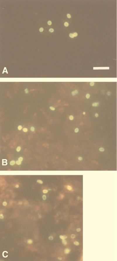
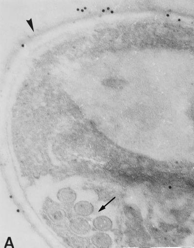
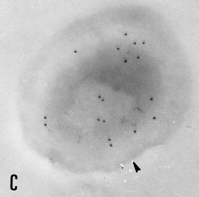
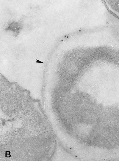
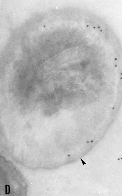
Several hybridomas producing antibodies detected by indirect immunofluorescence antibody test (IFAT) were established by fusion of mouse myeloma SP2/O with spleen cells from BALB/c mice immunized against whole spores (protocol 1) or chitinase-treated spores (protocol 2) of Enterocytozoon bieneusi and were cloned twice by limiting dilutions. Two monoclonal antibodies (MAbs), 3B82H2 from protocol 1, isotyped as immunoglobulin M (IgM), and 6E52D9 from protocol 2, isotyped as IgG, were expanded in both ascites and culture. IFAT with the MAbs showed that both MAbs reacted exclusively with the walls of the spores of E. bieneusi, strongly staining the surface of mature spores, and produced titers of greater than 4,096. Immunogold electron microscopy confirmed the specific reactivities of both antibodies. No cross-reaction, either with the spores of the other intestinal microsporidium species Encephalitozoon intestinalis or with yeast cells, bacteria, or any other intestinal parasites, was observed. The MAbs were used to identify E. bieneusi spores in fecal specimens from patients suspected of having intestinal microsporidiosis. The IFAT was validated against standard staining methods (Chromotrope 2R and Uvitex 2B) and PCR. We report here the first description and characterization of two MAbs specific for the spore wall of E. bieneusi. These MAbs have great potential for the demonstration and species determination of E. bieneusi, and their application in immunofluorescence identification of E. bieneusi in stool samples could offer a new diagnostic tool for clinical laboratories.


Figures

FIG. 1
FIG. 1
Purified whole spores of E. bieneusi stained by indirect immunofluorescence with MAbs 6E52D9 (A) and 3B82H2 (B) in ascitic fluid. MAbs recognize antigens localized in the spores walls. (C) Formalin-fixed smear of a fecal sample, from one of the 14 patients with microsporidia, reacted with a 1:512 dilution of MAb 6E52D9. Note the bright fluorescence of spore walls. Bar = 5 μm.
FIG. 2
FIG. 2
Immunogold electron micrographs of E. bieneusi mature spores after incubation with MAb 6E52D9 and MAb 3B82H2. (A) Labelling of the outer layer of the spore wall (exospore [arrowhead]) with MAb 6E52D9. The arrow indicates the double row arrangement of E. bieneusi polar tube sections. Magnification, ×120,000. (B) The MAb selectively labels the exospore (arrowhead). No gold particles are visible at the surface or inside the bacteria (left). Magnification, ×100,000. (C) The tangential section of the spore treated with MAb 3B82H2 shows the distribution of gold particles in the internal layer of the wall (endospore [arrowhead]). Magnification, ×100,000. (D) Labelling of the endospore (arrowhead) with the same MAb. Magnification, ×100,000.
FIG. 2
FIG. 2
Immunogold electron micrographs of E. bieneusi mature spores after incubation with MAb 6E52D9 and MAb 3B82H2. (A) Labelling of the outer layer of the spore wall (exospore [arrowhead]) with MAb 6E52D9. The arrow indicates the double row arrangement of E. bieneusi polar tube sections. Magnification, ×120,000. (B) The MAb selectively labels the exospore (arrowhead). No gold particles are visible at the surface or inside the bacteria (left). Magnification, ×100,000. (C) The tangential section of the spore treated with MAb 3B82H2 shows the distribution of gold particles in the internal layer of the wall (endospore [arrowhead]). Magnification, ×100,000. (D) Labelling of the endospore (arrowhead) with the same MAb. Magnification, ×100,000.

References

    1. Accoceberry I, Carrière J, Thellier M, Biligui S, Danis M, Datry A. Rat model for the human intestinal microsporidian Enterocytozoon bieneusi. J Eukaryot Microbiol. 1997;44:83S. - PubMed
    1. Accoceberry I, Greiner P, Thellier M, Achbarou A, Biligui S, Danis M, Datry A. Rabbit model for human intestinal microsporidia. J Eukaryot Microbiol. 1997;44:82S. - PubMed
    1. Aldras A M, Orenstein J M, Kotler D P, Shadduck J A, Didier E S. Detection of microsporidia by indirect immunofluorescence antibody test using polyclonal and monoclonal antibodies. J Clin Microbiol. 1994;32:608–612. - PMC - PubMed
    1. Asmuth D M, DeGirolami P C, Federman M, Ezratty C R, Pleskow D K, Desai G, Wanke C A. Clinical features of microsporidiosis in patients with AIDS. Clin Infect Dis. 1994;18:819–825. - PubMed
    1. Beckers P J A, Derks G J M M, van Gool T, Rietveld F J R, Sauerwein R W. Encephalitozoon intestinalis-specific monoclonal antibodies for laboratory diagnosis of microsporidiosis. J Clin Microbiol. 1996;34:282–285. - PMC - PubMed

Publication types

MeSH terms