. 1999 Dec;19(12):8191-200.
doi: 10.1128/MCB.19.12.8191.

Flagellar morphogenesis: protein targeting and assembly in the paraflagellar rod of trypanosomes

Affiliations

Flagellar morphogenesis: protein targeting and assembly in the paraflagellar rod of trypanosomes

P Bastin et al. Mol Cell Biol. 1999 Dec.

Abstract

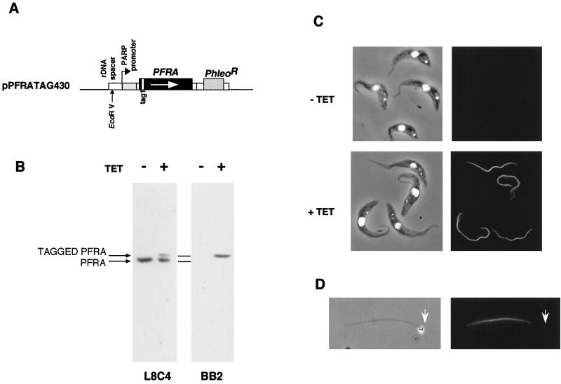
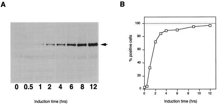
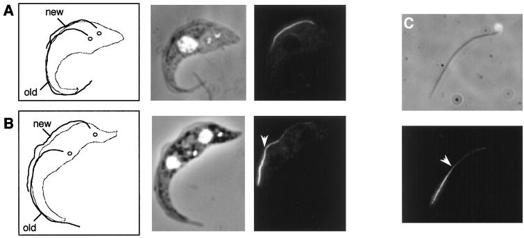
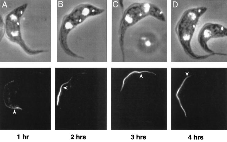
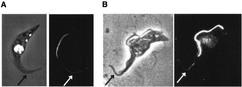
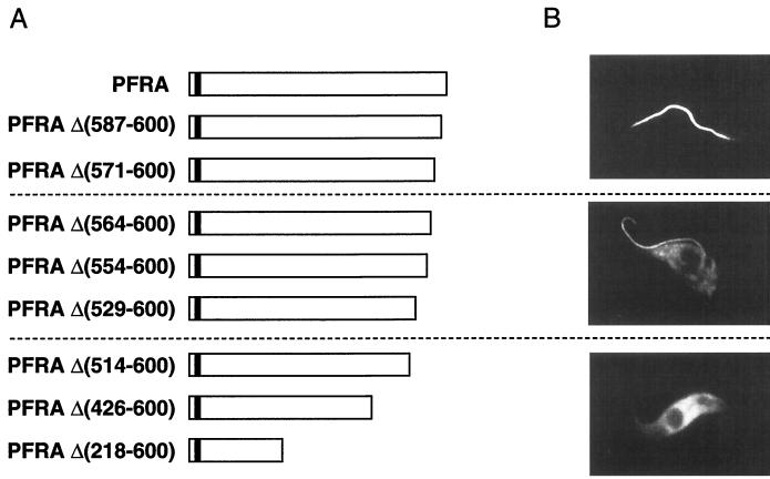
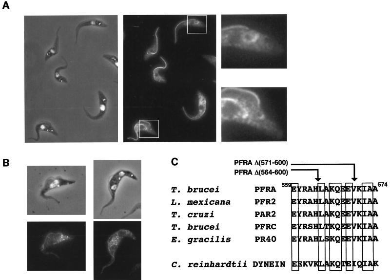
The paraflagellar rod (PFR) of the African trypanosome Trypanosoma brucei represents an excellent model to study flagellum assembly. The PFR is an intraflagellar structure present alongside the axoneme and is composed of two major proteins, PFRA and PFRC. By inducible expression of a functional epitope-tagged PFRA protein, we have been able to monitor PFR assembly in vivo. As T. brucei cells progress through their cell cycle, they possess both an old and a new flagellum. The induction of expression of tagged PFRA in trypanosomes growing a new flagellum provided an excellent marker of newly synthesized subunits. This procedure showed two different sites of addition: a major, polar site at the distal tip of the flagellum and a minor, nonpolar site along the length of the partially assembled PFR. Moreover, we have observed turnover of epitope-tagged PFRA in old flagella that takes place throughout the length of the PFR structure. Expression of truncated PFRA mutant proteins identified a sequence necessary for flagellum localization by import or binding. This sequence was not sufficient to confer full flagellum localization to a green fluorescent protein reporter. A second sequence, necessary for the addition of PFRA protein to the distal tip, was also identified. In the absence of this sequence, the mutant PFRA proteins were localized both in the cytosol and in the flagellum where they could still be added along the length of the PFR. This seven-amino-acid sequence is conserved in all PFRA and PFRC proteins and shows homology to a sequence in the flagellar dynein heavy chain of Chlamydomonas reinhardtii.


Figures

FIG. 1
FIG. 1
Expression of full-length, epitope-tagged PFRA in PFRAtag trypanosomes. (A) Construct used for inducible expression of full-length, epitope-tagged PFRA. Large boxes represent coding sequences, small boxes represent promoters, 5′ or 3′ untranslated regions, and targeting sequences. Thin lines represent the pGEM bacterial sequence. The EcoRV restriction site used for linearization and insertion into the trypanosome genome (12, 25, 52) is indicated by a vertical arrow. (B) Immunoblot of PFRAtag trypanosome total protein samples grown with or without tetracycline. Membranes were probed with either the anti-PFRA L8C4 monoclonal antibody (left) or with the antitag BB2 monoclonal antibody (right). (C) Immunofluorescence of PFRAtag trypanosomes grown with or without tetracycline. Left, DAPI images (white), merged to phase contrast images; right, immunofluorescence signal. (D) Detergent and salt-extracted flagellum from PFRAtag trypanosomes grown with tetracycline and stained with the BB2 monoclonal antibody. Left, DAPI image (white), merged to phase contrast image, showing the presence of the kinetoplast (arrowhead) that remains tightly connected to the basal body (37); right, immunofluorescence signal.
FIG. 2
FIG. 2
Induction kinetics of expression of tagged PFRA. PFRAtag trypanosomes were grown for the indicated periods in the presence of tetracycline. (A) Immunoblotting analysis with the antitag BB2 monoclonal antibody, showing the presence of the epitope-tagged PFRA protein at the expected molecular weight as early as 1 h after addition of the inducer. (B) Immunofluorescence analysis with the antitag BB2. Positive cells were scored and plotted versus time of induction (at least 1,000 cells for each time point).
FIG. 3
FIG. 3
The pattern of PFR assembly. PFRAtag trypanosomes were grown for 2 h in the presence of tetracycline and were analyzed by immunofluorescence with the antitag BB2 monoclonal antibody. The left panels of A and B show drawings of the trypanosome cell indicating the new and old flagella and the separated kinetoplasts (47). Note that the PFR is only present inside the flagellum from the point where the axoneme exits the cell body (2). The central panels show the DAPI images (white) merged to phase contrast images, and the right panels show the immunofluorescence signal. (A) Trypanosome with a short new flagellum whose assembly was initiated during the period of induction of tagged PFRA protein. This exhibits homogeneous PFR staining along the length of the PFR. (B) Trypanosome whose growth of the new flagellum was initiated before the induction show bipartite staining. The distal end is brightly labelled, but the proximal end is not. (C) Detergent and salt-extracted flagellum from a PFRAtag trypanosome induced for 2 h with tetracycline. The proximal end of the flagellum can be identified by the attached mitochondrial DNA (37). The bipartite staining is still present, with more intense labelling at the distal end. The arrowhead indicates the break point between the bright distal and the less intense proximal staining.
FIG. 4
FIG. 4
Pattern and timing of PFR assembly. PFRAtag trypanosomes were grown for 1 (A), 2 (B), 3 (C) or 4 h (D) in the presence of tetracycline and were analyzed by immunofluorescence with the antitag BB2 monoclonal antibody. The top panels show the DAPI images (white) merged to the phase contrast images, and the bottom panels show the immunofluorescence signal. The arrowhead indicates the break point between the bright distal and the less intense proximal staining.
FIG. 5
FIG. 5
PFRA incorporation into the old flagellum of PFRAtag trypanosomes. (A) PFRAtag trypanosomes were grown for 2 h in the presence of tetracycline and were analyzed by immunofluorescence with the antitag BB2 monoclonal antibody. The left panel shows the DAPI image (white) merged to the phase contrast image, and the right panel shows the immunofluorescence signal. The tagged PFRA protein is present mostly in the new flagellum as expected but also in the old flagellum (indicated by the arrow). (B) Wild-type trypanosomes were transiently transfected with the plasmid pKMPFRATAG (3) to express the tagged PFRA protein and were then fixed 2 h after electroporation. Positive cells show the presence of the tagged PFRA protein in the new flagellum but also in the old flagellum (arrow). Transient transfection often leads to considerable overexpression, explaining the difference in signal intensity between the two experiments.
FIG. 6
FIG. 6
Immunolocalization of truncated PFRA proteins. A series of epitope-tagged truncated PFRA proteins were expressed in wild-type trypanosomes. (A) Map of the different PFRA truncations. The deleted segment is shown by the numbering of amino acids in the PFRA sequence (43). White boxes represent PFRA sequences, and the small black box indicates the sequence of the epitope tag. (B) Immunofluorescence localization of the epitope-tagged truncated proteins revealed by the antitag BB2 monoclonal antibody. Only one representative image is shown for each group.
FIG. 7
FIG. 7
The truncated PFRAΔ(554-600) shows dual localization and is only added along the length of the growing PFR. (A) Trypanosomes were induced to express the truncated and epitope-tagged PFRAΔ(554-600) for 2 days and were processed for immunofluorescence with the anti-tag BB2. The left panel illustrates DAPI staining (white) merged to the phase contrast image, and the central panel shows the immunofluorescence signal. There is a concentration of truncated protein around the basal body and kinetoplast area of both growing and old flagella (areas enlarged on the right panel). (B) Time course induction of PFRAΔ(554-600) expression. Cells were grown for 2 h in the presence of tetracycline. The truncated protein is localized in both the new and the old flagellum but without any polarity. (C) Sequence comparison of the PFRA region required for addition at the distal tip of the flagellum. The arrows indicate the carboxy-terminal end of the truncated PFRA that is not added at the distal tip [PFRAΔ(564-600)] and of the PFRAΔ(571-600) that behaves normally. Indicated numbers correspond to the T. brucei PFRA sequence (43). Conserved residues are boxed. A high degree of conservation is observed in PFRA homologues in T. cruzi (PAR2 [5]), L. mexicana (PFR2 [28]), E. gracilis (PR-40 [30]), and PFRC (10), but homology is also found with the Chlamydomonas heavy chain of axonemal β-dynein (27).

References

    1. Baccetti B. Evolutionary trends in sperm structure. Comp Biochem Physiol. 1986;85:29–36. - PubMed
    1. Bastin P, Matthews K R, Gull K. The paraflagellar rod of Kinetoplastida: solved and unsolved questions. Parasitol Today. 1996;12:302–307. - PubMed
    1. Bastin P, Bagherzadeh A, Matthews K R, Gull K. A novel epitope tag system to study protein targeting and organelle biogenesis in Trypanosoma brucei. Mol Biochem Parasitol. 1996;77:235–239. - PubMed
    1. Bastin P, Sherwin T, Gull K. Paraflagellar rod is vital for trypanosome motility. Nature. 1998;391:548. - PubMed
    1. Bastin P, Gull K. Assembly and function of complex flagellar structures illustrated by the paraflagellar rod of trypanosomes. Protist. 1999;150:113–123. - PubMed

Publication types

MeSH terms

Substances