. 1999 Dec;19(12):8292-301.
doi: 10.1128/MCB.19.12.8292.

Mismatch repair processing of carcinogen-DNA adducts triggers apoptosis

Affiliations

Mismatch repair processing of carcinogen-DNA adducts triggers apoptosis

J Wu et al. Mol Cell Biol. 1999 Dec.

Abstract

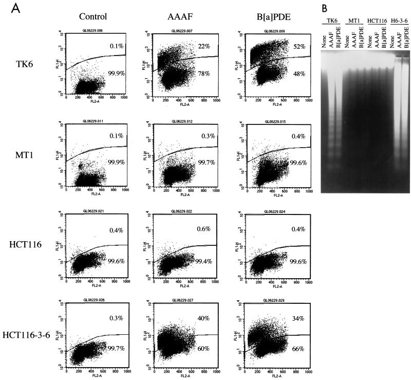
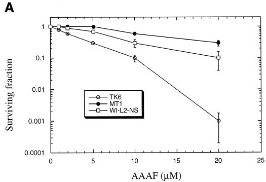
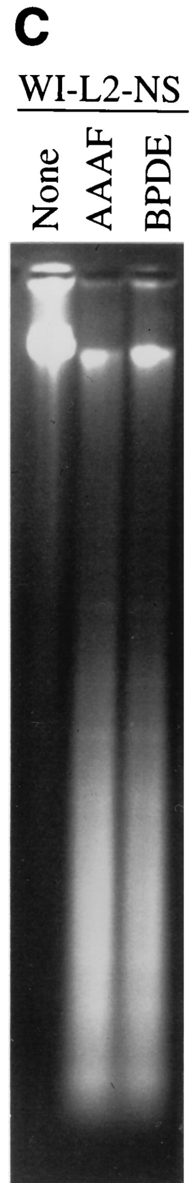
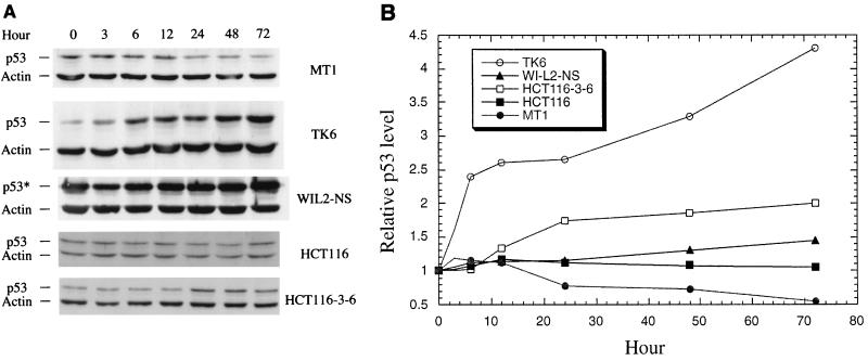
The DNA mismatch repair pathway is well known for its role in correcting biosynthetic errors of DNA replication. We report here a novel role for mismatch repair in signaling programmed cell death in response to DNA damage induced by chemical carcinogens. Cells proficient in mismatch repair were highly sensitive to the cytotoxic effects of chemical carcinogens, while cells defective in either human MutS or MutL homologs were relatively insensitive. Since wild-type cells but not mutant cells underwent apoptosis upon treatment with chemical carcinogens, the apoptotic response is dependent on a functional mismatch repair system. By analyzing p53 expression in several pairs of cell lines, we found that the mismatch repair-dependent apoptotic response was mediated through both p53-dependent and p53-independent pathways. In vitro biochemical studies demonstrated that the human mismatch recognition proteins hMutSalpha and hMutSbeta efficiently recognized DNA damage induced by chemical carcinogens, suggesting a direct participation of mismatch repair proteins in mediating the apoptotic response. Taken together, these studies further elucidate the mechanism by which mismatch repair deficiency predisposes to cancer, i.e., the deficiency not only causes a failure to repair mismatches generated during DNA metabolism but also fails to direct damaged and mutation-prone cells to commit suicide.


Figures

FIG. 1
FIG. 1
DNA substrates. (A) Construction of a 50-mer oligonucleotide containing carcinogen adducts. A 32P-labeled B[a]PDE-containing 11-mer (oligonucleotide A [5′-CCATCG*CTACC-3′]) was ligated with oligonucleotide B (5′-GACTACGTACTGTTACGGCT-3′) after they were annealed to a complementary 50-mer (oligomer C [5′-GCAGATCTGGCCTGATTGCGGTAGCGATGGAGCCGTAACAGTACGTAGTC-3′). The ligated oligomer was then elongated to 50 nucleotides by using oligonucleotide C as the template. The 50-mer homoduplex (G-C) and heteroduplex (A-C) were also constructed by using oligonucleotides 5′-CCATCGCTACC-3′ and 5′-CCATCACTACC-3′, respectively, as oligonucleotide A. dNTPs, deoxynucleoside triphosphates. (B) Circular DNA substrates. Each 6,440-bp circular substrate contained a strand break at the Sau96I site in the complementary (C) strand. Substrate G-T was a heteroduplex containing a single G-T mismatch, substrate G-C was a homoduplex, and substrate G-C/* was a homoduplex modified by chemical carcinogens in the complementary strand. The small circles represent carcinogen-DNA adducts. Several restriction sites relevant to this study are also shown.
FIG. 2
FIG. 2
Binding of hMutSα and hMutSβ to DNA containing B[a]PDE. Unless otherwise indicated, gel shift assays were performed in 25-μl reaction mixtures containing 0.5 pmol of 32P-labeled oligonucleotide duplexes, 0.25 pmol of purified hMutSα or hMutSβ, 0.4 mg of double-stranded-f1MR3 DNA (competitor DNA), 10 mM HEPES-KOH (pH 7.5), 110 mM KCl, 1 mM EDTA, 1 mM dithiothreitol, and 4% glycerol. After 20 min of incubation on ice, 5 μl of 50% sucrose was added. The samples were fractionated at room temperature through a 6% nondenaturing polyacrylamide gel in 6.7 mM Tris-acetate (pH 7.5)–1 mM EDTA with buffer recirculation. NT, nucleotide.
FIG. 3
FIG. 3
Inhibition of MMR reaction by DNA-containing carcinogen adducts. G-T mismatch repair was performed as described previously (30) by using 50 μg of HeLa nuclear extracts and 100 ng of a heteroduplex containing a single G-T mismatch. Repair assays were performed in reaction mixtures containing 100 ng of the G-T mismatched heteroduplex, the indicated competitor DNA, and 50 μg of HeLa nuclear extracts. The reaction mixtures were incubated at 37°C for 15 min. After phenol and ether extraction, DNA substrates were recovered by ethanol precipitation and digested with restriction endonucleases to score the repair of the G-T heteroduplex. For the homoduplex (G-C) or carcinogen-modified homoduplexes (G-C/B[a]PDE, and G-C/AAAF), restriction endonucleases HindIII and Bsp106 were used to detect the repair of the G-T substrate, producing 3.1- and 3.3-kb fragments (see Fig. 1). For the M13-fd heteroduplex, HindIII and BseRI (producing 2.8- and 3.6-kb fragments) were used since Bsp106 cleaves the M13-fd substrate into fragments of the same size as those obtained by HindIII and Bsp106 cleavage of the repaired G-T substrate.
FIG. 4
FIG. 4
Sensitivity of MMR-proficient cells to the cytotoxicity of chemical carcinogens. Graphs show the clonogenic survival of MMR-proficient (TK6 and HCT116-3-6) and MMR-deficient (MT1 and HCT116) cells following exposure to AAAF (A) or B[a]PDE (B). Standard deviations were calculated from three independent experiments.
FIG. 5
FIG. 5
Chemical carcinogens induce apoptosis. (A) TUNEL analysis. Cells were incubated with medium alone (control) or with medium containing 10 μM AAAF or 0.3 μM B[a]PDE at 37°C for 1 h and were then cultured in fresh medium for 24 h before being harvested for flow cytometry analysis as described in Materials and Methods. The cells above the gate are apoptotic (TUNEL positive), while those below the gate are nonapoptotic (TUNEL negative). The percentages of cells in each category are shown. FL2-A (horizontal axis) indicates PI staining, and FL1H (vertical axis) indicates FITC-dUTP staining. (B) DNA fragmentation in TK6 and HCT116-3-6 (H6-3-6) cells induced by AAAF and B[a]PDE. Cells were treated as described above. Genomic DNA was isolated by protease K digestion and phenol-chloroform extraction, fractionated through 1.6% agarose gels, and visualized under UV light in the presence of ethidium bromide. None, control.
FIG. 6
FIG. 6
Response of p53-defective WI-L2-NS cells to treatments with chemical carcinogens. (A) clonogenic survival; (B) TUNEL analysis; (C) DNA fragmentation analysis. None, control.
FIG. 6
FIG. 6
Response of p53-defective WI-L2-NS cells to treatments with chemical carcinogens. (A) clonogenic survival; (B) TUNEL analysis; (C) DNA fragmentation analysis. None, control.
FIG. 6
FIG. 6
Response of p53-defective WI-L2-NS cells to treatments with chemical carcinogens. (A) clonogenic survival; (B) TUNEL analysis; (C) DNA fragmentation analysis. None, control.
FIG. 7
FIG. 7
Expression of p53 in MMR-proficient and -deficient cells after treatment with AAAF. (A) Western blot analysis. After AAAF treatment, cells were cultured in fresh medium for the indicated times. Actin was used as an internal control, and equal amounts of protein were loaded for each sample. Both p53 protein and actin were detected by chemiluminescence on a Western blot by using antibodies against p53 and actin (Sigma), respectively. p53*, mutant p53 protein. (B) Relative amounts of p53. The p53 level at each point was normalized by its corresponding actin level as well as by the amount of p53 at time zero.

References

    1. Acharaya S, Wilson T, Gradia S, Kane M F, Guerrette S, Marsischky G T, Kolodner R, Fishel R. hMSH2 forms specific mispair-binding complexes with hMSH3 and hMSH6. Proc Natl Acad Sci USA. 1996;93:13629–13634. - PMC - PubMed
    1. Alani E, Lee S, Kane M F, Griffith J, Kolodner R D. Saccharomyces cerevisiae MSH2, a mispaired base recognition protein, also recognizes Holliday junctions in DNA. J Mol Biol. 1997;265:289–301. - PubMed
    1. Alani E, Reenan R A, Kolodner R D. Interaction between mismatch repair and genetic recombination in Saccharomyces cerevisiae. Genetics. 1994;137:19–39. - PMC - PubMed
    1. Allen D J, Makhov A, Grilley M, Taylor J, Thresher R, Modrich P, Griffith J D. MutS mediates heteroduplex loop formation by a translocation mechanism. EMBO J. 1997;16:4467–4476. - PMC - PubMed
    1. Anthoney D A, McIlwrath A J, Gallagher W M, Edlin A R M, Brown R. Microsatellite instability, apoptosis, and loss of p53 function in drug-resistant tumor cells. Cancer Res. 1996;56:1374–1381. - PubMed

Publication types

Substances