. 1999 Dec;19(12):8314-25.
doi: 10.1128/MCB.19.12.8314.

Characterization of a novel member of the DOK family that binds and modulates Abl signaling

Affiliations

Characterization of a novel member of the DOK family that binds and modulates Abl signaling

F Cong et al. Mol Cell Biol. 1999 Dec.

Abstract

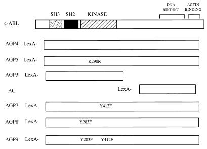
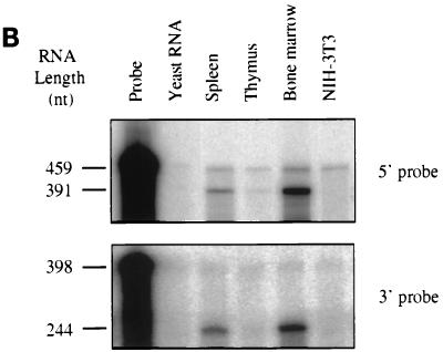
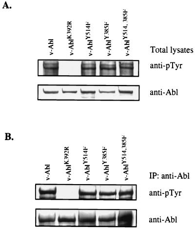
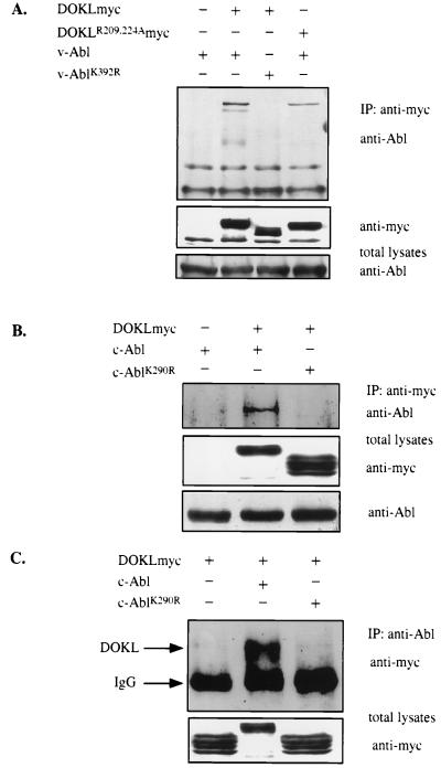
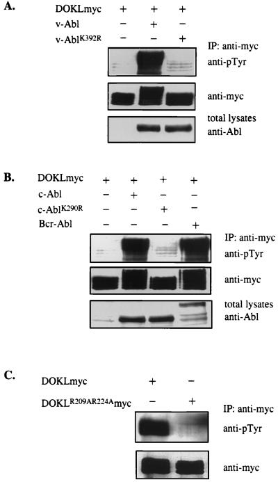
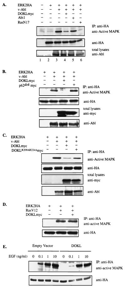
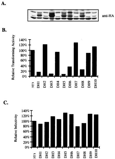
A novel member of the p62(dok) family of proteins, termed DOKL, is described. DOKL contains features of intracellular signaling molecules, including an N-terminal PH (pleckstrin homology) domain, a central PTB (phosphotyrosine binding) domain, and a C-terminal domain with multiple potential tyrosine phosphorylation sites and proline-rich regions, which might serve as docking sites for SH2- and SH3-containing proteins. The DOKL gene is predominantly expressed in bone marrow, spleen, and lung, although low-level expression of the RNA can also be detected in other tissues. DOKL and p62(dok) bind through their PTB domains to the Abelson tyrosine kinase in a kinase-dependent manner in both yeast and mammalian cells. DOKL is phosphorylated by the Abl tyrosine kinase in vivo. In contrast to p62(dok), DOKL lacks YxxP motifs in the C terminus and does not bind to Ras GTPase-activating protein (RasGAP) upon phosphorylation. Overexpression of DOKL, but not p62(dok), suppresses v-Abl-induced mitogen-activated protein (MAP) kinase activation but has no effect on constitutively activated Ras- and epidermal growth factor-induced MAP kinase activation. The inhibitory effect requires the PTB domain of DOKL. Finally, overexpression of DOKL in NIH 3T3 cells inhibits the transforming activity of v-Abl. These results suggest that DOKL may modulate Abl function.


Figures

FIG. 1
FIG. 1
Schematic representation of Abl baits used in the yeast two-hybrid assays. The positions of the SH3, SH2, tyrosine kinase, DNA-binding, and actin-binding domains on Abl are indicated. Yeast two-hybrid constructs AGP3, AGP4, AGP5, and AC were made by cloning different Abl gene fragments into the yeast two-hybrid vector SH2-1 in frame with the LexA DNA binding domain. The K290R mutation renders AGP5 kinase inactive. The sequences in AGP4 coding for two previously identified Abl autophosphorylation sites, Tyr283 and Tyr412, were mutated to generate yeast two-hybrid constructs AGP7, AGP8, and AGP9. The stable expression of all these yeast two-hybrid constructs was confirmed by Western blotting.
FIG. 2
FIG. 2
Sequence analysis of Abl-interacting protein DOKL. (A) Deduced amino acid sequence of DOKL. The potential PH domain is boxed with dashed lines. The region with homology to the IRS-1 PTB domain is underlined. Arg209 and Arg224, two Arg residues that are conserved in IRS-1 and that directly interact with phosphotyrosine residues of other molecules are indicated with asterisks. Three potential SH2 domain binding sites (YxxV motif) are circled. Two potential SH3 binding sites (PxxP motif) are boxed with solid lines. (B) Amino acid sequence homology between DOKL and p62dok. The similarities and identities between homologous regions are indicated. The degree of similarity between DOKL and p62dok is high at the N-terminal halves of the molecules but low at the C-terminal halves.
FIG. 3
FIG. 3
Distribution of DOKL mRNA expression in tissue and subcellular localization of DOKL protein. (A) Northern blot analysis of DOKL gene expression. A filter containing poly(A)+-selected RNA prepared from multiple mouse tissues was hybridized with a radiolabeled DOKL probe (top) or actin probe (bottom). The positions of migration of RNA molecular weight markers are indicated at left. (B) RNase protection analysis of DOKL gene expression. Two antisense probes corresponding to either a 5′ region (nucleotides 21 to 412) (top) or a 3′ region (nucleotides 1057 to 1301) (bottom) of DOKL cDNA were used in RNase protection assays. Equal amounts of total RNAs from the indicated mouse tissues and cells were used to protect DOKL antisense RNA probes from RNase digestion; products were analyzed by electrophoresis and autoradiography. Similar results were obtained when different preparations of total RNA were used in RNase protection assays. Sizes of RNA probes and protected RNA fragments are indicated at left. Probe, 32P-labeled RNA without RNase; yeast RNA, negative control RNA. (C) DOKL is localized in the cytoplasm. NIH 3T3 cells were transiently transfected with myc-tagged DOKL, and the expressed proteins were localized by indirect immunofluorescence with anti-myc antibodies. Cells were examined with a Nikon immunofluorescence microscope. Cells with different DOKL expression levels are shown in this field. Many cells in this field were not transfected and are not visible; the background staining is very faint.
FIG. 3
FIG. 3
Distribution of DOKL mRNA expression in tissue and subcellular localization of DOKL protein. (A) Northern blot analysis of DOKL gene expression. A filter containing poly(A)+-selected RNA prepared from multiple mouse tissues was hybridized with a radiolabeled DOKL probe (top) or actin probe (bottom). The positions of migration of RNA molecular weight markers are indicated at left. (B) RNase protection analysis of DOKL gene expression. Two antisense probes corresponding to either a 5′ region (nucleotides 21 to 412) (top) or a 3′ region (nucleotides 1057 to 1301) (bottom) of DOKL cDNA were used in RNase protection assays. Equal amounts of total RNAs from the indicated mouse tissues and cells were used to protect DOKL antisense RNA probes from RNase digestion; products were analyzed by electrophoresis and autoradiography. Similar results were obtained when different preparations of total RNA were used in RNase protection assays. Sizes of RNA probes and protected RNA fragments are indicated at left. Probe, 32P-labeled RNA without RNase; yeast RNA, negative control RNA. (C) DOKL is localized in the cytoplasm. NIH 3T3 cells were transiently transfected with myc-tagged DOKL, and the expressed proteins were localized by indirect immunofluorescence with anti-myc antibodies. Cells were examined with a Nikon immunofluorescence microscope. Cells with different DOKL expression levels are shown in this field. Many cells in this field were not transfected and are not visible; the background staining is very faint.
FIG. 4
FIG. 4
Tyrosine phosphorylation levels of v-Abl mutants. v-Abl mutant viruses were generated by transiently transfecting the packaging cell line. NIH 3T3 cells were infected with v-Abl viruses and selected in G418-containing medium. Cell lysates were directly separated by SDS-PAGE (A) or they were immunoprecipitated (IP) with anti-Abl antibodies and the immunoprecipitates were separated by SDS-PAGE (B). Blots were probed with anti-phosphotyrosine (anti-pTyr) antibodies (top sections) and reprobed with anti-Abl antibodies (bottom sections). The v-Abl mutant with both previously identified autophosphorylation sites mutated still contains a significant level of phosphotyrosine.
FIG. 5
FIG. 5
DOKL and Abl form a complex in vivo. (A) DOKL binding to v-Abl requires Abl kinase activity and the DOKL PTB domain. 293 cells were transfected with the indicated expression constructs, and cell lysates were immunoprecipitated (IP) with anti-myc antibodies. Immunoprecipitated proteins were separated by SDS-PAGE, transferred to a nitrocellulose membrane, and probed with anti-Abl antibodies (top). The membrane was reprobed with anti-myc antibodies to examine the expression of DOKL (middle). The expression levels of Abl were examined by probing total lysates with anti-Abl antibodies (bottom). That a lower amount of v-Abl is associated with DOKLR209, 224A than with the wild-type DOKL has been repeatedly observed. DOKL migration becomes slower with coexpression of wild-type v-Abl. (B) DOKL binding to c-Abl requires Abl kinase activity. 293 cells were transfected with the indicated expression constructs. Cell lysates were immunoprecipitated with anti-myc antibodies, and immunoprecipitates were fractionated and probed with anti-Abl antibodies (top). The expression levels of DOKL and Abl in total lysates were examined with anti-myc (middle) and anti-Abl antibodies (bottom). (C) DOKL binding to c-Abl can be detected with anti-Abl antisera. 293 cells were transfected with the indicated expression constructs. Cell lysates were immunoprecipitated with anti-Abl antibodies, and immunoprecipitates were fractionated and probed with anti-myc antibodies (top). The expression levels of DOKL and Abl in total lysates were examined with anti-myc (middle) and anti-Abl antibodies (bottom). IgG, immunoglobulin G.
FIG. 6
FIG. 6
p62dok, but not DOKL, binds to RasGAP upon phosphorylation. 293 cells were transfected with the indicated expression constructs, and cell lysates were immunoprecipitated (IP) with anti-RasGAP antibodies. Immunoprecipitates were fractionated and blotted with anti-myc antibodies (top). The expression levels of RasGAP and DOK proteins in total lysates were examined with anti-RasGAP (middle) and anti-myc antibodies (bottom).
FIG. 7
FIG. 7
DOKL is a substrate of activated Abl tyrosine kinase. (A and B) Phosphorylation of DOKL by Abl kinases. 293 cells were transfected with the indicated expression constructs. Cell lysates were immunoprecipitated (IP) with anti-myc antibodies, and immunoprecipitates were fractionated and probed with anti-phosphotyrosine (anti-pTyr) antibodies (top sections). The expression levels of DOKL were examined with anti-myc antibodies (middle sections). The expression of Abl kinases were examined by blotting total lysates with anti-Abl antibodies (bottom sections). DOKL migration becomes slower due to phosphorylation upon coexpression of active Abl kinases. (C) Phosphorylation of DOKL by endogenous tyrosine kinases. NIH 3T3 cells were transfected with the indicated expression constructs. Cell lysates were immunoprecipitated with anti-myc antibodies, and immunoprecipitates were fractionated and probed with anti-pTyr antibodies (top). The expression levels of DOKL were examined with anti-myc antibodies (bottom). The reason that tyrosine phosphorylation of DOKL in the absence of exogenous kinases appears greater in panel C than in panels A and B is that the blot in panel C was exposed for a much longer time.
FIG. 8
FIG. 8
Overexpression of DOKL inhibits v-Abl-dependent MAP kinase (MAPK) activation. (A) Overexpression of DOKL represses v-Abl-induced MAP kinase activation. 293 cells were transfected with the indicated plasmids. Forty hours after transfection, cells were starved with 0.2% serum for 18 h. After starvation, cells were lysed with RIPA buffer and cell lysates were immunoprecipitated (IP) with anti-HA antibodies. Immunoprecipitates were fractionated, transferred, and probed with anti-active MAP kinase antibodies. The membrane was stripped and reprobed with anti-HA antibodies. The expression levels of v-Abl were checked with anti-Abl antibodies. (B) Overexpression of p62dok does not inhibit v-Abl-induced MAP kinase activation. Cells and lysates were prepared as for panel A; antisera used are indicated. (C) The PTB domain is required for inhibiting v-Abl-induced MAP kinase activation by DOKL. Cells and lysates were prepared as for panel A; antisera used are indicated. (D) Overexpression of DOKL does not affect constitutively active Ras-induced MAP kinase activation. Cells and lysates were prepared as for panel A; antisera used are indicated. (E) Overexpression of DOKL does not affect EGF-induced MAP kinase activation. 293 cells were cotransfected with the indicated plasmids. Forty hours after transfection, cells were starved in 0.2% serum for 18 h. Cells were then restimulated with different amounts of EGF for 10 min. Cell lysates were immunoprecipitated with anti-HA antibodies. Immunoprecipitates were fractionated, probed with anti-active MAP kinase antibodies (top) and reprobed with anti-HA antibodies (bottom).
FIG. 9
FIG. 9
Overexpression of DOKL in NIH 3T3 cells represses v-Abl transforming activity. (A) Western blot analysis of DOKL expression. NIH 3T3 cells were cotransfected with pCGN-DOKL and a puromycin selection marker. Puromycin-resistant clones were picked, expanded, and analyzed for DOKL expression with anti-HA antibodies. Extracts from parental NIH 3T3 cells are also shown. Several lines expressing HA-tagged DOKL protein were identified. Lane designations match those for bars in panels B and C. (B) Transforming efficiencies of v-Abl on DOKL-overexpressing cell lines. Cell lines were infected with serially diluted v-Abl virus and plated in low-concentration serum, and the number of transformed foci on plates were scored. The titer of v-Abl virus, in focus-forming units (FFU) per milliliter, was determined for each cell line. The apparent viral titers for the cell lines relative to the apparent viral titer for parental NIH 3T3 cells (100% corresponds to 1.1 × 10⁵ FFU/ml) are indicated. (C) Infectivities of pGDN virus on DOKL-overexpressing cell lines. Cell lines were infected with serially diluted pGDN virus and selected in G418-containing medium, and G418-resistant clones were scored. The apparent viral titers for the cell lines relative to the apparent viral titer for parental NIH 3T3 cells (100% corresponds to 7.7 × 10⁵ FFU/ml) are indicated.

References

    1. Alexandropoulos K, Baltimore D. Coordinate activation of c-Src by SH3- and SH2-binding sites on a novel p130Cas-related protein, Sin. Genes Dev. 1996;10:1341–1355. - PubMed
    1. Bhat A, Johnson K J, Oda T, Corbin A S, Druker B J. Interactions of p62(dok) with p210(bcr-abl) and bcr-abl-associated proteins. J Biol Chem. 1998;273:32360–32368. - PubMed
    1. Bhat A, Kolibaba K, Oda T, Ohno-Jones S, Heaney C, Druker B J. Interactions of CBL with BCR-ABL and CRKL in BCR-ABL-transformed myeloid cells. J Biol Chem. 1997;272:16170–16175. - PubMed
    1. Carlesso N, Frank D A, Griffin J D. Tyrosyl phosphorylation and DNA binding activity of signal transducers and activators of transcription (STAT) proteins in hematopoietic cell lines transformed by bcr/abl. J Exp Med. 1996;183:811–820. - PMC - PubMed
    1. Carpino N, Wisniewski D, Strife A, Marshak D, Kobayashi R, Stillman B, Clarkson B. p62(dok): a constitutively tyrosine-phosphorylated, GAP-associated protein in chronic myelogenous leukemia progenitor cells. Cell. 1997;88:197–204. - PubMed

Publication types

MeSH terms

Associated data