. 1999 Dec;19(12):8393-9.
doi: 10.1128/MCB.19.12.8393.

Coexamination of site-specific transcription factor binding and promoter activity in living cells

Affiliations

Coexamination of site-specific transcription factor binding and promoter activity in living cells

K E Boyd et al. Mol Cell Biol. 1999 Dec.

Abstract

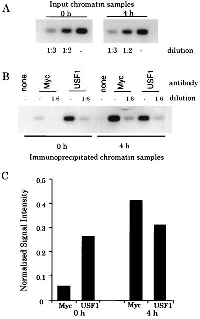
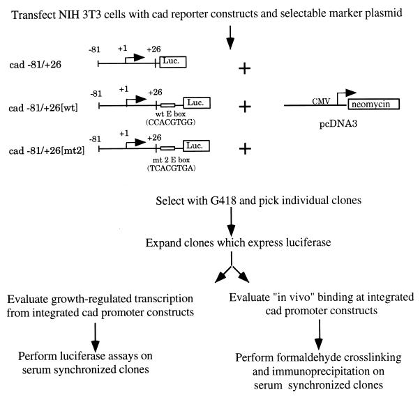
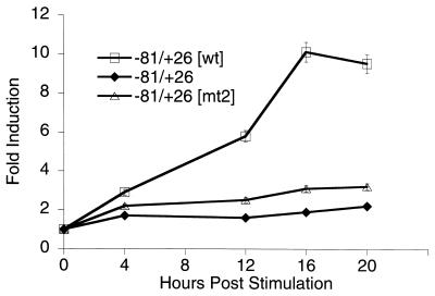
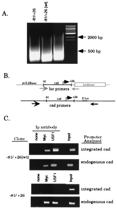
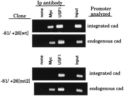
Previously, we have used a chromatin cross-linking and immunoprecipitation protocol for the analysis of Myc and USF binding to the cad promoter. The adaptation of this technique for the study of mammalian transcription factors was a big step forward in the analysis of transcription factor family member specificity, allowing for the first time a definitive knowledge of which factor binds to a promoter region under normal physiological conditions. However, due to limitations of the assay, our previous studies could not definitively prove that both Myc and USF bound to the exact same site on the cad promoter, nor could we directly correlate loss of in vivo binding of a particular factor with loss of transcriptional activity. Therefore, we have further modified the chromatin immunoprecipitation protocol to alleviate these problems. We have now shown that it is possible to coexamine growth-regulated transcriptional activity and promoter occupancy by using stably integrated promoter constructs. We show that both Myc and USF bind to the exact same E box on the cad promoter, suggesting that competition between these two factors for a single site occurs in living cells. We also find that cad promoter constructs that retain USF binding but lose Myc binding in vivo no longer display an increase in transcriptional activity in mid- to late G(1) phase of the cell cycle. Finally, we propose that cell cycle-regulated transcriptional activation of the cad promoter may be a stochastic, rather than a predetermined, process.


Figures

FIG. 1
FIG. 1
Myc does not displace USF from the cad promoter. (A) Fourteen cycles of PCR amplification were performed on chromatin from quiescent cells (0 h) or from cells which had been stimulated with serum for 4 h. Prior to PCR amplification, samples were diluted as indicated. PCR products were electrophoresed on an agarose gel, Southern blotted, hybridized with a radiolabeled cad probe, and analyzed with the phosphorimager with ImageQuant software. (B) PCRs were performed and analyzed as described above for immunoprecipitates from reactions containing no primary antibody (none), c-Myc antibody (Myc), or USF antibody (USF1). (C) Graphical analysis of cad PCR signals from the Southern blot shown in panel B. Normalized signal intensity is the quantitated numerical value of the signals from the diluted (1:6) anti-Myc and -USF1 lanes at 0 and 4 h normalized to the signal intensity of the input chromatin (1:2 diluted) for the 0- and 4-h samples, respectively. The signal intensity of the input chromatin (1:2 diluted) was arbitrarily set to a value of 1, and the normalized signals are presented as a fraction of this value.
FIG. 2
FIG. 2
USF1 does not bind the cad initiator element in vitro. Gel shift analysis of USF1 binding at the cad E box and cad initiator elements. Ten micrograms of HeLa cell nuclear extract was incubated with radiolabeled cad E-box and cad initiator probes. Binding complexes were supershifted by the addition of anti-USF1 antibody or competed by the addition of excess unlabeled probe. Samples were resolved on a 6% native polyacrylamide gel. The sequence of each gel shift probe is shown below the gel, along with the USF1 consensus binding sequence. The arrow indicates the nucleotide where transcription initiates. Y represents pyrimidine nucleotides, and R represents purine nucleotides. The E-box element is underlined.
FIG. 3
FIG. 3
Experimental approach to studying site-specific binding of Myc and USF1 to the E-box element in the cad promoter. See the text for details. Luc., luciferase; wt, wild type; CMV, cytomegalovirus.
FIG. 4
FIG. 4
Analysis of growth-regulated expression from integrated cad promoter constructs. Graphical representation of relative fold induction of stably integrated cad promoter-reporter activity throughout the growth cycle. NIH 3T3 cells were transfected with the indicated constructs, and stably transfected clones were selected with G418. Clones were serum starved for 48 h and then stimulated to grow by the addition of 10% serum to the culture medium. Cells were harvested for luciferase activity at the points indicated. Relative fold activation was calculated by normalizing the luciferase activity from serum-stimulated cells to the activity of the same clone prior to serum treatment. Data represents the average activity of one clone for each construct obtained from three independent time course experiments. Error bars represent the standard errors of the means.
FIG. 5
FIG. 5
Myc and USF1 bind specifically to the E box within the cad promoter. Clones of stably transfected cells (−81/+26[wt] clone 1 and −81/+26 clone 5) were serum starved for 48 h, then serum stimulated for 8 h, and cross-linked with formaldehyde. Cross-linked chromatin from each line was prepared and immunoprecipitated with antibodies against Myc and USF1. (A) EtBr-stained agarose gel showing the size of the DNA fragments following sonication. Lanes were loaded with 2.5% input chromatin from the specified clone after reversal of the cross-links and proteinase K treatment. DNA size markers are as indicated. (B) Schematic of the annealing position of PCR primers used to specifically amplify either integrated or endogenous cad promoters. The luciferase and cad primers amplify products of 200 and 350 bp, respectively. (C) PCR analysis of immunoprecipitation reactions with luciferase or cad primers to amplify the integrated or endogenous cad promoter, respectively. For all stable clones, immunoprecipitates were resuspended in 30 μl of H2O and input chromatin samples were diluted to 1% in 100 μl of H2O. The copy number of the integrated plasmid was normalized by utilizing appropriate sample volumes such that the input chromatin signal for all clones analyzed was approximately equivalent (last lane of each upper panel, integrated cad). Volumes used for both input chromatin and the immunoprecipitates (Ip) were as follows: 3 μl of the −81/+26-5 samples and 2 μl of −81/+26[wt]-1 samples. PCR mixtures with the cad primers (endogenous cad) contained equivalent sample volumes (2 μl) for all clones examined. PCR products were electrophoresed on a 1.5% agarose gel and stained with EtBr.
FIG. 6
FIG. 6
USF1, but not Myc, binds the 5′ TCACGTGA E box in intact cells. The stable cell lines −81/+26[wt] clone 1 (containing the CCACGTGG E box) and −81/+26[mt2] clone 2 (containing the TCACGTGA E box) were serum synchronized, cross-linked, and immunoprecipitated as described in the legend to Fig. 5. PCR analysis was performed with luciferase primers (integrated cad) and cad primers (endogenous cad). The copy number of the integrated plasmids was normalized by utilizing appropriate sample volumes so that the input signals for each clone were equivalent, as described in the legend to Fig. 5. Volumes used were as follows: 2 μl of the −81/+26[wt] samples and 1 μl of the −81/+26[mt2] samples. PCRs with the cad primers were performed with equivalent sample volumes for all clones examined. PCR products were electrophoresed on a 1.5% agarose gel and stained with EtBr. Ip, immunoprecipitation.

References

    1. Aksan I, Goding C R. Targeting the microphthalmia basic helix-loop-helix–leucine zipper transcription factor to a subset of E-box elements in vitro and in vivo. Mol Cell Biol. 1998;18:6930–6938. - PMC - PubMed
    1. Alberts A S, Geneste O, Treisman R. Activation of SRF-regulated chromosomal templates by Rho-family GTPases requires a signal that also induces H4 hyperacetylation. Cell. 1998;92:475–487. - PubMed
    1. Bendall A J, Molloy P L. Base preferences for DNA binding by the bHLH-Zip protein USF: effects of MgCl2 on specificity and comparison with binding of Myc family members. Nucleic Acids Res. 1994;22:2801–2810. - PMC - PubMed
    1. Blackwell T K, Huang J, Ma A, Kretzner L, Alt F, Eisenman R, Weintraub H. Binding of Myc proteins to canonical and noncanonical DNA sequences. Mol Cell Biol. 1993;13:5216–5224. - PMC - PubMed
    1. Blackwell T K, Kretzner L, Blackwood E M, Eisenman R N, Weintraub H. Sequence-specific DNA-binding by the c-Myc protein. Science. 1990;250:1149–1151. - PubMed

Publication types

LinkOut - more resources