. 1999 Dec;19(12):8442-50.
doi: 10.1128/MCB.19.12.8442.

Distinct cellular factors regulate the c-myb promoter through its E2F element

Affiliations

Distinct cellular factors regulate the c-myb promoter through its E2F element

M R Campanero et al. Mol Cell Biol. 1999 Dec.

Abstract

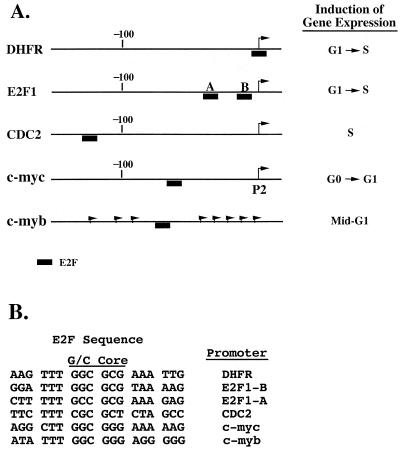
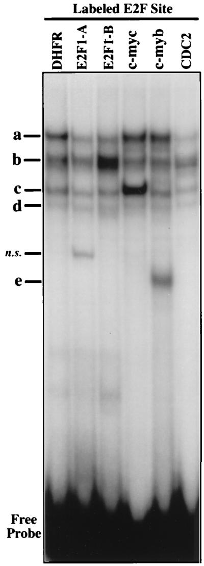
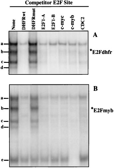
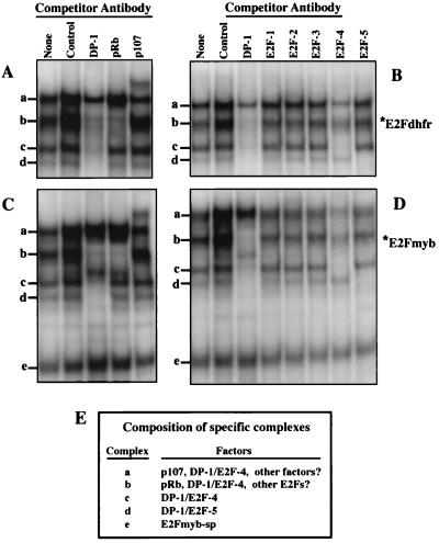
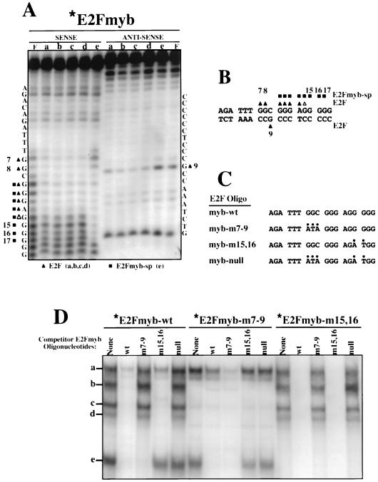
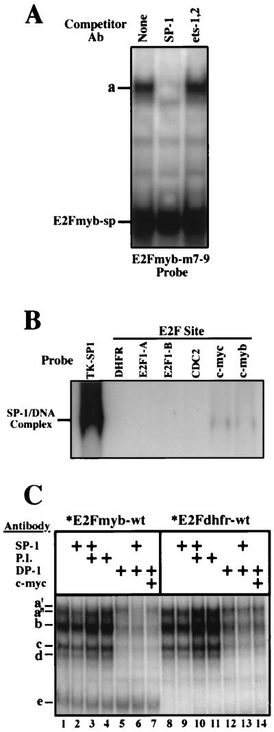
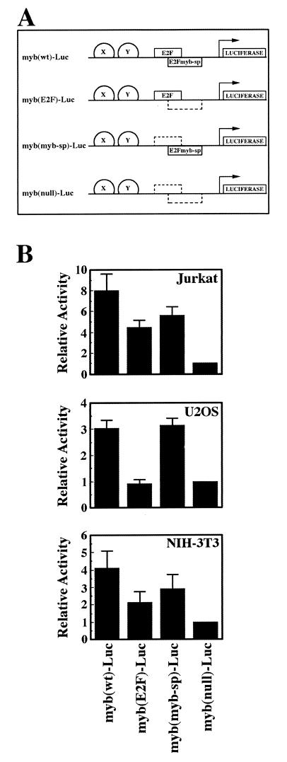
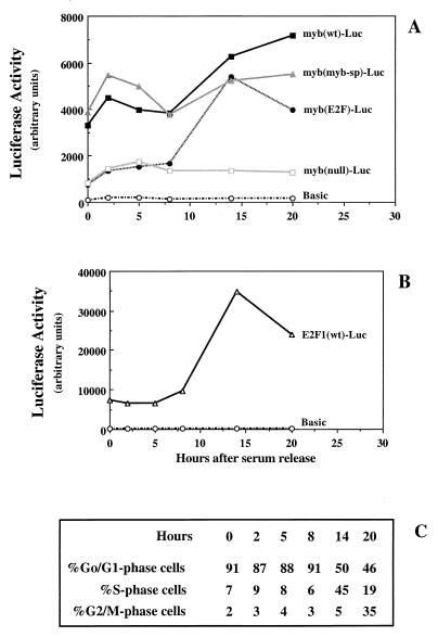
Most E2F-driven promoters are transiently activated around the G(1)/S transition. Although the promoter for the c-myb proto-oncogene harbors an E2F element, it is induced early in G(1) following entry into the cell cycle. Furthermore, this promoter remains active throughout subsequent cell cycles. Since E2F sites function as repressor elements during G(1) (due to the association of pRb with E2F factors), we investigated whether the E2F element in the c-myb promoter is regulated differently than E2F elements in promoters that are repressed during G(1). By gel shift analysis, the E2F element from the c-myb promoter was found to form a unique complex, referred to as E2Fmyb-sp, which was not observed with E2F elements from several other promoters. Antibodies to DP-1, E2F1 to -5, p107, or pRb failed to either supershift or block E2Fmyb-sp complex formation. Methylation interference experiments indicate that the DNA contact residues for the E2Fmyb-sp complex are distinct from but overlapping with residues required for the binding of E2F proteins. In addition to the identification of E2Fmyb-sp, we have found that SP-1 binds to the c-myb E2F element. Functional studies revealed that E2Fmyb-sp and/or SP-1 are required to achieve full activation of the c-myb promoter in different cell types and to maintain elevated expression of the c-myb promoter during G(1) in NIH 3T3 cells. These studies demonstrate that E2F elements can be regulated differently through the binding of unique sets of proteins.


Figures

FIG. 1
FIG. 1
(A) Schematic representation of several E2F-regulated cellular promoters. Arrows indicate transcription initiation sites. (B) Alignment of E2F elements from the DHFR, E2F1, cdc2, c-myc, and c-myb promoters.
FIG. 2
FIG. 2
EMSA of E2F elements from different promoters. Complex formation in nuclear extracts from the lymphoblastoid cell line X50-7 and the indicated radiolabeled E2F elements (Fig. 1) was analyzed by EMSA as described in Materials and Methods. Protein-DNA complexes are indicated as a to e. DHFR, c-myc, c-myb, and CDC2, oligonucleotides corresponding to the E2F sites within the DHFR, c-myc, c-myb, and cdc2 promoters, respectively; E2F1-A and E2F1-B, oligonucleotides corresponding to the two E2F sites found in the E2F1 promoter; n.s., a nonspecific complex.
FIG. 3
FIG. 3
Identification of a DNA-binding species which interact solely with the E2F elements within the c-myb promoter. Complexes formed with X50-7 nuclear extracts and radiolabeled E2F elements from the DHFR (A) and c-myb (B) promoters were analyzed by EMSA. Reaction mixtures were preincubated in the absence (None) or in the presence of a 100-fold excess of the indicated unlabeled competitor oligonucleotides. Positions of complexes a to e are indicated. DHFRwt, c-myc, c-myb, and CDC2, oligonucleotides corresponding to the E2F sites within the DHFR, c-myc, c-myb, and cdc2 promoters, respectively; E2F1-A and E2F1-B, oligonucleotides corresponding to the two E2F sites found in the E2F1 promoter; DHFRmut, mutant DHFR probe.
FIG. 4
FIG. 4
Identification of E2F or pRb family members in E2Fdhfr and E2Fmyb protein-DNA complexes. Binding reactions were performed with X50-7 nuclear extracts and either DHFR (A and B) or c-myb (C and D) E2F site probes. Extracts were preincubated in the absence (None) or in the presence of antibodies against the indicated proteins for 20 min prior to addition of the labeled probes. Control denotes a DP-1 preimmune rabbit polyclonal antibody. A nonspecific increase in overall binding was noticed in the presence of crude rabbit polyclonal antibodies (Control, DP-1, and pRb). Positions of complexes a to e are indicated. (E) Summary of the results shown in panels A to D detailing the composition of each of the specific complexes.
FIG. 5
FIG. 5
E2Fmyb-sp and E2F complexes interact with overlapping DNA sequences. (A) Methylation interference analysis of complexes a to e was performed with X50-7 nuclear extracts and the E2Fmyb probe. Sequences of the sense and antisense strands are indicated adjacent to the gels. Triangles and squares indicate bases whose methylation completely (filled) or partially (empty) blocked formation of complexes a to d (triangles) or e (squares). The cleavage pattern observed with an unbound probe (F) is also shown. (B) Summary of methylation interference analysis shown in panel A. (C) Sequence of wild-type and mutant E2Fmyb probes. For each mutant, alterations relative to the wild-type sequences are indicated by dots. (D) EMSA analysis of complexes formed between X50-7 nuclear factors and radiolabeled E2Fmyb-wt, E2Fmyb-m7-9, and E2Fmyb-m15,16 probes. Reaction mixtures were incubated in the absence (None) or in the presence of a 100-fold excess of the indicated unlabeled competitor oligonucleotides. Positions of complexes a to e are marked.
FIG. 6
FIG. 6
SP-1 binds to the c-myc and c-myb E2F elements. (A) Complex formation in assays employing X50-7 nuclear extracts and a radiolabeled E2Fmyb-m7-9 probe. Reaction mixtures were incubated in the absence (None) or in the presence of the indicated antibodies (Ab). (B) Binding of recombinant SP-1 to a TK-SP1 or to the indicated E2F elements. Binding reactions were performed with 0.5 footprint units of baculovirus-generated SP-1 (Promega). (C) Competition analysis of complexes formation in assays using X50-7 nuclear extracts and either an E2Fmyb-wt (lanes 1 to 7) or E2Fdhfr (lanes 8 to 14) probe. Reaction mixtures were incubated in the absence or in the presence of the indicated antibodies: anti-SP-1 (lanes 2, 3, 5, 9, 10, and 13), preimmune anti-DP-1 (P.I.; lanes 3, 4, 10, and 11), anti-DP-1 (lanes 5 to 7 and 12 to 14), and anti-c-Myc (lanes 7 and 14).
FIG. 7
FIG. 7
Functional regulation of the c-myb promoter through the E2F element in asynchronous cells. (A) Schematic representation of the luciferase reporter plasmids used in panel B and Fig. 8. Wild-type (solid box) and mutant (dashed box) E2F and E2Fmyb-sp binding sites are shown as boxes; the presence of other elements in the c-myb promoter which were left unmodified is represented as semicircles labeled X and Y. (B) The indicated myb-Luc reporter plasmids containing wild-type or mutant E2F elements were cotransfected with pCMV-βgal into asynchronously growing Jurkat, U2OS, and NIH 3T3 cells; 40 h later, cell extracts were prepared and luciferase and β-galactosidase assays were performed. Luciferase values were normalized for β-galactosidase activity and represent the means of at least four different experiments. Luciferase activities of the indicated promoter reporter plasmids relative to cells transfected with myb(null)-Luc ± standard deviation (error bars) are shown.
FIG. 8
FIG. 8
Activation of the c-myb promoter by E2Fmyb-sp and/or SP-1 through the E2F element during the G1 phase of the cell cycle. (A) The indicated myb-Luc reporter plasmids containing wild-type or mutant E2F elements were cotransfected with pCMV-βgal into NIH 3T3 cells. The cells were placed in low (0.5%) serum 4 h after the removal of the calcium phosphate precipitates. The cells remained in low serum for >48 h to induce quiescence, at which point serum was added (time zero). At the indicated time points, cells were removed for cell cycle analysis by flow cytometry and for determination of luciferase and β-galactosidase activities. (B) An E2F1 luciferase reporter plasmid containing wild-type E2F elements was analyzed in parallel as a control. Luciferase values from a representative experiment (normalized for β-galactosidase activity) (A and B) and percentages of cells in each phase of the cell cycle at the indicated time points (C) are shown.

References

    1. Adams P D, Kaelin W K. The cellular effects of E2F overexpression. Curr Top Microbiol Immunol. 1996;208:79–93. - PubMed
    1. Alitalo K, Winqvist R, Lin C C, De La Chapelle A, Schwab M, Bishop J M. Aberrant expression of an amplified c-myb oncogene in two cell lines from a colon carcinoma. Proc Natl Acad Sci USA. 1984;81:4534–4538. - PMC - PubMed
    1. Allen K E, de la Luna S, Kerkhoven R M, Bernards R, La Thangue N B. Distinct mechanisms of nuclear accumulation regulate the functional consequence of E2F transcription factors. J Cell Sci. 1997;110:2819–2831. - PubMed
    1. Ausubel F M, Brent R, Kingston R E, Moore D D, Seidman J G, Smith J A, Struhl K, editors. Short protocols in molecular biology. New York, N.Y: John Wiley & Sons, Inc.; 1995.
    1. Bandara L R, Buck V M, Zamanian M, Johnston L H, La Thangue N B. Functional synergy between DP-1 and E2F-1 in the cell cycle-regulating transcription factor DRTF1/E2F. EMBO J. 1993;12:4317–4324. - PMC - PubMed

Publication types

MeSH terms

LinkOut - more resources