. 1999 Dec;19(12):8451-60.
doi: 10.1128/MCB.19.12.8451.

The Drosophila polycomb protein interacts with nucleosomal core particles In vitro via its repression domain

Affiliations

The Drosophila polycomb protein interacts with nucleosomal core particles In vitro via its repression domain

A Breiling et al. Mol Cell Biol. 1999 Dec.

Abstract

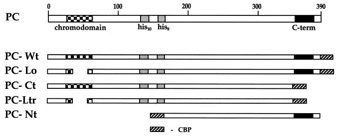
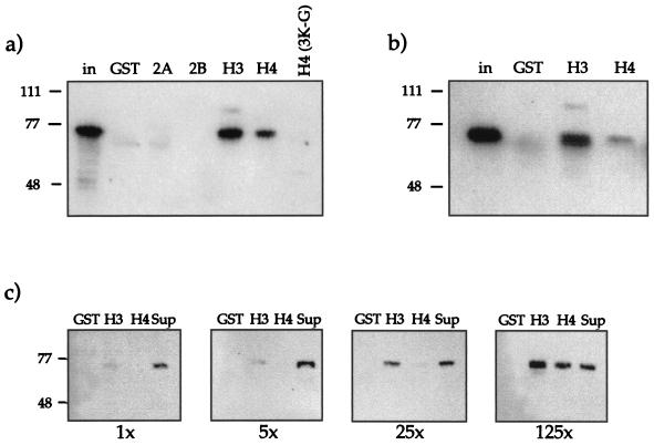
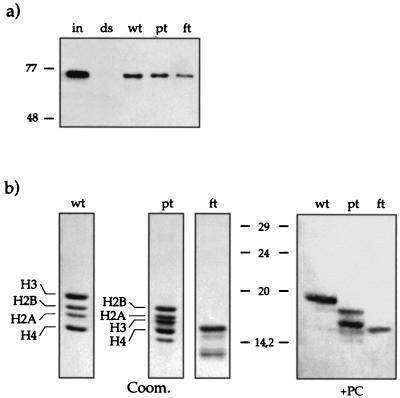
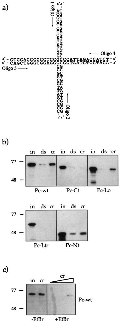
The proteins of the Polycomb group (PcG) are required for maintaining regulator genes, such as the homeotic selectors, stably and heritably repressed in appropriate developmental domains. It has been suggested that PcG proteins silence genes by creating higher-order chromatin structures at their chromosomal targets, thus preventing the interaction of components of the transcriptional machinery with their cis-regulatory elements. An unresolved issue is how higher order-structures are anchored at the chromatin base, the nucleosomal fiber. Here we show a direct biochemical interaction of a PcG protein-the Polycomb (PC) protein-with nucleosomal core particles in vitro. The main nucleosome-binding domain coincides with a region in the C-terminal part of PC previously identified as the repression domain. Our results suggest that PC, by binding to the core particle, recruits other PcG proteins to chromatin. This interaction could provide a key step in the establishment or regulation of higher-order chromatin structures.


Figures

FIG. 1
FIG. 1
The conserved domain of the PC protein and the protein fusions used for the analysis. The three prominent regions of PC are indicated: the chromodomain, the two repeats of 10 and 8 histidines, and the C terminus, which is involved in repression. PC-Wt features the whole sequence of PC. PC-Lo carries a deletion of 24 amino acids in the chromodomain, PC-Ct has a C-terminal truncation of 42 amino acids, and PC-Ltr is a combination of both. PC-Nt has a large N-terminal truncation, deleting the chromodomain and the His repeats. All fusion proteins carry the 35 amino acids of CBP either as C-terminal fusions (PC-Wt, PC-Lo, PC-Ct, and PC-Ltr) or as N-terminal fusions (PC-Nt).
FIG. 2
FIG. 2
Far-Western blot analysis with Drosophila core histones and recombinant PC (Pc-Wt). (a) Histones (H1, 1 μg; H2, 5 μg) were separated on an SDS–15% PAGE gel, electroblotted, and renatured. The figure shows the resolved histones in a Coomassie blue-stained gel (Coom.), the blot stained with Ponceau red (Ponceau), and the blot after incubation with PC-Wt (150 ng/cm2) (+PC). After extensive washing, PC signals were detected with PC antiserum and visualized by ECL. (b) Purified core histones were treated as for panel a but were incubated with 50 ng of PC-Wt/cm2 (+PC) or without addition of protein (−PC). The positions of the four core histones and the sizes of the molecular weight standards (M1 and M2) in kilodaltons are indicated.
FIG. 3
FIG. 3
Reconstitution of nucleosomes. (a) Scheme illustrating the reconstitution protocol. Purified core histones (untreated, partially trypsinized, or fully trypsinized) and DNA template were mixed in a reconstitution buffer containing 2 M NaCl. During gradual reduction of the NaCl concentrations, histone octamers formed on the DNA fragment (see Materials and Methods). (b) The completeness of reconstitution was tested by analysis of the reconstituted nucleosomes on a native (band shift) gel. The 146-bp DNA fragment was radiolabeled with [γ-32P]ATP. The signals for the free DNA (dsDNA) and the nucleosomal DNA (wt, untreated; pt, partially trypsinized; ft, fully trypsinized) are indicated. Only nucleosome preparations that did not contain free DNA were coupled to paramagnetic beads and used for further experiments. An autoradiogram of the gel is shown. (c) Histone contents of the input material and the reconstituted nucleosomes. A silver-stained SDS–15% PAGE gel with 50 ng of input material (purified core histones) and 80 ng of coupled reconstituted nucleosomes is shown (abbreviations for the nucleosome preparations are as explained for panel b). (d) Reconstituted nucleosomes remain stable in the presence of EtBr. Two aliquots (80 ng) of nucleosomes (nu) on beads were incubated for 1 h in the presence of 50 μg of EtBr/ml (+EtBr) or without EtBr (−EtBr) in binding buffer. After a wash, both samples were resolved by SDS-PAGE and silver stained. For reference an aliquot of the input material (50 ng) was included (in).
FIG. 4
FIG. 4
PC binding to reconstituted nucleosomes. (a) PC and PSC are coisolated with reconstituted nucleosomes. Portions (20 μl) of nuclear extract were incubated with immobilized nucleosomes (nu) reconstituted on 60 ng of DNA or an equivalent amount of free DNA template (ds). After extensive washing, the bound material was analyzed by SDS-PAGE. Bound proteins were visualized by Western blot analyses with antisera against the indicated proteins. On the left, 5 μl of extract was loaded (in) to indicate the presence of the relevant proteins in the input material. (b) Interaction of recombinant PC (Pc-Wt) with reconstituted nucleosomes. Portions (300 ng) of PC-Wt (input) were incubated with 20 ng of nucleosome-carrying DNA (nu) or a free DNA fragment (ds) coupled to paramagnetic beads in the presence or absence of EtBr (50 μg/ml). The assay was performed as for panel a. (c) Interaction of mutant PC derivatives with reconstituted nucleosomes. The assay was performed as for panel b. A 300-ng portion of each mutant PC derivative was used. (d) Interaction of mutant PC derivatives as in panel c with reconstituted nucleosomes in the presence of EtBr (50 μg/ml).
FIG. 5
FIG. 5
Binding of PC to N-terminal “tail” domains of histones. (a) PC-Wt (300 ng) was incubated with GST or with different GST fusion proteins (GST-H2A, -H2B, -H3, and -H4) immobilized on Sepharose beads. GST fusion proteins were used at a 125-fold excess with respect to PC. For comparison the input is shown (in). After extensive washing, the bound material was analyzed by SDS-PAGE and Western blotting with antisera against PC. (b) Tail interaction assay with PC-Wt as in panel a, but in the presence of EtBr (50 μg/ml). PC-Wt (300 ng) was incubated with a 100-fold molar excess of GST, GST-H3, or GST-H4 immobilized on Sepharose beads. (c) PC interacts preferentially with the histone H3 N-terminal domain. PC-Wt was incubated with GST, GST-H3, or GST-H4 at a 1-, 5-, 25-, or 125-fold molar excess of fusion protein over PC (1×, 5×, 25×, and 125×, respectively). In the rightmost lanes, the supernatant (Sup) (unbound fraction) of the second sample (GST-H3) was loaded for comparison. After washing, the bound material was analyzed by SDS-PAGE and Western blotting as before.
FIG. 6
FIG. 6
Interaction of recombinant PC with reconstituted nucleosomes by using trypsinized histones. (a) The assay was performed like those described for Fig. 4 except that PC-Wt was also incubated with trypsinized nucleosomes (wt, untreated; pt, partially trypsinized; ft, fully trypsinized). Portions (300 ng) of PC-Wt (input [in]) were incubated with 20 ng of nucleosome-wrapped DNA (wt, pt, ft) or a free DNA fragment (ds) coupled to paramagnetic beads in the presence of EtBr (50 μg/ml). (b) Far-Western blot analysis of the interaction of recombinant PC with trypsinized histones. Nontrypsinized (wt), partially trypsinized (pt), and fully trypsinized (ft) histones were separated on an SDS–15% PAGE gel, electroblotted, and renatured. Left three panels, Coomassie stain of the input material. Note the band of digested H3 in the pt fraction below H2A. Right panel, blot after incubation with PC-Wt. PC interacts mainly with H3 and weakly with H4 and H2B (as shown in Fig. 2) when full-length histones are present (wt). After partial trypsin treatment, PC still interacts with the shortened H3 and interacts more efficiently with H2B and H4 (pt). PC can still interact with fully trypsinized proteins, though it is no longer possible to determine with which of the histones it interacts (ft).
FIG. 7
FIG. 7
Interaction of recombinant Pc (Pc-Wt) with crDNA. (a) The four-way junction DNA molecule shown in the open conformation. Under the conditions of our experiment (5 mM MgCl2), the molecule had a distorted, stacked X-like structure. crDNA was formed by annealing the four oligonucleotides indicated. Oligonucleotide 1 (Oligo 1) was biotinylated for coupling to paramagnetic beads. (b) Binding assay with crDNA and PC. Equal amounts of crDNA (cr) and dsDNA (ds) (1.25 pmol of each) immobilized on paramagnetic beads were incubated with 300 ng (input [in]) of the different PC derivatives (PC-Wt, PC-Ct, PC-Lo, PC-Ltr, and PC-Nt). After extensive washing, the bound material was analyzed by using SDS-PAGE and Western blotting as before. PC proteins were used at a fivefold molar excess in this experiment. (c) Influence of EtBr on the PC-crDNA interaction. Left two lanes (−EtBr), input of PC-Wt (300 ng) (in) and bound PC after incubation with 0.125 pmol of crDNA (cr). Right lanes (+EtBr), bound PC after incubation with 0.125, 0.3, and 1.5 pmol of crDNA in the presence of EtBr (50 μg/ml).

References

    1. Alkema M J, Jacobs J, Voncken J W, Jenkins N A, Copeland N G, Satijn D P E, Otte A P, Berns A, van Lohuizen M. MPc2, a new murine homologue of the Drosophila Polycomb protein, is a member of the mouse Polycomb transcriptional repressor complex. J Mol Biol. 1997;273:993–1003. - PubMed
    1. Ausio J, Dong F, van Holde K E. Use of selectively trypsinized nucleosome core particles to analyze the role of histone “tails” in the stabilization of the nucleosome. J Mol Biol. 1989;206:451–463. - PubMed
    1. Ball L J, Murzina N V, Broadhurst R W, Raine A R C, Archer S J, Stott F J, Murzin A G, Singh P B, Domaille P J, Laue E D. Structure of the chromatin binding (chromo) domain from mouse modifier protein 1. EMBO J. 1997;16:2473–2481. - PMC - PubMed
    1. Bianchi M E. Interaction of a protein from rat liver nuclei with cruciform DNA. EMBO J. 1988;7:843–849. - PMC - PubMed
    1. Blank T A, Becker P B. Electrostatic mechanisms of nucleosome spacing. J Mol Biol. 1995;252:305–313. - PubMed

Publication types

LinkOut - more resources