. 1999 Dec;19(12):8492-504.
doi: 10.1128/MCB.19.12.8492.

Bridge-1, a novel PDZ-domain coactivator of E2A-mediated regulation of insulin gene transcription

Affiliations

Bridge-1, a novel PDZ-domain coactivator of E2A-mediated regulation of insulin gene transcription

M K Thomas et al. Mol Cell Biol. 1999 Dec.

Abstract

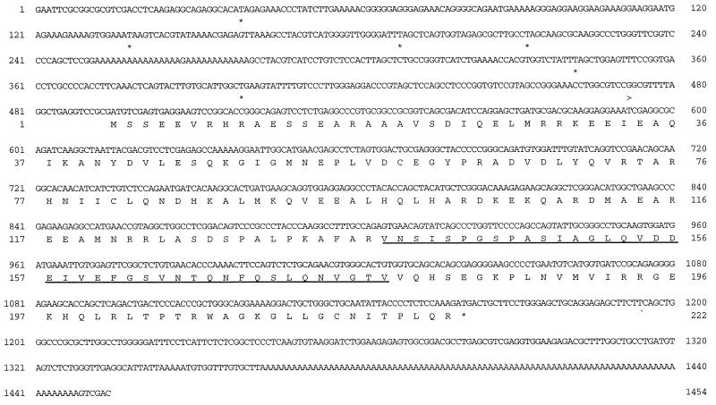
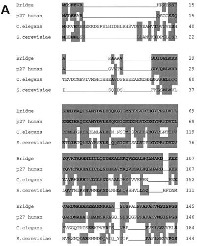
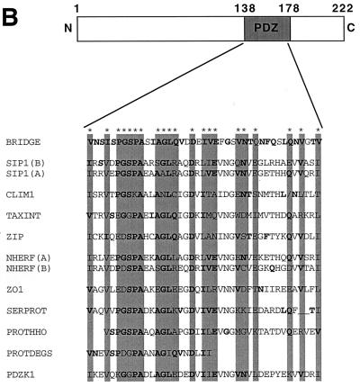
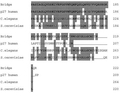
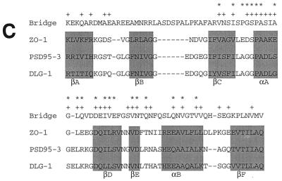
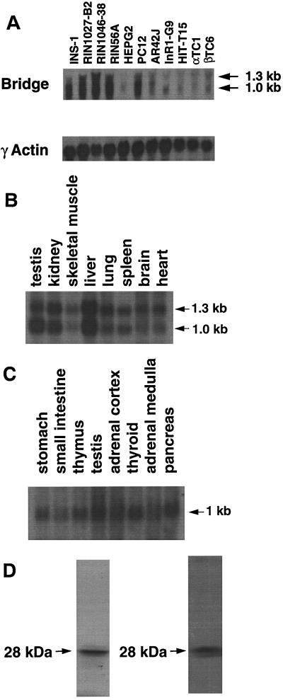
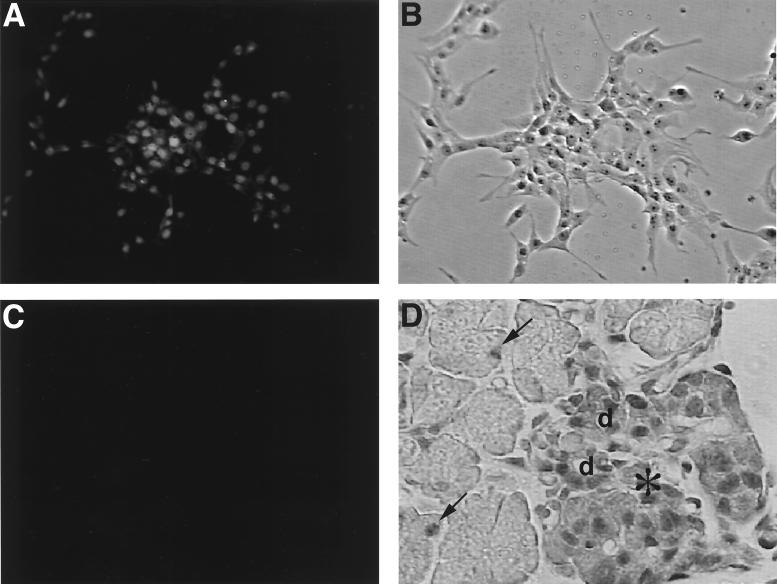
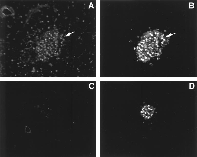
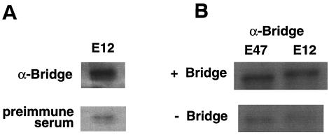
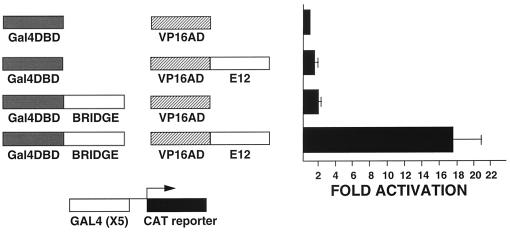
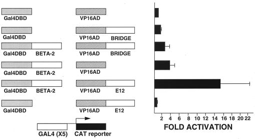
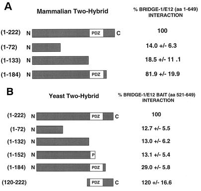
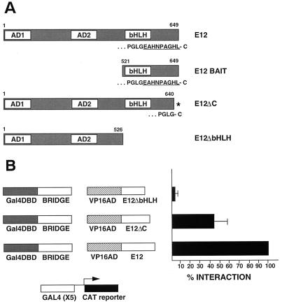
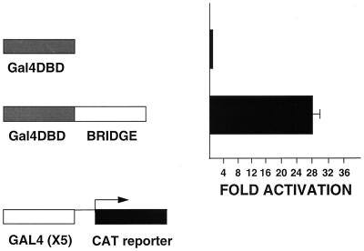
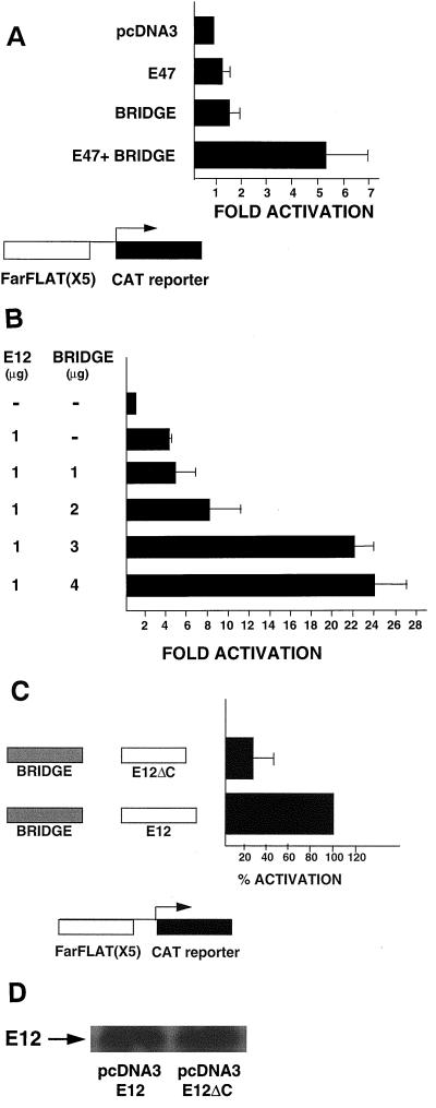
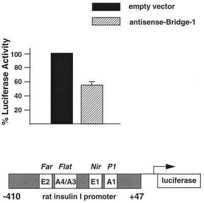
Proteins in the E2A family of basic helix-loop-helix transcription factors are important in a wide spectrum of physiologic processes as diverse as neurogenesis, myogenesis, lymphopoeisis, and sex determination. In the pancreatic beta cell, E2A proteins, in combination with tissue-specific transcription factors, regulate expression of the insulin gene and other genes critical for beta-cell function. By yeast two-hybrid screening of a cDNA library prepared from rat insulinoma (INS-1) cells, we identified a novel protein, Bridge-1, that interacts with E2A proteins and functions as a coactivator of gene transcription mediated by E12 and E47. Bridge-1 contains a PDZ-like domain, a domain known to be involved in protein-protein interactions. Bridge-1 is highly expressed in pancreatic islets and islet cell lines and the expression pattern is primarily nuclear. The interaction of Bridge-1 with E2A proteins is further demonstrated by coimmunoprecipitation of in vitro-translated Bridge-1 with E12 or E47 and by mammalian two-hybrid studies. The PDZ-like domain of Bridge-1 is required for interaction with the carboxy terminus of E12. In both yeast and mammalian two-hybrid interaction studies, Bridge-1 mutants lacking an intact PDZ-like domain interact poorly with E12. An E12 mutant (E12DeltaC) lacking the carboxy-terminal nine amino acids shows impaired interaction with Bridge-1. Bridge-1 has direct transactivational activity, since a Gal4 DNA-binding domain-Bridge-1 fusion protein transactivates a Gal4CAT reporter. Bridge-1 also functions as a coactivator by enhancing E12- or E47-mediated activation of a rat insulin I gene minienhancer promoter-reporter construct in transient-transfection experiments. Substitution of the mutant E12DeltaC for E12 reduces the coactivation of the rat insulin I minienhancer by Bridge-1. Inactivation of endogenous Bridge-1 in insulinoma (INS-1) cells by expression of a Bridge-1 antisense RNA diminishes rat insulin I promoter activity. Bridge-1, by utilizing its PDZ-like domain to interact with E12, may provide a new mechanism for the coactivation and regulation of transcription of the insulin gene.


Figures

FIG. 1
FIG. 1
Sequence of rat Bridge-1 cDNA and encoded protein. Positions of in-frame stop codons are designated by asterisks under the corresponding nucleotide sequences. The arrowhead is placed below the corresponding nucleotide sequence to indicate the starting position of the two-hybrid clone 18. The PDZ-like domain is underlined.
FIG. 2
FIG. 2
(A) Homologies between Bridge-1 protein sequence and sequences from other species. Sequences used for alignment are as follows: Bridge, rat Bridge-1 sequence; p27 human, human proteasomal modulator subunit p27 (GenBank number AB003177); C. elegans, sequence from chromosome III of C. elegans (46) (GenBank number U23453); S. cerevisiae, hypothetical 24.8-kDa protein in FAA3-BET1 intergenic region from S. cerevisiae (GenBank number P40555). Amino acid similarities as determined by BLAST analysis are shown as gray boxes, and identities are in boldface. (B) PDZ-like domain homologies between Bridge-1 and other PDZ domain-containing proteins. The Bridge-1 protein is schematically depicted to illustrate the PDZ-like domain (PDZ) identified by homology with other proteins. Alignment positions with conserved similar or identical residues by BLAST analysis in at least 50% of the aligned sequences are indicated with asterisks and gray boxes. Identical amino acids are in boldface. Protein sequences represented are as follows: BRIDGE, rat Bridge-1, amino acids 138 to 178; SIP1 A and B, interacting protein with human SRY (34) (GenBank number U82108), PDZ-domain A, amino acids 36 to 76, and PDZ-domain B, amino acids 176 to 216; CLIM1, human carboxyl-terminal LIM domain protein (GenBank number U90878), amino acids 30 to 65 and 76 to 80; TAXINT, human Tax interaction protein 1 (36) (GenBank number AF028823), amino acids 49 to 89; ZIP, human zipper containing protein (9) (GenBank number 631508), amino acids 76 to 116; NHERF, rabbit protein cofactor that mediates protein kinase A regulation of the renal brush border membrane Na+-H+ exchanger (45, 48) (GenBank number U19815) PDZ-domain A, amino acids 39 to 79, and PDZ-domain B, amino acids 179 to 219; ZO-1, mouse tight junction protein ZO-1 (17) (GenBank number P39447), amino acids 447 to 487; SERPROT, Aquifex aeolicus periplasmic serine protease (8) (GenBank number AE000741), amino acids 280 to 318; PROTHHO, Synechocystis sp. protease HhoB (GenBank number D90911), amino acids 346 to 384; PROTDEGS, E. coli protease DEGS precursor (GenBank number P31137), amino acids 284 to 305; and PDZK1, human PDZ domain containing-protein (22) (GenBank number AF012281), amino acids 403 to 443. (C) Comparison of the Bridge-1 PDZ-like domain with typical PDZ domain sequences. Alignment of typical PDZ domains and designation of regions of secondary structure (boxed) are depicted as described by Doyle et al. (10). A segment of Bridge-1 sequence is aligned for comparison. Amino acids within the Bridge-1 sequence identified as conserved among sequences aligned in Fig. 2B are designated with an asterisk for reference. For each aligned position, similarities or identities determined by BLAST analysis between the Bridge-1 sequence and any of the three ZO-1, PSD95-3, or DLG-1 sequences are indicated (+). Amino acid sequences illustrated are as follows: Bridge-1, amino acids 105 to 190; ZO-1, mouse tight junction protein ZO-1 (17) (GenBank number P39447), amino acids 423 to 501; PSD95-3, rat presynaptic density protein 95, PDZ domain 3 (5) (GenBank number P31016), amino acids 312 to 391; and DLG-1, D. melanogaster lethal (1) discs-Large-1 tumor suppressor protein (47) (GenBank number P31007), amino acids 485 to 564.
FIG. 2
FIG. 2
(A) Homologies between Bridge-1 protein sequence and sequences from other species. Sequences used for alignment are as follows: Bridge, rat Bridge-1 sequence; p27 human, human proteasomal modulator subunit p27 (GenBank number AB003177); C. elegans, sequence from chromosome III of C. elegans (46) (GenBank number U23453); S. cerevisiae, hypothetical 24.8-kDa protein in FAA3-BET1 intergenic region from S. cerevisiae (GenBank number P40555). Amino acid similarities as determined by BLAST analysis are shown as gray boxes, and identities are in boldface. (B) PDZ-like domain homologies between Bridge-1 and other PDZ domain-containing proteins. The Bridge-1 protein is schematically depicted to illustrate the PDZ-like domain (PDZ) identified by homology with other proteins. Alignment positions with conserved similar or identical residues by BLAST analysis in at least 50% of the aligned sequences are indicated with asterisks and gray boxes. Identical amino acids are in boldface. Protein sequences represented are as follows: BRIDGE, rat Bridge-1, amino acids 138 to 178; SIP1 A and B, interacting protein with human SRY (34) (GenBank number U82108), PDZ-domain A, amino acids 36 to 76, and PDZ-domain B, amino acids 176 to 216; CLIM1, human carboxyl-terminal LIM domain protein (GenBank number U90878), amino acids 30 to 65 and 76 to 80; TAXINT, human Tax interaction protein 1 (36) (GenBank number AF028823), amino acids 49 to 89; ZIP, human zipper containing protein (9) (GenBank number 631508), amino acids 76 to 116; NHERF, rabbit protein cofactor that mediates protein kinase A regulation of the renal brush border membrane Na+-H+ exchanger (45, 48) (GenBank number U19815) PDZ-domain A, amino acids 39 to 79, and PDZ-domain B, amino acids 179 to 219; ZO-1, mouse tight junction protein ZO-1 (17) (GenBank number P39447), amino acids 447 to 487; SERPROT, Aquifex aeolicus periplasmic serine protease (8) (GenBank number AE000741), amino acids 280 to 318; PROTHHO, Synechocystis sp. protease HhoB (GenBank number D90911), amino acids 346 to 384; PROTDEGS, E. coli protease DEGS precursor (GenBank number P31137), amino acids 284 to 305; and PDZK1, human PDZ domain containing-protein (22) (GenBank number AF012281), amino acids 403 to 443. (C) Comparison of the Bridge-1 PDZ-like domain with typical PDZ domain sequences. Alignment of typical PDZ domains and designation of regions of secondary structure (boxed) are depicted as described by Doyle et al. (10). A segment of Bridge-1 sequence is aligned for comparison. Amino acids within the Bridge-1 sequence identified as conserved among sequences aligned in Fig. 2B are designated with an asterisk for reference. For each aligned position, similarities or identities determined by BLAST analysis between the Bridge-1 sequence and any of the three ZO-1, PSD95-3, or DLG-1 sequences are indicated (+). Amino acid sequences illustrated are as follows: Bridge-1, amino acids 105 to 190; ZO-1, mouse tight junction protein ZO-1 (17) (GenBank number P39447), amino acids 423 to 501; PSD95-3, rat presynaptic density protein 95, PDZ domain 3 (5) (GenBank number P31016), amino acids 312 to 391; and DLG-1, D. melanogaster lethal (1) discs-Large-1 tumor suppressor protein (47) (GenBank number P31007), amino acids 485 to 564.
FIG. 3
FIG. 3
Characterization of Bridge-1 expression. (A) Northern blot of Bridge-1 transcript in total RNA from rodent cell lines (upper panel). For each lane, 20 μg of total RNA derived from the following cell lines was hybridized with 32P-labeled Bridge-1 cDNA: INS-1, rat insulinoma cells; RIN1027-B2, rat islet tumor somatostatin-secreting cells; RIN1046-38, rat insulinoma cells; RIN56A, rat insulinoma cells; HepG2, human hepatoblastoma cells; PC12, rat pheochromocytoma cells; AR42J, rat exocrine pancreatic tumor cells; InR1-G9, hamster islet tumor glucagon-secreting cells; HIT-T15, hamster insulinoma cells; αTC1, mouse islet tumor glucagon-secreting cells; βTC6, mouse islet tumor insulin-secreting cells. The blot was stripped and reprobed with a gamma-actin cDNA as a loading control (lower panel). (B) Northern blot of Bridge-1 transcript in mouse tissues. A commercially generated Northern blot (Clontech) containing 2 μg of poly(A)+ RNA per lane from the murine tissues indicated was hybridized with 32P-labeled Bridge-1 cDNA. (C) Northern blot of Bridge-1 transcript in human endocrine tissues. A commercially generated Northern blot (Clontech) containing 2 μg of poly(A)+ RNA per lane from the human tissues indicated was hybridized with 32P-labeled Bridge-1 cDNA. (D) Autoradiogram after SDS-PAGE fractionation of 35S-labeled in vitro-translated Bridge-1 protein (left panel). Western blot analysis of in vitro-translated Bridge-1 after SDS-PAGE fractionation (right panel).
FIG. 4
FIG. 4
Immunocytochemical staining of Bridge-1 in RIN1027-B2 cells and mouse pancreas. Fluorescent immunostaining of RIN1027-B2 cells was conducted with rabbit polyclonal anti-Bridge-1 antisera (A) or preimmune antisera (C) and photographed under identical conditions. (B) A phase-contrast view of the field of cells stained with anti-Bridge-1 antisera is shown for comparison. (D) Mouse embryonic pancreas at day 19 was stained with rabbit polyclonal anti-Bridge-1 antisera. An islet (∗), ducts (d), and an adjacent exocrine pancreas are shown. Examples of exocrine cell nuclei with positive Bridge-1 immunostaining are indicated with arrows.
FIG. 5
FIG. 5
Bridge-1 is coexpressed with insulin in murine pancreas. Fluorescent immunostaining of adult murine pancreas was conducted by costaining with rabbit polyclonal anti-Bridge-1 antiserum (A) and guinea pig anti-insulin antiserum (B). Costaining of an adjacent section of murine pancreas with rabbit preimmune antiserum (C) and guinea pig anti-insulin antiserum (D), photographed under identical conditions, is shown for comparison. Arrows point to β cells that coexpress Bridge-1 and insulin within an Islet of Langerhans.
FIG. 6
FIG. 6
Coimmunoprecipitation of Bridge-1 and E2A proteins. (A) 35S-labeled in vitro-translated E12 was incubated with cold in vitro-translated Bridge-1 prior to immunoprecipitation with anti-Bridge-1 rabbit polyclonal antisera (upper panel) or preimmune antisera (lower panel). (B) Immunoprecipitation reactions with anti-Bridge-1 antisera were conducted with 35S-labeled in vitro-translated E12 or E47 in the presence (upper panel) or absence (lower panel) of cold in vitro-translated Bridge-1. Autoradiograms of the immunoprecipitation reactions after SDS-PAGE fractionation are shown. In vitro-translated E12 and E47 migrated on SDS-PAGE at approximately 69 and 68 kDa, respectively.
FIG. 7
FIG. 7
Bridge-1 interacts with E12 in a mammalian two-hybrid system. HeLa cells were transiently transfected with 5 μg of Gal4CAT reporter and 10 μg of pM (Gal4DBD), pM-Bridge (Gal4DBD–Bridge-1), pVP16 (VP16AD), or pVP16-E12 (VP16AD-E12) as indicated. Results shown are the means ± the standard error of the mean (SEM) of five transfections (n = 5), each conducted in duplicate.
FIG. 8
FIG. 8
Bridge-1 and Beta-2/NeuroD do not interact in a mammalian two-hybrid system. HeLa cells were transiently transfected with 5 μg of Gal4CAT reporter and 10 μg of pM (Gal4DBD), pM–Beta-2 (Gal4DBD BETA-2), pVP16 (VP16AD), pVP16-E12 (VP16AD E12), or pVP16–Bridge-1 (VP16AD BRIDGE) as indicated. Results shown are the means ± the SEM of two transfections conducted in duplicate.
FIG. 9
FIG. 9
The Bridge-1–E12 interaction requires the PDZ-like domain of Bridge-1. (A) In a mammalian two-hybrid system, HeLa cells were transiently transfected with Gal4CAT reporter, pM, pM-Bridge-1, pVP16, or pVP16-E12, as in Figure 7. pM–Bridge-1(1-72), pM–Bridge-1(1-133), and pM–Bridge-1(1-184) were substituted for pM–Bridge-1. The interaction of each mutant with pVP16-E12 was assessed and normalized to the interaction seen for the full-length pM–Bridge-1 and pVP16-E12. Aliquots of the transfected cell extracts were assessed by Western blotting to confirm comparable expression of the pM–Bridge-1 constructs tested. Results shown are the mean ± the SEM of four transfections (n = 4), conducted in duplicate. (B) By yeast two-hybrid screening, Bridge-1 mutant fusion proteins (amino acids 1 to 222, amino acids 1 to 72, amino acids 1 to 132, amino acids 1 to 152, amino acids 1 to 184, and amino acids 120 to 122) were tested for the strength of their interaction with rat E12(521-649) fusion proteins by measurement of β-galactosidase levels extracted from transformants in a yeast two-hybrid interaction assay. Results shown are the mean ± the SEM of three independent experiments (n = 3), conducted with two yeast transformants per construct. Expression levels of each of the Bridge-1 fusion constructs were comparable, as assessed by Western blots.
FIG. 10
FIG. 10
The carboxy terminus of E12 mediates the Bridge-1–E12 interaction. (A) Schematic diagrams of E12 (amino acids 1 to 649), the E12 fragment utilized as bait in the yeast two-hybrid analysis that identified Bridge-1 (amino acids 521 to 649), the E12 mutant E12ΔC (amino acids 1 to 640), and the E12 mutant E12ΔbHLH (amino acids 1 to 526). The two activation domains are designated as AD1 and AD2 and the bHLH domain as indicated (modeled after a published schematic diagram [39]). The carboxy-terminal sequences of E12, E12 bait, and E12ΔC are shown below the respective schematic diagrams. The asterisk designates the carboxy terminus of E12ΔC, truncated by nine amino acids (EAHNPAGHL), due to the introduction of a TAG stop codon corresponding to amino acid position 641 in E12. (B) HeLa cells were transiently transfected with Gal4CAT reporter, pM, pM–Bridge-1, pVP16 (VP16AD), or pVP16-E12 (VP16AD E12), as in Fig. 7. pVP16-E12ΔC or pVP16-E12ΔbHLH were substituted for pVP16-E12 and assessed for interaction with pM–Bridge-1. The observed interaction was normalized to that seen for pVP16-E12. Aliquots of transfected cell extracts were assessed by Western blotting to confirm comparable expression of E12 and E12 mutant fusion proteins. Results shown are the means ± the SEM of three to five transfections (n = 3 to 5) conducted in duplicate.
FIG. 11
FIG. 11
Bridge-1 has intrinsic transactivation potential. BHK cells were transiently transfected with 1 μg of Gal4CAT reporter and 4 μg of pM (Gal4DBD) or 4 μg of pM–Bridge-1 (Gal4DBD-Bridge), as indicated. Results shown are the means ± the SEM of three transfections (n = 3) conducted in triplicate.
FIG. 12
FIG. 12
Bridge-1 coactivation of rat insulin I minienhancer FarFlat reporter with E47 or E12. (A) Bridge-1 coactivation of FarFlat with E47. HeLa cells were transiently transfected with 2 μg of 5FF1CAT reporter and 2 μg of pcDNA3-E47, and/or 2 μg of pcDNA3-Bridge-1 as indicated. pcDNA3 was added to normalize the amount of pcDNA3 vectors across all transfections. pBluescript was included to provide a total of 25 μg of DNA per transfection. Results shown are the means ± the SEM of six transfections conducted in triplicate (n = 5) or duplicate (n = 1). (B) Dose-dependent Bridge-1 coactivation of FarFlat with E12. HeLa cells were transiently transfected with 2 μg of 5FF1CAT reporter in the presence or absence of 1 μg of pcDNA3-E12 and/or 1 to 4 μg of pcDNA3-Bridge-1 as indicated. pcDNA3 was added to normalize the amount of pcDNA3 vectors across all transfections. pBluescript was included to provide a total of 25 μg of DNA per transfection. Results shown are the means ± the SEM of triplicate samples. (C) The E12ΔC mutant diminishes Bridge-1 coactivation of FarFlat. HeLa cells were transiently transfected with 2 μg of 5FF1CAT reporter in the presence of 4 μg of pcDNA3–Bridge-1 and 1 μg of pcDNA3-E12 or 1 μg of pcDNA3-E12ΔC. pBluescript was included to provide a total of 25 μg of DNA per transfection. Results shown are the means ± the SEM of four transfections (n = 4) conducted in duplicate and normalized to the activity of pcDNA3–Bridge-1 with pcDNA3-E12. (D) Representative Western blot demonstrating comparable expression of pcDNA3-E12 and pcDNA3-E12ΔC. Aliquots of extracts from cells transfected in a representative experiment as described in panel C were subjected to SDS-PAGE, followed by Western blotting with rabbit polyclonal antisera directed against E12.
FIG. 13
FIG. 13
Expression of antisense Bridge-1 in transfected INS-1 cells decreases rat insulin I promoter activity. INS-1 cells were transiently transfected with 1 μg of AS–Bridge-1–pcDNA3 (antisense Bridge-1), a construct encoding the 1.4-kb Bridge-1 cDNA in the antisense orientation, or 1 μg of pcDNA3 (empty vector) and 1 μg of −410INS-LUC, a rat insulin I promoter-reporter construct spanning residues −410 to +47 of the rat insulin I promoter sequence (25). This portion of the rat insulin I promoter contains two sets of tandem E and A boxes as designated in the schematic diagram, including the minienhancer FarFlat. pBluescript was included to provide a total of 5 μg of DNA per transfection. Results shown are the means ± the SEM of two transfections (n = 2) conducted in duplicate and normalized for cellular extract protein concentrations. Data are expressed relative to the activity of pcDNA3 and −410INS-LUC.

References

    1. Anand G, Yin X, Shahidi A K, Grove L, Prochownik E V. Novel regulation of the helix-loop-helix protein Id1 by S5a, a subunit of the 26S proteasome. J Biol Chem. 1997;272:19140–19151. - PubMed
    1. Asfari M, Janjic D, Meda P, Li G, Halban P A, Wollheim C B. Establishment of 2-mercaptoethanol-dependent differentiated insulin-secreting cell lines. Endocrinology. 1992;130:167–178. - PubMed
    1. Bain G, Maandag E C, Izon D J, Amsen D, Kruisbeek A M, Weintraub B C, Krop I, Schlisssel M S, Feeney A J, van Room M, et al. E2A proteins are required for proper B cell development and initiation of immunoglobulin gene rearrangements. Cell. 1994;79:885–892. - PubMed
    1. Cabral J H M, Petosa C, Sutcliffe M J, Raza S, Byron O, Poy F, Marfatia S M, Chishti A H, Liddington R C. Crystal structure of a PDZ domain. Nature. 1996;382:649–652. - PubMed
    1. Cho K-O, Hunt C A, Kennedy M B. The rat brain postsynaptic density fraction contains a homolog of the Drosophila discs-large tumor suppressor protein. Neuron. 1992;9:929–942. - PubMed

Publication types

MeSH terms

Associated data