. 1999 Dec;19(12):8513-25.
doi: 10.1128/MCB.19.12.8513.

Functional analysis of the DXPas34 locus, a 3' regulator of Xist expression

Affiliations

Functional analysis of the DXPas34 locus, a 3' regulator of Xist expression

E Debrand et al. Mol Cell Biol. 1999 Dec.

Abstract

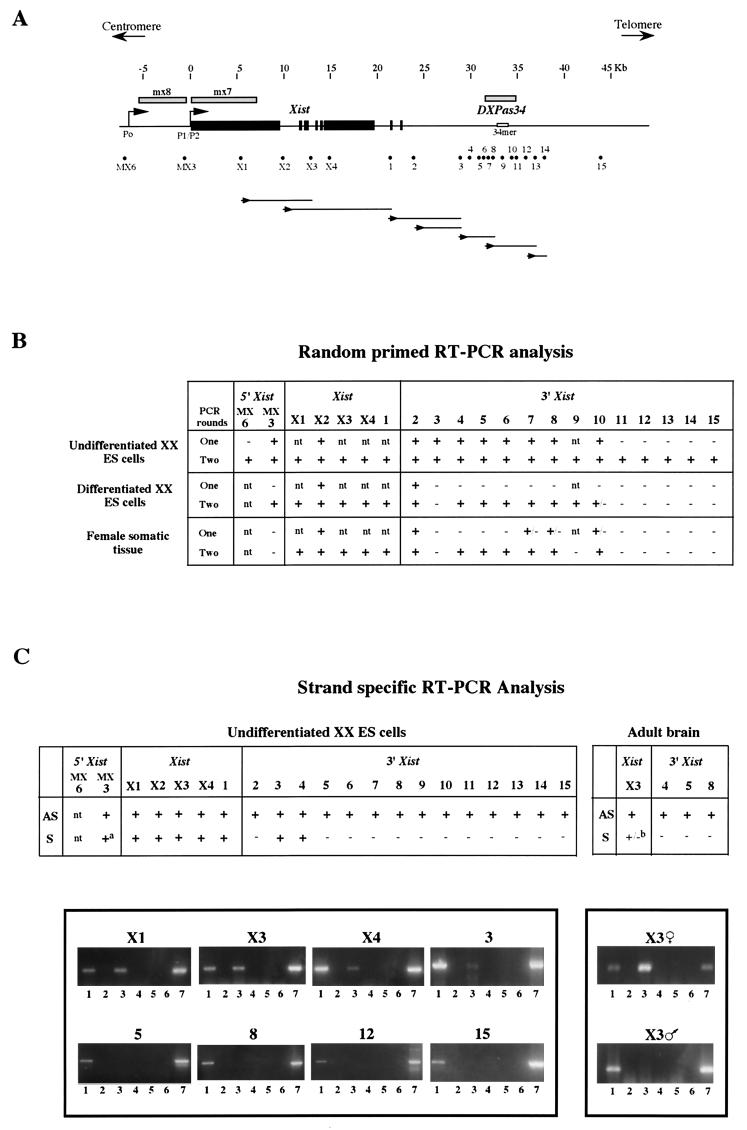
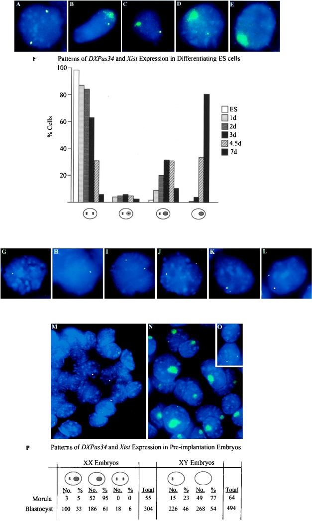
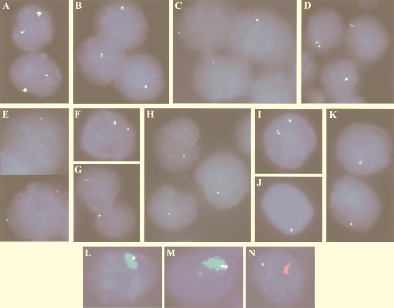
X inactivation in female mammals is controlled by a key locus on the X chromosome, the X-inactivation center (Xic). The Xic controls the initiation and propagation of inactivation in cis. It also ensures that the correct number of X chromosomes undergo inactivation (counting) and determines which X chromosome becomes inactivated (choice). The Xist gene maps to the Xic region and is essential for the initiation of X inactivation in cis. Regulatory elements of X inactivation have been proposed to lie 3' to Xist. One such element, lying 15 kb downstream of Xist, is the DXPas34 locus, which was first identified as a result of its hypermethylation on the active X chromosome and the correlation of its methylation level with allelism at the X-controlling element (Xce), a locus known to affect choice. In this study, we have tested the potential function of the DXPas34 locus in Xist regulation and X-inactivation initiation by deleting it in the context of large Xist-containing yeast artificial chromosome transgenes. Deletion of DXPas34 eliminates both Xist expression and antisense transcription present in this region in undifferentiated ES cells. It also leads to nonrandom inactivation of the deleted transgene upon differentiation. DXPas34 thus appears to be a critical regulator of Xist activity and X inactivation. The expression pattern of DXPas34 during early embryonic development, which we report here, further suggests that it could be implicated in the regulation of imprinted Xist expression.


Figures

FIG. 1
FIG. 1
(A) Map of the 50-kb region containing the Xist gene (exons shown as solid boxes) and the DXPas34 locus (which includes the 34-mer minisatellite repeat shown). Shaded bars above the map represent probes mx8, mx7, and DXPas34, used for RNA FISH. The positions of all the sites tested by RT-PCR are shown beneath the map as dots. Each dot represents the primer pairs used for RT-PCR analysis (for sequences of both outer and inner nested primer pairs, see Table 1). Lines below the map represent antisense transcript continuity: first-strand antisense cDNA was synthesized by using a sense primer (shown as an arrowhead below the appropriate site) and then amplified with primer pairs at sites downstream. Each line represents the extent of positive amplification obtained for the cDNA in question. (B) Summary of the RT-PCR data obtained by using random hexamers to prime cDNA synthesis. Control samples where reverse transcriptase was omitted were always included to rule out possible genomic DNA contamination. The results were assessed by using ethidium bromide-stained gels. Undifferentiated male and female ES cells gave identical results. Differentiated female ES cells represent 10-day EBs. Differentiated male ES cells gave identical results, apart from sites within Xist and site 2, which were negative after a single round of PCR. Somatic tissues tested were adult brain and in some cases liver. Male and female adult tissues gave identical results apart from site 2 (as above) and site MX3, which was positive in male but not in female cells. (C) Strand-specific RT-PCR analysis. (Top) Summary of the data; (bottom) representative sample of the data is shown. Antisense transcription was detected at all 20 sites from MX3 through to site 15 in undifferentiated ES cells. Sites X1 through to 1 within Xist also showed sense transcription. Sense transcripts were also detected at sites 3 and 4. (a) At MX3, sense transcript was only faintly, and not systematically, detected. In adult brain, sites X3, 4, 5, and 8 were tested and all showed antisense transcription. (b) Sense transcript was detected within Xist at X3 in females but not in males. cDNA synthesized from antisense transcript (AS) at each site was primed by using the sense primer, and cDNA synthesized from sense transcript (S) was primed by using an antisense primer. Lanes: 1, AS plus reverse transcriptase; 2, AS minus reverse transcriptase; 3, S plus reverse transcriptase; 4, S minus reverse transcriptase; 5, reverse transcriptase without any primer; 6, H2O control; 7, genomic DNA control. The specificity of the RT reactions was controlled by including an RNA sample where no primer was present (lane 5).
FIG. 2
FIG. 2
Two-color RNA FISH analysis of ES cells and preimplantation mouse embryos. (A to E) RNA FISH on XX ES cells with a Xist (λ 510) probe (green) and the 3.2-kb DXPas34 probe (red). Overlapping green and red signals are seen as yellow. (A) Undifferentiated XX ES cell showing colocalization of Xist and DXPas34 punctate signals. (B) Differentiating XX ES cell displaying an immature Xist RNA domain containing a DXPas34 signal on one X and a Xist/DXPas34 punctate signal on the other. (C) Differentiating XX ES cell displaying an immature Xist RNA domain without a DXPas34 signal. (D) Differentiating XX ES cell with a mature Xist RNA domain and no DXPas34 signal within it. (E) Fully differentiated XX ES cell with a mature Xist RNA domain and no DXPas34 signal on either X. (F) Frequency of cells exhibiting different patterns of Xist and DXPas34 RNA signals in XX ES cells and EBs at different times of differentiation in days (d). Xist RNA punctate signals (detected by λ 510) are shown as white pinpoints, and DXPas34 RNA signals are shown as black pinpoints. Immature Xist RNA domains are shown as light grey ovals. Mature Xist RNA domains are shown as dark grey ovals. Cell numbers scored were 1 day (n = 56), 2 days (n = 116), 3 days (n = 100), 4.5 days (n = 85), and 7 days (n = 78). A small proportion of cells (<5%), showing either two Xist pinpoints and only one DXPas34 pinpoint or vice versa (and no Xist RNA domain), which were detected at every stage, are not included in the histogram. (G to L) RNA FISH on XY and transgenic XY ES cells by using strand-specific Xist probes mx8 or exon I oligonucleotides (green) and the 3.2-kb DXPas34 probe (red). (G) Undifferentiated male ES cell. The Xist sense transcript colocalizes with the DXPas34 transcript. (H) Undifferentiated male ES cell. The Xist antisense transcript colocalizes with the DXPas34 transcript. (I) Undifferenitated female ES cell. Sense transcripts at both Xist alleles colocalize with DXPas34 transcripts. (J) Undifferentiated female ES cell. Antisense transcripts at both Xist alleles colocalize with DXPas34 transcripts. (K) Undifferentiated male ES cell containing a two-copy YAC PA-2 transgene (L412). The Xist sense transcript colocalizes with the DXPas34 transcript. (L) Undifferentiated L412 ES cell. The Xist antisense transcript colocalizes with the DXPas34 transcript. Note that in panels K and L, one allele (previously shown to be the two-copy transgene [20]) gives a slightly larger signal than the other. (M to P) RNA FISH on male and female blastocysts by using a Xist (λ 510) probe (green) and the 3.2-kb DXPas34 probe (red). Smaller probes from the DXPas34 region (sites 2, 3, 6, and 9) gave similar profiles to the larger DXPas34 probe. (M) Male blastocysts. In a proportion of cells, a Xist/DXPas34 RNA pinpoint signal is detected on the maternal X chromosome. (N) Female blastocysts. Most cells contain a Xist RNA domain corresponding to the paternal X with no sign of DXPas34 RNA within such domains. A proportion of these cells also contain a Xist/DXPas34 RNA pinpoint signal. (O) A small proportion of female blastocyst cells, probably corresponding to the ICM, contains no Xist RNA domain, and either one or two Xist/DXPas34 RNA pinpoint signals. (P) Frequency of the different patterns of Xist/DXPas34 expression observed in morulas and blastocysts.
FIG. 3
FIG. 3
Structural analysis of the DXPas34 deletion in YACs PA-2 and PA-3 F1n. (A) Structures of the two YACs used for the deletion of DXPas34, PA-2 (460 kb), and PA-3F1n (320 kb). The DNA probes, in and around the Xist gene, used to characterize the YACs and transgenic clones are shown as solid boxes along with the I-PpoI (PA-2 only) and SalI (Sa) sites that were informative in the analysis of the transgenes. (B) Structure of the DXPas34 locus before and after deletion. The 4.6-kb EcoRI fragment containing DXPas34 in the undeleted YACs and the structure of the same region after the deletion of DXPas34 and replacement with the URA3 yeast gene are indicated. A solid box above the map represents the extent of the 3-kb DXPas34 deletion, and dashed lines indicate the correspondence between the EcoRI sites of the intact and the deleted (ΔDXPas34) sequences. EcoRI (E), HpaII (H), MluI (M), EagI (Ea), and SalI (Sa) indicate restriction sites used for the structural analysis of this region. The sizes of informative EcoRI-SalI and EcoRI-EagI fragments are shown. Probes (a, b, c, and URA) used to assess the structure of the region, following homologous recombination, are shown as open boxes below each map. The region containing probe c disappears following the replacement of DXPas34 by the yeast URA3 gene (open box). Transcriptional orientation of URA3 is shown by an arrow (5′-3′). (C) Example of Southern blot analysis with probes a, b, c, and URA on YAC PA-2 Δ34.1 DNA compared to the parental undeleted YAC PA-2 DNA. EcoRI (E), EcoRI-SalI (E/Sa), and EcoRI-EagI (E/Ea) informative digests were used to demonstrate the disappearance of a SalI site and the region containing probe c, concomitant with the replacement by the URA3 and a new EagI site (B). Probe a detects 4.6-kb (undeleted), 2.9-kb (ΔDXPas34), and 1.7-kb EcoRI fragments, the last of these corresponding to the additional restriction fragment recognized by this probe which overlaps an EcoRI site (B). Similar results were obtained when this analysis was performed on YAC PA-3 F1n Δ34.3 and parental YAC PA-3 F1n DNAs (data not shown).
FIG. 4
FIG. 4
Xist and DXPas34 expression in ES cell lines with intact or ΔDXPas34 YAC transgenes. (A to D) Dual-color RNA FISH, with Xist (λ 510, green) and DXPas34 (red) probes, was performed on undifferentiated ES cells without denaturation of nuclei. Colocalization of green and red signals results in the appearance of yellow signals. (A) Control transgenic male line L412, which carries two copies of YAC PA-2. Xist and DXPas34 probes detect two colocalized signals, the larger one having been previously shown to be associated with the multicopy transgenic locus (20). (B to D) Line LP5 (which contains two or three copies of YAC PA-2 Δ34.1) (B), line LP6 (which contains one copy of YAC PA-2 Δ34.1) (C), and line LF1 (which contains three or four copies of YAC PA-3 F1n Δ34.3) (C) all exhibit a unique Xist/DXPas34 RNA signal. The presence of two closely associated pinpoint signals represents a replicated locus. (E) An example of the X chromosomal origin of the unique Xist RNA pinpoint in the LP6 representative undifferentiated ES cell line. The Xist RNA FISH signals in positioned nuclei were photographed, and the slides were then denatured and DNA FISH with a transgene-specific (pYAC4) probe was performed. This experiment clearly demonstrates that the single Xist RNA signal (green) observed in ΔDXPas34 transgenic cell lines prior to differentiation is not associated with the transgenic locus (red) and must therefore correspond to the X chromosome. (F to K) Study of the transcription in the region 3′ to Xist with probes corresponding to sites 2 (F to H) and 3 (I to K) (see Fig. 1A for probe positions) in control and ΔDXPas34 lines. Double RNA FISH with a Xist probe (green) and probes from site 2 or 3 (red) was performed. Line L412 (F) exhibits two Xist-site 2 colocalized signals per nucleus, whereas two representative ΔDXPas34 transgenic ES cell lines, LP6 (YAC PA-2 Δ34.1) (G) and LF1 (YAC PA-3F1n Δ34.3) (H), exhibit only a single Xist/site 2 signal. Identical results were obtained with the Xist/site 3 probe combination, on the same lines: L412 (I), LP6 (J), and LF1. (K). (L and M) Capacity of ΔDXPas34 multicopy YAC transgenes to trigger Xist RNA coating of an autosome in cis. Simultaneous RNA and DNA FISH was performed on differentiated cells from the LP5 (L) and LF1 (M) lines with a Xist probe (λ 510; green) and a transgene-specific probe (pYAC4). Formation of Xist RNA domains is observed on the autosome carrying the transgene in both lines. (N) Upon differentiation, transition to Xist RNA coating of an autosome in cis appears to be correctly regulated in the multicopy LF1 line: an mx8 probe (green), specific for Xist transcripts initiating upstream of P1/P2 (from the putative P0 promoter), detects only a single punctate signal per cell, while Xist RNA domain formation on the transgene is associated only with an mx7 (Xist exonI) signal (red).

References

    1. Ashe H L, Monks J, Wijgerde M, Fraser P, Proudfoot N J. Intergenic transcription and transinduction of the human beta-globin locus. Genes Dev. 1997;11:2494–2509. - PMC - PubMed
    1. Avner P. X chromosome inactivation: Xce and the candidate region for the X inactivation centre. In: Russo E, Riggs A, Martienssen R, editors. Epigenetic mechanisms of gene regulation. Cold Spring Harbor, N.Y: Cold Spring Harbor Laboratory Press; 1996. pp. 249–265.
    1. Avner P, Prissette M, Arnaud D, Courtier B, Cecchi C, Heard E. Molecular correlates of the murine Xce locus. Genet Res. 1998;72:217–224. - PubMed
    1. Beddington R S P. Isolation, culture and manipulation of postimplantation mouse embryos. Oxford, England: IRL Press; 1987.
    1. Borsani B, Tonlorenzi R, Simmler M-C, Dandolo L, Arnaud D, Capra V, Grompe M, Pizzuti A, Muzni D, Lawrence C, Willard H F, Avner P, Ballabio A. Characterization of a murine gene expressed from the inactive X chromosome. Nature. 1991;351:325–329. - PubMed

Publication types

MeSH terms

LinkOut - more resources