. 1999 Dec;19(12):8570-80.
doi: 10.1128/MCB.19.12.8570.

Premature expression of the winged helix transcription factor HFH-11B in regenerating mouse liver accelerates hepatocyte entry into S phase

Affiliations

Premature expression of the winged helix transcription factor HFH-11B in regenerating mouse liver accelerates hepatocyte entry into S phase

H Ye et al. Mol Cell Biol. 1999 Dec.

Abstract

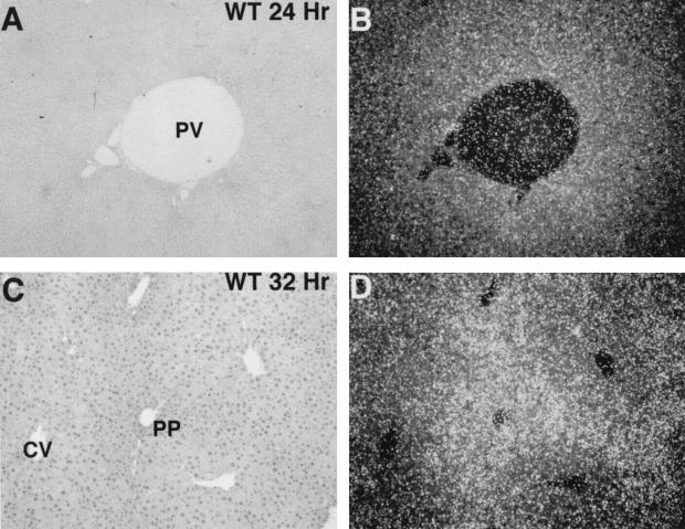
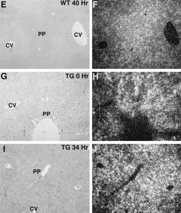
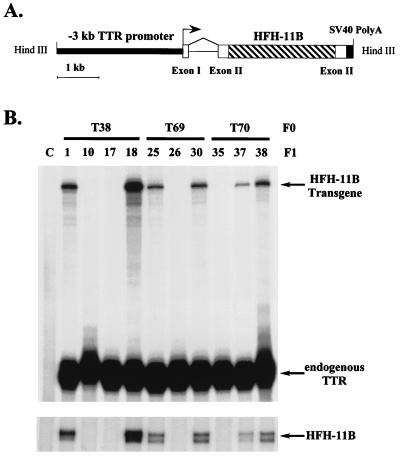
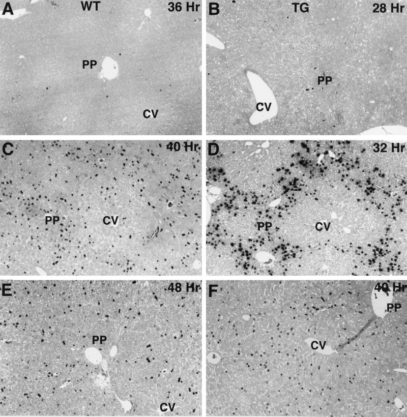
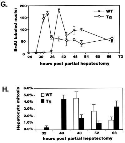
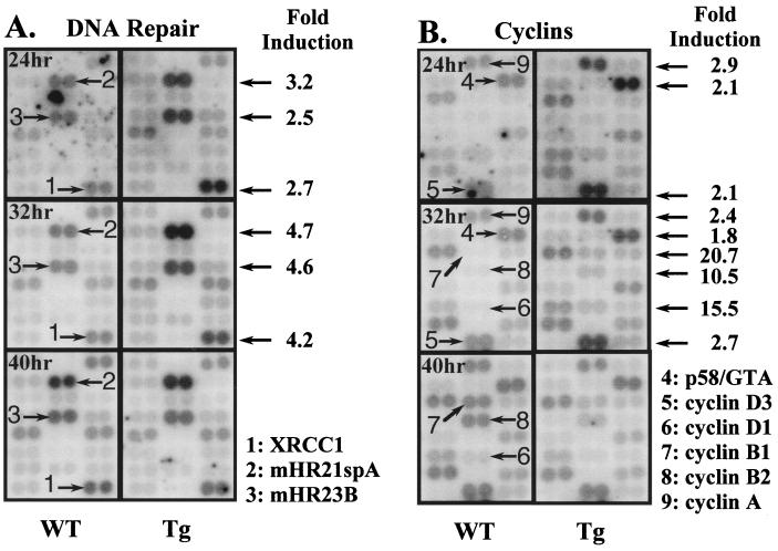
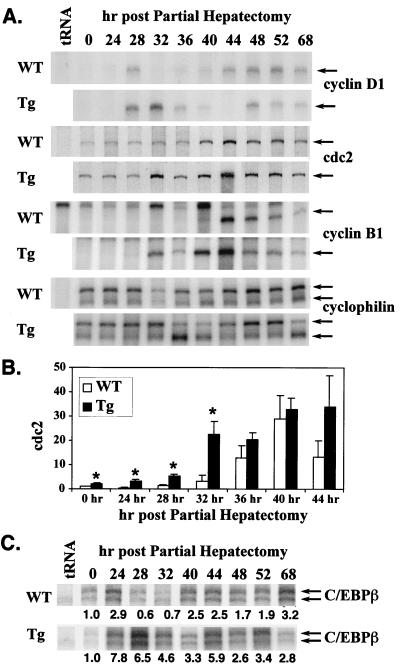
Two-thirds partial hepatectomy (PH) induces differentiated cells in the liver remnant to proliferate and regenerate to its original size. The proliferation-specific HNF-3/fork head homolog-11B protein (HFH-11B; also known as Trident and Win) is a family member of the winged helix/fork head transcription factors and in regenerating liver its expression is reactivated prior to hepatocyte entry into DNA replication (S phase). To examine whether HFH-11B regulates hepatocyte proliferation during liver regeneration, we used the -3-kb transthyretin (TTR) promoter to create transgenic mice that displayed ectopic hepatocyte expression of HFH-11B. Liver regeneration studies with the TTR-HFH-11B mice demonstrate that its premature expression resulted in an 8-h acceleration in the onset of hepatocyte DNA replication and mitosis. This liver regeneration phenotype is associated with protracted expression of cyclin D1 and C/EBPbeta, which are involved in stimulating DNA replication and premature expression of M phase promoting cyclin B1 and cdc2. Consistent with the early hepatocyte entry into S phase, regenerating transgenic livers exhibited earlier expression of DNA repair genes (XRCC1, mHR21spA, and mHR23B). Furthermore, in nonregenerating transgenic livers, ectopic HFH-11B expression did not elicit abnormal hepatocyte proliferation, a finding consistent with the retention of the HFH-11B transgene protein in the cytoplasm. We found that nuclear translocation of the HFH-11B transgene protein requires mitogenic signalling induced by PH and that its premature availability in regenerating transgenic liver allowed nuclear translocation to occur 8 h earlier than in wild type.


Figures

FIG. 1
FIG. 1
Induction of HFH-11 expression in regenerating mouse liver precedes DNA replication and is localized to hepatocytes of the periportal region. To determine the temporal and spatial expression pattern of HFH-11, paraffin sections of regenerating wild-type (WT) mouse liver were prepared at various times (hours) after PH and used for in situ hybridization with 33P-labeled antisense HFH-11 RNA probe. Shown in the left panels are the bright-field illuminations and in the right panels are the dark-field illuminations depicting the hybridization signals. Indicated on the liver sections are the portal vein (PV), the periportal region (PP) comprising the hepatocytes surrounding the portal vein, and the central vein (CV). In regenerating mouse liver, HFH-11 expression initiates at 24 h post-PH (A and B) and is maximally expressed at 32 h post-PH (C and D), exhibiting more-intense HFH-11 labeling in hepatocytes of the periportal region. (E and F) The HFH-11 periportal expression pattern continues in regenerating mouse liver at 40 h post-PH. (G and H) Nonregenerating hepatocytes from the transgenic (TG) T38 line (see Fig. 2) reveal ectopic HFH-11B expression throughout the liver parenchyma, but HFH-11 hybridization signals are more abundant in the periportal region. (I and J) Expression of the HFH-11 transgene continues in replicating hepatocytes at 34 h post-PH. Note that the in situ hybridization for regenerating wild-type liver is exposed for 1 week, which is three times longer than for the transgenic mouse livers.
FIG. 1
FIG. 1
Induction of HFH-11 expression in regenerating mouse liver precedes DNA replication and is localized to hepatocytes of the periportal region. To determine the temporal and spatial expression pattern of HFH-11, paraffin sections of regenerating wild-type (WT) mouse liver were prepared at various times (hours) after PH and used for in situ hybridization with 33P-labeled antisense HFH-11 RNA probe. Shown in the left panels are the bright-field illuminations and in the right panels are the dark-field illuminations depicting the hybridization signals. Indicated on the liver sections are the portal vein (PV), the periportal region (PP) comprising the hepatocytes surrounding the portal vein, and the central vein (CV). In regenerating mouse liver, HFH-11 expression initiates at 24 h post-PH (A and B) and is maximally expressed at 32 h post-PH (C and D), exhibiting more-intense HFH-11 labeling in hepatocytes of the periportal region. (E and F) The HFH-11 periportal expression pattern continues in regenerating mouse liver at 40 h post-PH. (G and H) Nonregenerating hepatocytes from the transgenic (TG) T38 line (see Fig. 2) reveal ectopic HFH-11B expression throughout the liver parenchyma, but HFH-11 hybridization signals are more abundant in the periportal region. (I and J) Expression of the HFH-11 transgene continues in replicating hepatocytes at 34 h post-PH. Note that the in situ hybridization for regenerating wild-type liver is exposed for 1 week, which is three times longer than for the transgenic mouse livers.
FIG. 2
FIG. 2
Hepatocyte nuclear localization of HFH-11 protein in regenerating wild-type and transgenic livers. To determine nuclear localization of the HFH-11 protein, paraffin sections of regenerating wild-type (WT; A to E) or transgenic (TG; F to K) mouse livers were prepared at various times (hours) after PH and used for immunohistochemical staining with the HFH-11 antibody (57). The HFH-11 antibody was detected with the horseradish peroxidase AEC substrate (stains red), followed by hematoxylin counterstaining (stains nuclei blue). (A to D) Nuclear localization of the HFH-11 protein in regenerating wild-type hepatocytes is observed at 32 and 36 h post-PH (red nuclei [indicated by arrows]), while perinuclear staining is found at 28 h post-PH, and their nuclei are counterstained blue by hematoxylin (indicated by asterisks). (E to I) Hepatocyte nuclear localization of the HFH-11B transgene protein requires mitotic signalling induced during liver regeneration. HFH-11B transgene protein is diffusely distributed in the cytoplasm of nonregenerating transgenic hepatocytes, and hematoxylin counterstains the nuclei blue (F [indicated by an asterisk]). In regenerating transgenic hepatocytes, HFH-11B nuclear localization (red, indicated by arrows) is observed at 24, 28, 32, and 36 h post-PH (H and I), while perinuclear and nuclear staining is found at 16 h post-PH (G). Hepatocyte nuclear translocation of the HFH-11B protein therefore occurs 8 h earlier in regenerating transgenic liver.
FIG. 3
FIG. 3
The −3-kb TTR promoter directs HFH-11B transgene expression in the adult liver. (A) Diagrammatical representation of the mouse −3-kb TTR promoter–HFH-11B transgene construct. Transgenic mice were created with the −3-kb TTR promoter region (small open box) driving expression of the human HFH-11B cDNA (striped box), which was cloned into the TTR second exon (large open box) that contains the SV40 polyadenylation signal (black box) (5, 6, 55). (B) Analysis of HFH-11B transgene expression in transgenic mouse livers. Total liver RNA was isolated from F1 transgenic mouse lines T38 (lanes 1 and 18), T60 (lanes 25 and 30), and T70 (lanes 37 and 38) and nontransgenic litter mates (lanes 10, 17, 26, and 35) and used for RNase protection assays with either the TTR-SV40 transgene or HFH-11B antisense-labeled RNA probes (see Materials and Methods). As reported previously, expression of the transgene produces RNase-resistant 310 nucleotide product (HFH-11B transgene), whereas expression of the endogenous TTR gene elicits an RNase protected fragment of 90 nucleotides (6, 55). Note that RNase protection with adult liver RNA and the HFH-11B probe only detected HFH-11B expression in the transgenic mouse lines. Lane numbers correspond to mouse numbers in each transgenic line.
FIG. 4
FIG. 4
Premature hepatocyte expression of HFH-11B accelerates DNA synthesis in regenerating transgenic mouse liver. Wild-type and TTR–HFH-11B transgenic mice were subjected to two-thirds PH and, 2 h prior to harvesting the regenerating livers, the mice received an intraperitoneal injection of BrdU to be detected by a monoclonal antibody to BrdU (see Materials and Methods). Immunohistochemical detection of hepatocytes incorporating BrdU is shown with regenerating wild-type (left panels) and transgenic (right panels) mouse livers. Maximal hepatocyte DNA replication for regenerating wild-type liver occurs at 40 h post-PH, whereas the peak of transgenic hepatocyte replication is observed 8 h earlier at 32 h post-PH. Note that wild-type regenerating livers also initiate DNA replication in hepatocytes surrounding the periportal region. Comparable BrdU incorporation is observed in regenerating wild-type and transgenic livers at 48 (E) and 40 (F) h post-PH, respectively. (G) Kinetics of BrdU incorporation into hepatocyte DNA during wild-type (WT) and transgenic (Tg) liver regeneration between 24 and 68 h post-PH. BrdU-positive hepatocytes (per 1,000 nuclei) from each sample were counted among at least 3,000 total hepatocytes, and the mean and standard deviation for each time point was calculated by using three to six regenerating livers. In regenerating transgenic livers, we observed an 8-h-earlier peak in hepatocyte DNA replication and no increase in the total number of replicating hepatocytes. An additional BrdU labeling time point (34 h post-PH) was used for regenerating transgenic liver. (H) Kinetics of hepatocyte mitosis during wild-type and transgenic liver regeneration between 32 and 68 h post-PH. Hepatocyte mitotic figures were counted in 10 high-power fields at the indicated time post-PH and are presented as a percentage of the total number of hepatocytes. The means from three regenerating mouse livers with the corresponding standard deviations are shown.
FIG. 4
FIG. 4
Premature hepatocyte expression of HFH-11B accelerates DNA synthesis in regenerating transgenic mouse liver. Wild-type and TTR–HFH-11B transgenic mice were subjected to two-thirds PH and, 2 h prior to harvesting the regenerating livers, the mice received an intraperitoneal injection of BrdU to be detected by a monoclonal antibody to BrdU (see Materials and Methods). Immunohistochemical detection of hepatocytes incorporating BrdU is shown with regenerating wild-type (left panels) and transgenic (right panels) mouse livers. Maximal hepatocyte DNA replication for regenerating wild-type liver occurs at 40 h post-PH, whereas the peak of transgenic hepatocyte replication is observed 8 h earlier at 32 h post-PH. Note that wild-type regenerating livers also initiate DNA replication in hepatocytes surrounding the periportal region. Comparable BrdU incorporation is observed in regenerating wild-type and transgenic livers at 48 (E) and 40 (F) h post-PH, respectively. (G) Kinetics of BrdU incorporation into hepatocyte DNA during wild-type (WT) and transgenic (Tg) liver regeneration between 24 and 68 h post-PH. BrdU-positive hepatocytes (per 1,000 nuclei) from each sample were counted among at least 3,000 total hepatocytes, and the mean and standard deviation for each time point was calculated by using three to six regenerating livers. In regenerating transgenic livers, we observed an 8-h-earlier peak in hepatocyte DNA replication and no increase in the total number of replicating hepatocytes. An additional BrdU labeling time point (34 h post-PH) was used for regenerating transgenic liver. (H) Kinetics of hepatocyte mitosis during wild-type and transgenic liver regeneration between 32 and 68 h post-PH. Hepatocyte mitotic figures were counted in 10 high-power fields at the indicated time post-PH and are presented as a percentage of the total number of hepatocytes. The means from three regenerating mouse livers with the corresponding standard deviations are shown.
FIG. 5
FIG. 5
Earlier expression of DNA repair and cyclin genes in regenerating transgenic livers. Radioactive cDNA was prepared from regenerating wild-type and transgenic livers at 24, 32, and 40 h post-PH and hybridized to six distinct Atlas mouse cDNA expression array blots. Composite images of two columns comparing hybridization signals of the DNA repair (A) or cyclin (B) genes in regenerating wild-type (WT) or transgenic (Tg) livers at the indicated hours post-PH are shown. Blots are normalized to the nine housekeeping cDNAs prior to comparing signals of other cDNAs which are spotted in duplicate on the blot. Arrows indicate transgenic regenerating cDNA signals which are increased compared to regenerating wild-type livers, and quantitation of the cDNA hybridization signals allowed determination of the fold induction. The numbering of cDNAs are as follows: 1, double-stranded break repair gene XRCC1; 2, mHR21spA (mouse homolog of the yeast rad21 gene); 3, the nucleotide excision repair mHR23B (mouse homolog of the yeast rad23 gene); 4, p58/GTA (galactosyltransferase-associated protein kinase or cdc2-related protein kinase); 5, cyclin D3; 7, cyclin B1; 8, cyclin B2; and 9, cyclin A.
FIG. 6
FIG. 6
Regenerating transgenic livers exhibit premature and prolonged expression of genes involved in DNA replication and mitosis. Total RNA was prepared from regenerating wild-type and transgenic mouse livers, and RNase protection assays were used to analyze for expression of cyclin D1, p34cdc2 (cdc2), cyclin B1, C/EBPβ, and cyclophilin. Both cyclophilin RNase-protected bands were used as a normalization internal control (indicated by arrows), and representative RNase protection assays displaying the expression levels of these genes at various times after PH are shown. (A) Regenerating transgenic livers exhibit protracted expression of S phase-promoting cyclin D1 and premature expression of M phase promoting cdc2 and cyclin B1. In regenerating transgenic livers, protracted cyclin D1 expression occurs during hepatocyte DNA replication (32 and 36 h post-PH), and cyclin B1 expression is induced at 32 h after PH, which precedes detectable wild-type expression by 8 h. Maximal transgenic expression of cdc2 is observed at 32 h post-PH, which precedes induced wild-type expression by 8 h (see panel B). (B) Increased expression of cdc2 RNA in regenerating transgenic mouse liver. Shown is the mean induction of cdc2 expression in regenerating wild-type and transgenic livers with the standard deviation derived from three to six mice per time point (the asterisk indicates P < 0.05). (C) Regenerating transgenic livers exhibit protracted expression of S phase-promoting C/EBPβ transcription factor. RNase protection assay demonstrates protracted expression of the C/EBPβ mRNA during transgenic hepatocyte DNA replication which occurs 24 and 32 h post-PH.

References

    1. Brenner D A. Signal transduction during liver regeneration. J Gastroenterol Hepatol. 1998;13:S93–S95. - PubMed
    1. Brunet A, Bonni A, Zigmond M J, Lin M Z, Juo P, Hu L S, Anderson M J, Arden K C, Blenis J, Greenberg M E. Akt promotes cell survival by phosphorylating and inhibiting a Forkhead transcription factor. Cell. 1999;96:857–868. - PubMed
    1. Clark K L, Halay E D, Lai E, Burley S K. Co-crystal structure of the HNF-3/fork head DNA-recognition motif resembles histone H5. Nature. 1993;364:412–420. - PubMed
    1. Corral M, Paris B, Guguen-Guillouzo C, Corcos D, Kruh J, Defer N. Increased expression of the N-myc gene during normal and neoplastic rat liver growth. Exp Cell Res. 1988;174:107–115. - PubMed
    1. Costa R H, Grayson D R, Darnell J E., Jr Multiple hepatocyte-enriched nuclear factors function in the regulation of transthyretin and alpha 1-antitrypsin genes. Mol Cell Biol. 1989;9:1415–1425. - PMC - PubMed

Publication types

MeSH terms