. 1999 Dec;19(12):8591-603.
doi: 10.1128/MCB.19.12.8591.

NF-Y associates with H3-H4 tetramers and octamers by multiple mechanisms

Affiliations

NF-Y associates with H3-H4 tetramers and octamers by multiple mechanisms

G Caretti et al. Mol Cell Biol. 1999 Dec.

Abstract

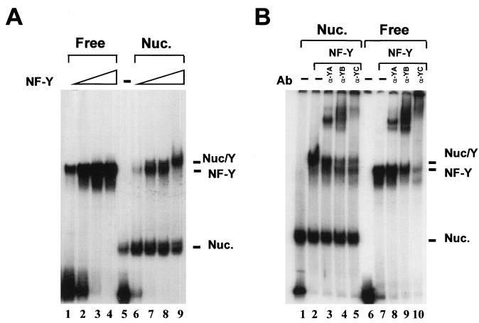
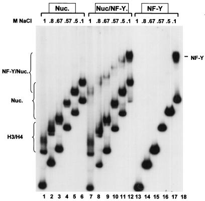
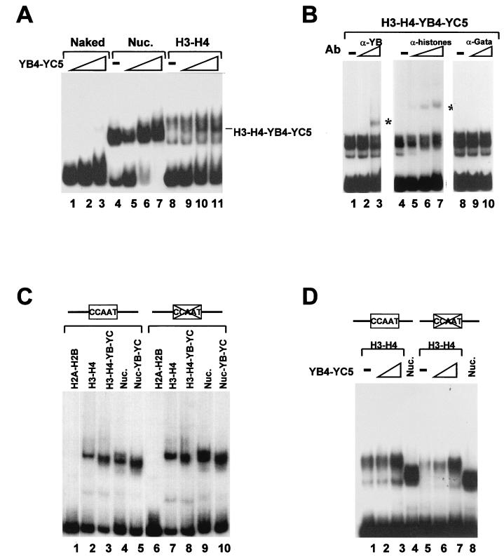
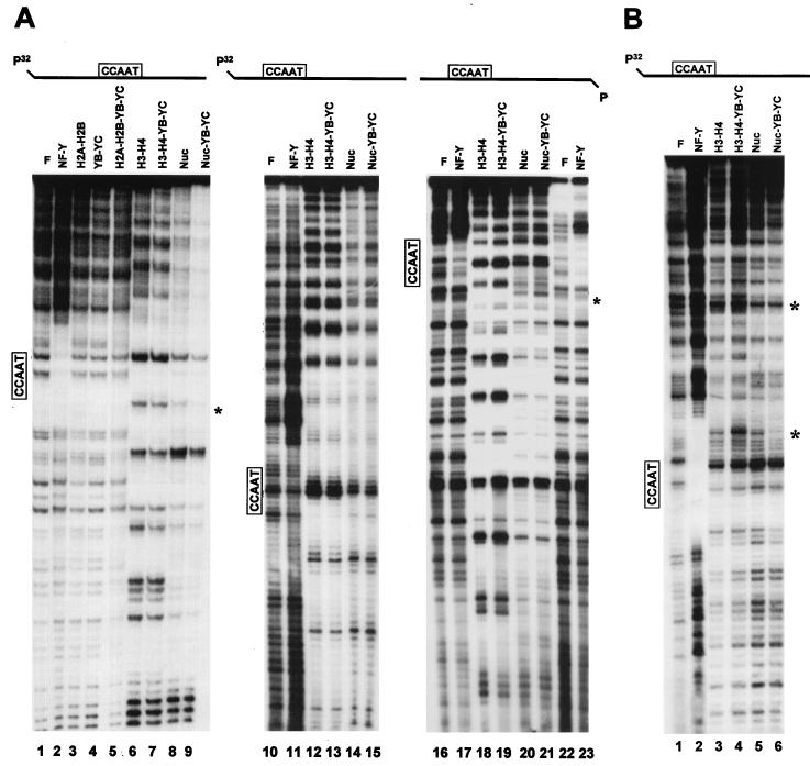
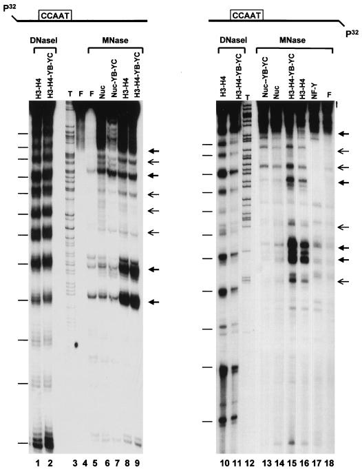
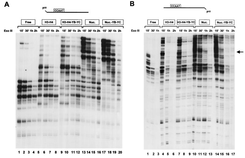
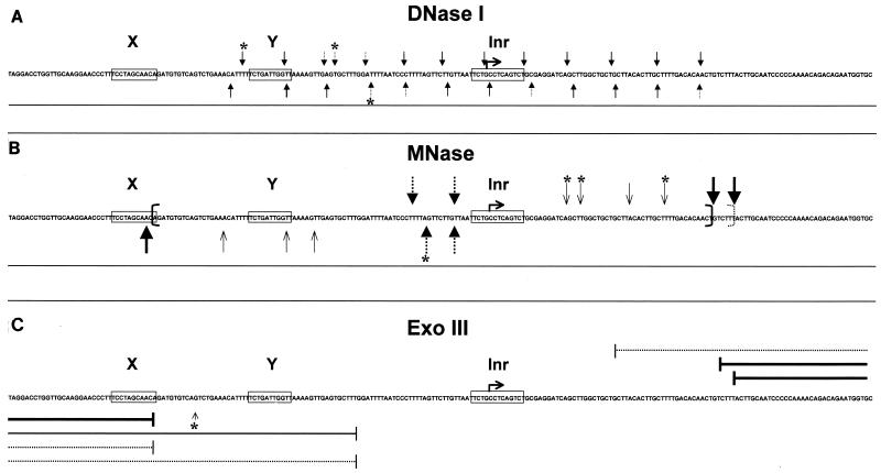
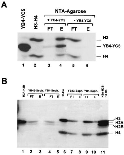
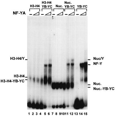
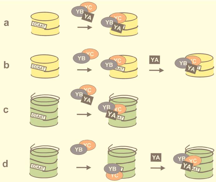
NF-Y is a CCAAT-binding trimer with two histonic subunits, NF-YB and NF-YC, resembling H2A-H2B. We previously showed that the short conserved domains of NF-Y efficiently bind to the major histocompatibility complex class II Ea Y box in DNA nucleosomized with purified chicken histones. Using wild-type NF-Y and recombinant histones, we find that NF-Y associates with H3-H4 early during nucleosome assembly, under conditions in which binding to naked DNA is not observed. In such assays, the NF-YB-NF-YC dimer forms complexes with H3-H4, for whose formation the CCAAT box is not required. We investigated whether they represent octamer-like structures, using DNase I, micrococcal nuclease, and exonuclease III, and found a highly positioned nucleosome on Ea, whose boundaries were mapped; addition of NF-YB-NF-YC does not lead to the formation of octameric structures, but changes in the digestion patterns are observed. NF-YA can bind to such preformed DNA complexes in a CCAAT-dependent way. In the absence of DNA, NF-YB-NF-YC subunits bind to H3-H4, but not to H2A-H2B, through the NF-YB histone fold. These results indicate that (i) the NF-Y histone fold dimer can efficiently associate DNA during nucleosome formation; (ii) it has an intrinsic affinity for H3-H4 but does not form octamers; and (iii) the interactions between NF-YA, NF-YB-NF-YC, and H3-H4 or nucleosomes are not mutually exclusive. Thus, NF-Y can intervene at different steps during nucleosome formation, and this scenario might be paradigmatic for other histone fold proteins involved in gene regulation.


Figures

FIG. 1
FIG. 1
wt NF-Y associates a nucleosome-bound Ea Y box. (A) EMSA of a dose response of wt NF-Y on mock-reconstituted (0.5, 1, 3, and 10 ng; lanes 1 to 4) or nucleosome (NUC.)-reconstituted (lanes 6 to 9) Ea fragment 2 (−115 to +60 of Ea [36]). Lane 5, no NF-Y added to nucleosomal DNA. (B) A 70% nucleosomized Ea fragment 2 was run without NF-Y (lane 1), with 5 ng of NF-Y (lane 2), and with the same amount of NF-Y incubated with the indicated anti-NF-Y antibody (Ab; 200 ng of purified antibody; lanes 3 to 5). The same was in lanes 7 to 10 except that mock-reconstituted DNA was used.
FIG. 2
FIG. 2
Binding of NF-Y during nucleosome assembly. Stoichiometric amounts of histones were assembled in high salts with Ea fragment 2 and cold competitor DNA, in the absence (lanes 1 to 6) or presence (lanes 7 to 12) of 5 ng of wt NF-Y. In lanes 13 to 18, 5 ng of wt NF-Y was used as in lanes 7 to 12, in the absence of histones. Mixtures were progressively diluted, and at each NaCl concentration, aliquots were added to a running polyacrylamide gel. The NF-Y, H3-H4, nucleosome, and NF-Y–nucleosome bands are indicated.
FIG. 3
FIG. 3
EMSA of a NF-YB–NF-YC–H3–H4 complex on the Ea promoter DNA. (A) Reconstitution of fragment 2 with increasing amounts of NF-YB4–NF-YC5 alone (200 ng, 400 ng, and 1 μg; lanes 1 to 3), with all core histones (lanes 5 to 7), or with H3-H4 only (lanes 9 to 11). Control reconstitutions with core histones and H3-H4 alone are in lanes 4 and 8. The hybrid complex is indicated. Nuc., nucleosomes. (B) Antibody challenge of the hybrid NF-YB–NF-YC–H3–H4 complex. The hybrid complex was incubated for 30 min at 4°C with increasing amounts of anti-NF-YB (100 and 500 ng; lanes 2 and 3), antihistone (10 ng, 100 ng, and 1 μg; lanes 5 to 7), or control anti-Gata1 (100 and 500 ng; lanes 9 and 10) antibodies (Ab). The supershifted complexes are indicated by asterisks. (C) The indicated combinations of proteins (H2A-H2B [lanes 1 and 6], H3-H4 [lanes 2 and 7], H3–H4–NF-YB–NF-YC [lanes 3 and 8], H2A-H2B-H3-H4 [lanes 4 and 9], and H2A-H2B-H3-H4-NF-YB-NF-YC [lanes 5 and 10]) were incubated with wt Ea DNA (lanes 1 to 5) or with a Y-box mutant (lanes 6 to 10). (D) Same as panel C except that H3-H4 tetramers were incubated alone (lanes 1 and 5) or with NF-YB4-NF-YC5 (200 ng [lanes 2 and 6] and 1 μg [lanes 3 and 7]). Nucleosomes are in lanes 4 and 8.
FIG. 4
FIG. 4
DNase I footprint of histone-NF-YB–NF-YC combinations. (A) The DNAs used were Ea fragment 2 (labeled on the top strand; lanes 1 to 9) and fragment 6 (−145 to +35 of Ea [36]; labeled on the top strand [lanes 10 to 15] and bottom strand [lanes 16 to 23]). After reconstitutions with stoichiometric amounts of the indicated HFM protein combinations, aliquots were digested with DNase I and analyzed in sequencing gels. Asterisks denote bands that were diminished (lanes 8 and 9) or increased (lanes 20 and 21) when NF-YB–NF-YC was added to reconstitutions. To locate the position of the NF-Y footprinted area, samples in lanes 2, 11, 17, and 23 contained only the NF-Y trimer, without histones. Asterisks indicate protections (lane 9) and hypersensitivities (lanes 19 and 21) upon addition of NF-YB–NF-YC to histones. F, Free DNA; Nuc, nucleosomes. (B) The indicated complexes were DNase I digested, purified from gels (see Materials and Methods), eluted, and run on sequencing gels. Lane 1, free DNA; lane 2, DNA and NF-Y; lanes 3 to 6, H3-H4, H3–H4–NF-YB–NF-YC, nucleosome, and nucleosome–NF-YB–NF-YC, respectively. Asterisks indicate protections (lane 4) and hypersensitivity (lane 6).
FIG. 5
FIG. 5
MNase accessibility assay of histone–NF-YB–NF-YC combinations. Stoichiometric amounts of the indicated combinations of HFM proteins were reconstituted with fragment 2 (labeled on the top strand [lanes 4 to 9] and bottom strand [lanes 13 to 18]), cut with MNase, and analyzed on sequencing gels. In lane 17, the NF-Y trimer was used to show the NF-Y footprinted area. F refers to free, mock-reconstituted DNA (lanes 4 and 5; uncut and cut with MNase, respectively). Arrows correspond to the major and minor hypersensitive sites. Part of the H3-H4 and H3–H4–NF-YB–NF-YC reconstitutions were cut with DNase I and run in parallel (lanes 1, 2, 10, and 11). Bars correspond to the 10-bp cutting patterns of DNase I. Sequencing reactions (T; lanes 3 and 12) were run in parallel to precisely map the sites of MNase cuts.
FIG. 6
FIG. 6
Exo III assay of histone–NF-YB–NF-YC combinations. Stoichiometric amounts of the indicated combinations of HFM proteins were reconstituted with fragment 2 (labeled on the top strand [A] and bottom strand [B]), cut with Exo III for the indicated length of time, and analyzed on sequencing gels. Nuc., nucleosome.
FIG. 7
FIG. 7
Recapitulation of the data obtained with DNase I, MNase, and Exo III assays. The X, Y, and Inr elements are boxed, and the major +1 start site of the Ea promoter is outlined. (A) DNase I. Arrows indicate the cuts in the nucleosomal region. (B) MNase. Thick arrows indicate the cuts with nucleosomes and dotted arrows with H3-H4 tetramers, thin arrows represent minor cutting sites, and brackets delimit the boundaries of the nucleosome. (C) Exo III. Thick and dotted lines indicate major stops with nucleosomes and H3-H4 tetramers, respectively. The small arrow indicates a major stop which is bypassed when NF-YB–NF-YC is reconstituted with nucleosomes. Asterisks refer to differences in the pattern observed when the NF-YB and NF-YC subunits are added to histones.
FIG. 8
FIG. 8
Protein-protein interactions between NF-YB–NF-YC and core histone dimers. (A) NTA-agarose columns. Lanes: 1 and 2, NF-YB4–NF-YC5 and H3-H4, respectively; 3 and 4, flowthrough and eluted material from NTA-agarose supplemented with His-tagged NF-YB4–NF-YC5; 5 and 6, control NTA-agarose columns run without NF-YB4–NF-YB5. (B) NF-YB4 and NF-YB43–Sepharose (Seph.) columns. Load, flowthrough, and eluates of pure H2A-H2B (lanes 1 to 5) and H3-H4 (lanes 6 to 10) are indicated. The four histones are run together in lane 11.
FIG. 9
FIG. 9
NF-YA associates with histone-bound NF-YB–NF-YC. Increasing concentrations of wt NF-YA (1 ng [lanes 2, 5, 8, 11, and 14] and 5 ng [lanes 3, 6, 9, 12, and 15]) were incubated with mixed complexes previously reconstituted with the indicated combinations of HFM proteins: H3-H4 (lanes 1 to 3), H3–H4–NF-YB–NF-YC (lanes 4 to 6), H3-H4-H2A-H2B (lanes 7 to 9), H3–H4–H2A–H2B–NF-YB–NF-YC (lanes 10 to 12), and NF-YB–NF-YC (lanes 13 to 15). Nuc., nucleosome.
FIG. 10
FIG. 10
Models for NF-Y–histone association. The NF-Y trimer associates with H3-H4 tetramers (a) or nucleosomes (c). The NF-YB–NF-YC dimer associates with H3-H4 tetramers (b) and nucleosomes, presumably through H3-H4, and forms structures which can still be bound by NF-YA (d). H3-H4 tetramers are in yellow; nucleosomes are in green.

References

    1. Alevizopoulos A, Dusserre Y, Tsai-Pflugfelder M, von der Weid T, Whali W, Mermod N. A proline-rich TGF-β-responsive transcriptional activator interacts with histones H3. Genes Dev. 1995;9:3051–3066. - PubMed
    1. Arents G, Moudrianakis E N. The histone fold: a ubiquitous architectural motif utilized in DNA compaction and protein dimerization. Proc Natl Acad Sci USA. 1995;92:11170–11174. - PMC - PubMed
    1. Baxevanis A D, Arents G, Moudrianakis E N, Landsman D. A variety of DNA-binding and multimeric proteins contain the histone fold motif. Nucleic Acids Res. 1995;23:2685–2691. - PMC - PubMed
    1. Bellorini M, Lee D K, Dantonel J C, Zemzoumi K, Roeder R G, Tora L, Mantovani R. CCAAT binding NF-Y-TBP interactions: NF-YB and NF-YC require short domains adjacent to their histone fold motifs for association with TBP basic residues. Nucleic Acids Res. 1997;25:2174–2181. - PMC - PubMed
    1. Bellorini M, Zemzoumi K, Farina A, Berthelsen J, Piaggio G, Mantovani R. Cloning and expression of human NF-YC. Gene. 1997;193:119–125. - PubMed

Publication types

Grants and funding

LinkOut - more resources