. 1999 Dec;67(12):6293-302.
doi: 10.1128/IAI.67.12.6293-6302.1999.

Selective recruitment of T-cell subsets to the udder during staphylococcal and streptococcal mastitis: analysis of lymphocyte subsets and adhesion molecule expression

Affiliations

Selective recruitment of T-cell subsets to the udder during staphylococcal and streptococcal mastitis: analysis of lymphocyte subsets and adhesion molecule expression

J Soltys et al. Infect Immun. 1999 Dec.

Abstract

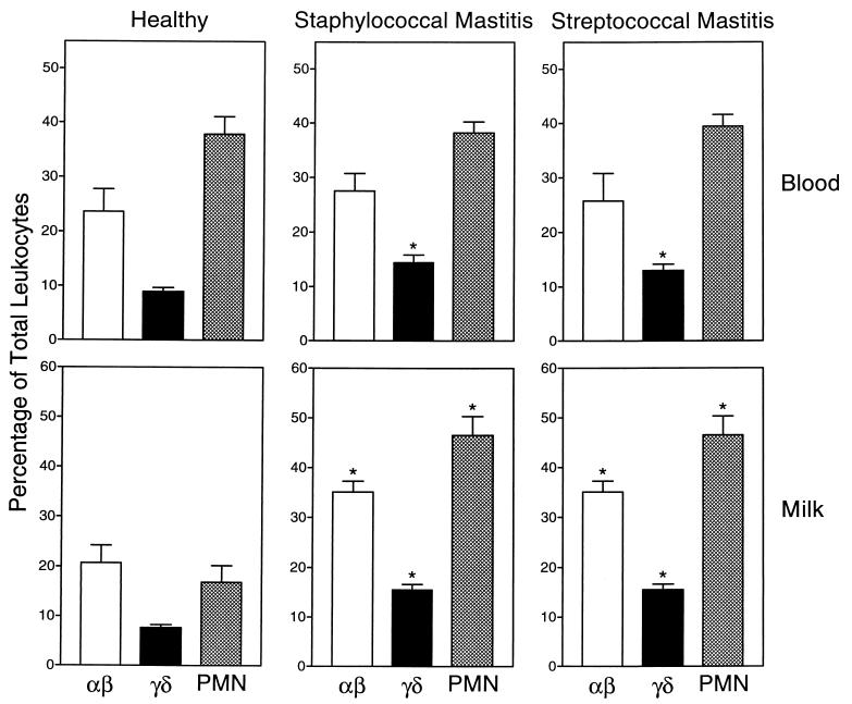
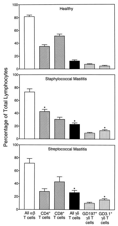
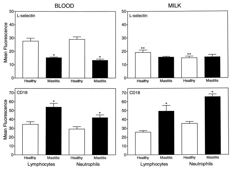
During bacterial infection of the bovine mammary gland, large numbers of leukocytes migrate into the udder, resulting in the establishment of a host response against the pathogen. Currently, the specific leukocyte populations mediating this immune response are not well defined. In the studies described here, we analyzed blood and milk from healthy cows and cows with naturally occurring mastitis to determine if distinct alphabeta and gammadelta T-lymphocyte subsets were involved in the response of the udder to a mastitis pathogen and if the type of mastitis pathogen influenced the subset composition of these responding leukocytes. Although blood samples from cows with confirmed staphylococcal and streptococcal mastitis were characterized by increased numbers of gammadelta T cells, the most dramatic changes in leukocyte distributions occurred in milk samples from these cows, with a 75% increase in alphabeta T-cell levels and a 100% increase in gammadelta T-cell levels relative to the levels in milk samples from healthy animals. Interestingly, the increase in alphabeta T-cell numbers observed in milk from cows with staphylococcal mastitis was primarily due to increased numbers of CD4(+) T cells, while the increase in alphabeta T-cell numbers observed in cows with streptococcal mastitis was due to a parallel increase in both CD4(+) and CD8(+) T-cell numbers. The increased numbers of gammadelta T cells in milk from cows with staphylococcal and streptococcal mastitis were due to a selective recruitment of a distinct gammadelta T-cell subset (GD3.1(+)), while no change in the numbers of GD197(+) gammadelta T cells was observed. We also analyzed adhesion protein expression on blood and milk leukocytes and found that, in comparison to the situation for healthy cows, L-selectin was down-regulated and CD18 was up-regulated on leukocytes from cows with mastitis. Thus, shedding of L-selectin and up-regulation of CD18 by neutrophils may provide a sensitive indicator of early inflammatory responses during bovine mastitis. Overall, these studies suggest that distinct alphabeta and gammadelta T-cell subsets are involved in the host defense of the udder against mastitis infection and that selective recruitment of these T-cell subsets depends on the infectious agent involved.


Figures

FIG. 1
FIG. 1
Leukocyte subset distribution in blood and milk of healthy animals. Mixed bovine leukocytes were isolated from the blood and milk of 10 healthy lactating cows (left panels), 10 cows with staphylococcal mastitis (center panels), and 10 cows with streptococcal mastitis (right panels). The cells were labeled with monoclonal antibodies against αβ T-cell antigen CD2 (CC42), pan-γδ T-cell receptor (GD3.8), and bovine neutrophils (BN15.6) and analyzed by flow cytometry as described in Materials and Methods. Lymphocytes and neutrophils (PMN) were identified by their distinctive forward and side light scatter profiles, and the percentage of total leukocytes staining above the background (secondary antibody only) for the specific antigens listed above was determined. The data are expressed as mean ± standard error of the mean (n = 10). The asterisk indicates statistically significant differences (P, <0.05) between mastitis and healthy samples.
FIG. 2
FIG. 2
Lymphocyte subset distribution in milk of cows with staphylococcal or streptococcal mastitis. Purified lymphocytes were isolated from the milk of 10 healthy lactating cows, 10 cows with staphylococcal mastitis, and 10 cows with streptococcal mastitis. The cells were labeled with monoclonal antibodies against CD2, CD4, CD8, pan-γδ TCR, GD3.1+ γδ TCR subset, and GD197+ γδ TCR subset and analyzed by flow cytometry as described in Materials and Methods. The percentage of total lymphocytes staining above the background (secondary antibody only) for the specific antigens listed above was determined. The data are expressed as mean ± standard error of the mean (N = 10). The asterisk indicates statistically significant differences (P, <0.05) between mastitis and healthy samples.
FIG. 3
FIG. 3
Two-color flow cytometric analysis of bovine blood and milk lymphocytes. Purified lymphocytes were isolated from the blood (A to C) and milk (D to F) of cows with mastitis, and two-color flow cytometric analysis was performed as described in Materials and Methods. (A and D) Staining of blood (A) and milk (D) lymphocytes with antibody GD3.8 (FL3, specific for γδ T cells) versus antibody CC58 (FL1, specific for bovine CD8). (B and E) Staining of blood (B) and milk (E) lymphocytes with antibody GD3.8 (FL3, specific for γδ T cells) versus antibody CC42 (FL1, specific for bovine CD2). (C and F) Control staining levels with secondary antibodies only in blood (C) and milk (F) samples. The data are representative of at least five independent experiments.
FIG. 4
FIG. 4
Adhesion molecule expression on blood and milk leukocytes obtained from healthy and infected cows. Purified lymphocytes and neutrophils isolated from the blood and milk of 10 healthy cows and 20 cows with acute mastitis were labeled with anti-L-selectin or anti-CD18 monoclonal antibodies followed by an FITC-labeled secondary antibody and then analyzed by flow cytometry as described in Materials and Methods. The results are expressed as mean fluorescence intensity ± standard error of the mean for cells staining positively for these antigens. ∗, statistically significant differences (P, <0.05) between mastitis and healthy samples; ∗∗, statistically significant differences (P, <0.05) between comparable milk and blood samples.
FIG. 5
FIG. 5
Three-color flow cytometric analysis of L-selectin expression on bovine blood and milk lymphocytes. Purified lymphocytes were isolated from the blood (A to C) and milk (D to F) of cows with mastitis, and three-color flow cytometric analysis was performed as described in Materials and Methods. (A and D) Staining of blood (A) and milk (D) lymphocytes with antibody Dreg-56 (FL2, specific for L-selectin) versus antibody CC42 (FL1, specific for bovine CD2). (B and E) Staining of blood (B) and milk (E) lymphocytes with antibody GD3.8 (FL3, specific for γδ T cells) versus antibody Dreg-56 (FL2, specific for L-selectin). (C and F) Control staining levels with an isotype control antibody (same isotype as Dreg-56) in blood (C) and milk (F) samples. The data are representative of at least 10 independent experiments.

References

    1. Ali H, Haribabu B, Richardson R M, Snyderman R. Mechanisms of inflammation and leukocyte activation. Med Clin N Am. 1997;81:1–28. - PubMed
    1. Boismenu R, Havran W L. Modulation of epithelial cell growth by intraepithelial γδ T cells. Science. 1994;266:1253–1255. - PubMed
    1. Boismenu R, Havran W L. An innate view of gamma delta T cells. Curr Opin Immunol. 1997;9:57–63. - PubMed
    1. Borregaard N, Kjeldsen L, Sengelov H, Diamond M S, Springer T A, Anderson H C, Kishimoto T K, Bainton D F. Changes in subcellular localization and surface expression of L-selectin, alkaline phosphatase, and Mac-1 in human neutrophils during stimulation with inflammatory mediators. J Leukoc Biol. 1994;56:80–87. - PubMed
    1. Chien Y H, Jores R, Crowley M P. Recognition by gamma/delta T cells. Annu Rev Immunol. 1996;14:511–532. - PubMed

Publication types