. 1999 Dec;67(12):6461-72.
doi: 10.1128/IAI.67.12.6461-6472.1999.

Regulation of human CD4(+) alphabeta T-cell-receptor-positive (TCR(+)) and gammadelta TCR(+) T-cell responses to Mycobacterium tuberculosis by interleukin-10 and transforming growth factor beta

Affiliations

Regulation of human CD4(+) alphabeta T-cell-receptor-positive (TCR(+)) and gammadelta TCR(+) T-cell responses to Mycobacterium tuberculosis by interleukin-10 and transforming growth factor beta

R E Rojas et al. Infect Immun. 1999 Dec.

Abstract

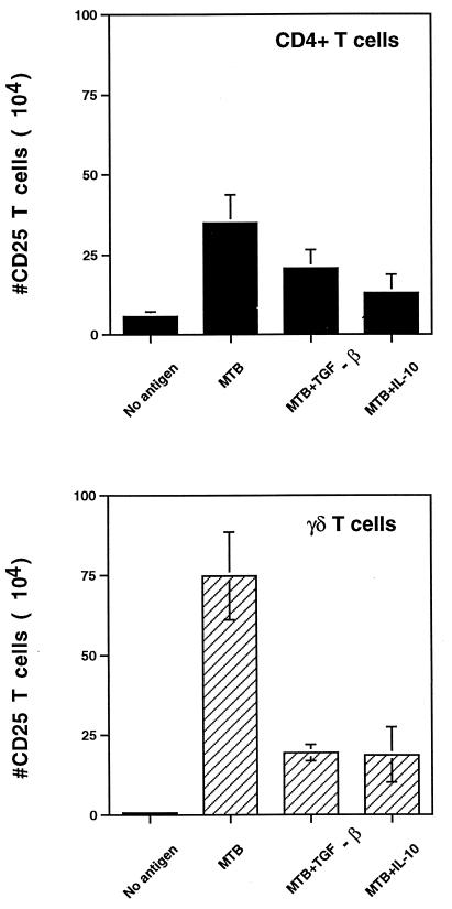
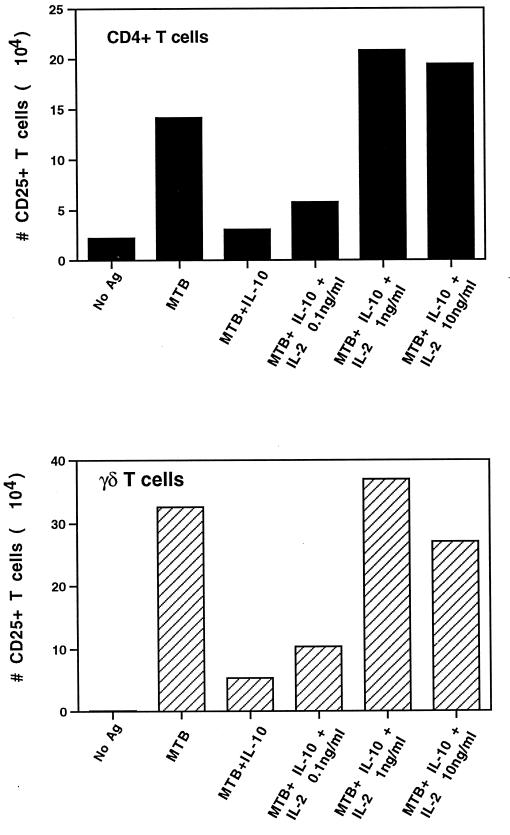
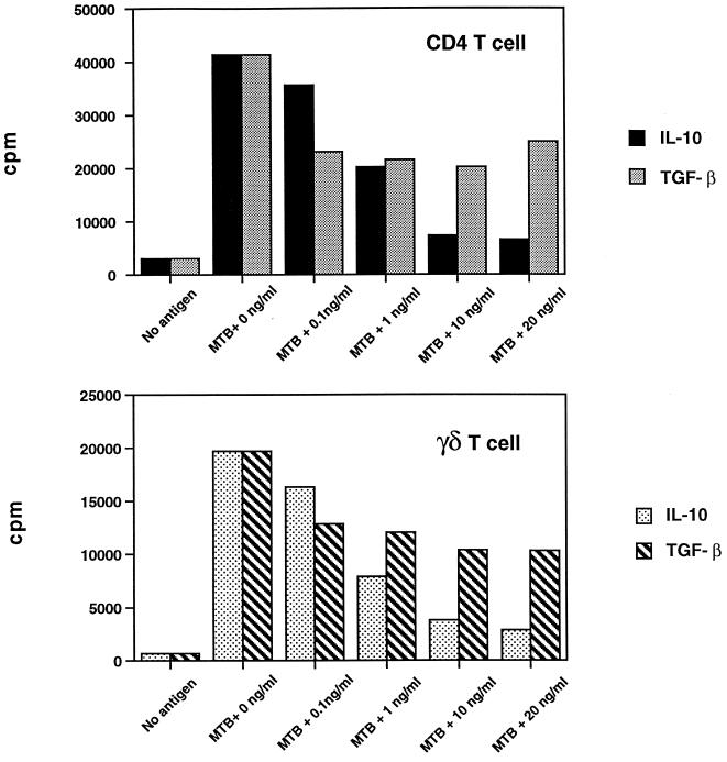
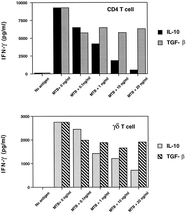
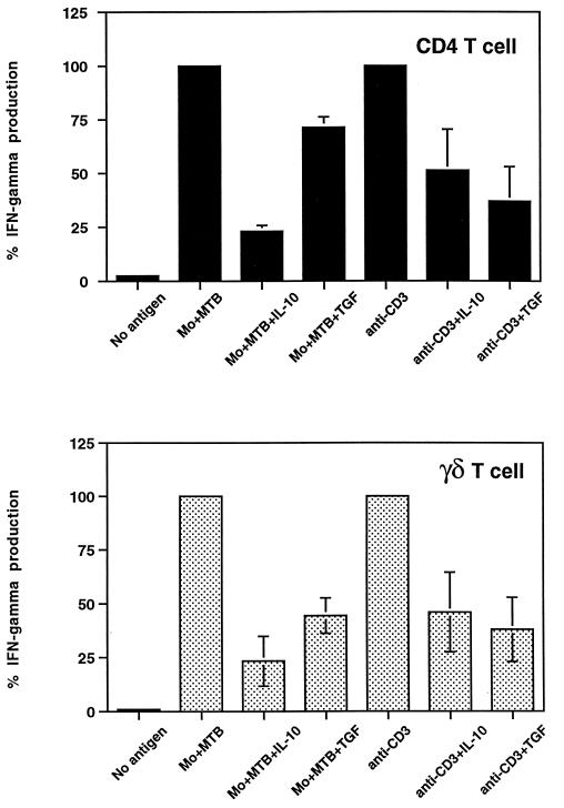
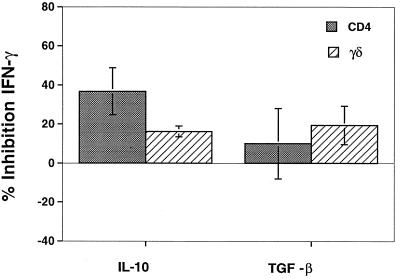
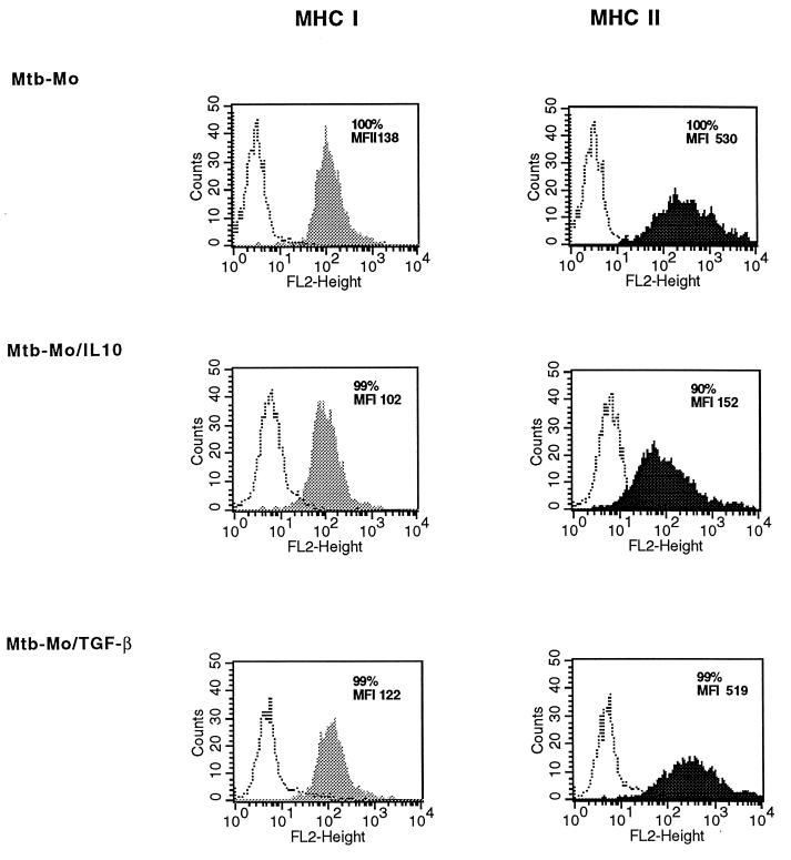
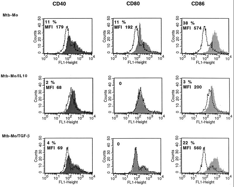
Mycobacterium tuberculosis is the etiologic agent of human tuberculosis and is estimated to infect one-third of the world's population. Control of M. tuberculosis requires T cells and macrophages. T-cell function is modulated by the cytokine environment, which in mycobacterial infection is a balance of proinflammatory (interleukin-1 [IL-1], IL-6, IL-8, IL-12, and tumor necrosis factor alpha) and inhibitory (IL-10 and transforming growth factor beta [TGF-beta]) cytokines. IL-10 and TGF-beta are produced by M. tuberculosis-infected macrophages. The effect of IL-10 and TGF-beta on M. tuberculosis-reactive human CD4(+) and gammadelta T cells, the two major human T-cell subsets activated by M. tuberculosis, was investigated. Both IL-10 and TGF-beta inhibited proliferation and gamma interferon production by CD4(+) and gammadelta T cells. IL-10 was a more potent inhibitor than TGF-beta for both T-cell subsets. Combinations of IL-10 and TGF-beta did not result in additive or synergistic inhibition. IL-10 inhibited gammadelta and CD4(+) T cells directly and inhibited monocyte antigen-presenting cell (APC) function for CD4(+) T cells and, to a lesser extent, for gammadelta T cells. TGF-beta inhibited both CD4(+) and gammadelta T cells directly and had little effect on APC function for gammadelta and CD4(+) T cells. IL-10 down-regulated major histocompatibility complex (MHC) class I, MHC class II, CD40, B7-1, and B7-2 expression on M. tuberculosis-infected monocytes to a greater extent than TGF-beta. Neither cytokine affected the uptake of M. tuberculosis by monocytes. Thus, IL-10 and TGF-beta both inhibited CD4(+) and gammadelta T cells but differed in the mechanism used to inhibit T-cell responses to M. tuberculosis.


Figures

FIG. 1
FIG. 1
CD25 expression on CD4+ and γδ T cells analyzed by two-color flow cytometry after M. tuberculosis stimulation. PBMC from purified protein derivative-positive donors (2 × 10⁶ cells/well) were stimulated with M. tuberculosis (MTB) (2.5 × 10⁶ bacilli/well) in the presence or absence of IL-10 or TGF-β. After 1 week, cells were harvested, counted, stained with CD25-PE and CD4-FITC or TCRδ1, and analyzed by two-color flow cytometry. Results represent the number (mean ± standard error of the mean) of CD25+ CD4+ and CD25+ γδ TCR+ T cells on day 7 (P value for cytokine-treated versus nontreated wells, <0.05).
FIG. 2
FIG. 2
CD25 expression on CD4+ and γδ T cells analyzed by two-color flow cytometry after M. tuberculosis stimulation and treatment with IL-10 and IL-2. PBMC from purified protein derivative-positive donors (2 × 10⁶ cells/well) were stimulated with M. tuberculosis (MTB) (2.5 × 10⁶ bacilli/well) in the presence or absence of IL-10 (10 ng/ml) and different doses of IL-2. After 1 week, cells were harvested, counted, stained with CD25-PE and CD4-FITC or TCRδ1, and analyzed by two-color flow cytometry. Results represent the number of CD25+ CD4+ and CD25+ γδ TCR+ T cells on day 7 in one representative experiment. A similar result was obtained when IL-2 was added to TGF-β-treated cultures. Standard error of the mean did not exceed 10%. Ag, antigen.
FIG. 3
FIG. 3
Effect of IL-10 and TGF-β on M. tuberculosis-stimulated CD4+ and γδ TCR+ T-cell proliferation. Positively selected CD4+ and γδ T cells (5 × 10⁴ cells/well) from M. tuberculosis-stimulated PBMC were cocultured with irradiated monocytes (105 cells/well), M. tuberculosis (MTB) (106 bacilli/well), and different concentrations of IL-10 or TGF-β. After 3 days, [3H]thymidine incorporation was measured and expressed as counts per minute. One representative experiment of four is shown. The standard error of the mean of triplicates did not exceed 10%. Both IL-10 and TGF-β inhibited CD4+ and γδ T-cell proliferation (n = 4; P, <0.05), but IL-10 was a more potent inhibitor than TGF-β for both CD4+ and γδ T cells.
FIG. 4
FIG. 4
Effect of IL-10 and TGF-β on IFN-γ production by M. tuberculosis-stimulated CD4+ and γδ TCR+ T cells. Positively selected CD4 and γδ T cells (5 × 10⁴ cells/well) from M. tuberculosis-stimulated PBMC were cocultured with irradiated monocytes (105 cells/well), M. tuberculosis (MTB) (106 bacilli/well), and different concentrations of IL-10 or TGF-β. After 3 days, supernatants were collected and IFN-γ levels were measured by an ELISA. Data represent the mean of triplicates from a representative experiment; the standard error of the mean did not exceed 10%. IL-10 inhibited IFN-γ production by both CD4+ and γδ T cells (n = 4; P, <0.05), but TGF-β was a less potent inhibitor than IL-10 for CD4+ and γδ T cells.
FIG. 5
FIG. 5
Direct inhibition by IL-10 and TGF-β of CD4+ and γδ TCR+ T-cell IFN-γ production. Positively selected CD4+ and γδ T cells (5 × 10⁴ or 5 × 10⁵ cells/well) were stimulated with either M. tuberculosis-infected macrophages (Mo+MTB) (105 cells/well) or plate-bound anti-CD3 antibodies (1 μg/ml). IL-10 or TGF-β (10 ng/ml) was added on day 0; on day 3, supernatants were harvested and IFN-γ levels were measured by an ELISA. Inhibition of IFN-γ production in the presence of cytokines is expressed as the percentage of maximal production in response to M. tuberculosis or anti-CD3 antibodies. Results represent the mean of three different experiments ± the standard error of the mean. Inhibition by IL-10 and TGF-β was significant for both M. tuberculosis- and anti-CD3 antibody-stimulated IFN-γ production by CD4+ and γδ T cells (n = 3; P, <0.05).
FIG. 6
FIG. 6
Effect of IL-10 and TGF-β on APC function. Monocytes were infected with M. tuberculosis in the presence or absence of IL-10 or TGF-β for 24 h and then fixed with 1% paraformaldehyde. Fixed APC were cocultured with CD4+ or γδ T cells for 3 days, and IFN-γ levels were measured by an ELISA. The percent inhibition of IFN-γ secretion by CD4+ or γδ T cells after stimulation with IL-10- or TGF-β-treated APC is shown. IL-10 inhibited IFN-γ production by CD4+ and γδ T cells (P value for cytokine-treated versus non-cytokine-treated monocytes, <0.05; n = 3), while TGF-β did not affect monocyte APC function (P >0.05; n = 3). Error bars show the standard error of the mean.
FIG. 7
FIG. 7
Class I and class II MHC expression on monocytes analyzed by flow cytometry. M. tuberculosis-infected macrophages (Mtb-Mo) were treated with IL-10 or TGF-β for 24 h. Cells were stained with rat anti-class I MHC or rat anti-class II MHC IgG followed by PE-conjugated goat anti-rat IgG and analyzed by one-color flow cytometry. Monocytes were gated according to their side scatter and forward angle scatter parameters and were 85 to 95% CD14+ CD3. The percentages of class I MHC- and class II MHC-positive cells and the MFI (mean fluorescence of cells with specific antibody − mean fluorescence of cells with matched isotype IgG) are indicated. One representative experiment of three is shown. FL2 height represents PE fluorescence (log scale). Open histograms represent fluorescence with matched isotype control antibody; shaded and closed histograms represent staining for MHC I and MHC II, respectively.
FIG. 8
FIG. 8
CD40, CD80 (B7-1), and CD86 (B7-2) surface expression on monocytes analyzed by flow cytometry. M. tuberculosis-infected macrophages (Mtb-Mo) were treated with IL-10, TGF-β or no cytokine for 24 h. Cells were stained with mouse anti-CD40, anti-CD80, or anti-CD86 IgG followed by PE-conjugated goat anti-mouse IgG and analyzed by one-color flow cytometry. Monocytes were gated according to their SSC and FSC parameters and were 85 to 95% CD14+ CD3. The percentages of CD40-, CD80-, and CD86-positive cells and the MFI (mean fluorescence of cells with specific antibody − mean fluorescence of cells with matched isotype IgG) are indicated. One representative experiment of three is shown. FL1 height represents FITC fluorescence. Open histograms represent fluorescence with isotype control antibody. Shaded histograms represent staining for CD40, CD80, or CD86.

References

    1. Ahuja S S, Paliogianni F, Yamada H, Balow J E, Boumpas D T. Effect of transforming growth factor-beta on early and late activation events in human T cells. J Immunol. 1993;150:3109–3118. - PubMed
    1. Balaji K N, Boom W H. Processing of Mycobacterium tuberculosis bacilli by human monocytes for CD4+αβ and γδ T cells: role of particulate antigen. Infect Immun. 1998;66:98–106. - PMC - PubMed
    1. Balaji K N, Schwander S, Rich E A, Boom W H. Alveolar macrophages as accessory cells for human γδ T cells activated by M. tuberculosis. J Immunol. 1995;154:5959–5968. - PubMed
    1. Barnes P F, Lu S, Abrams J S, Wang E, Yamamura M, Modlin R L. Cytokine production at the site of disease in human tuberculosis. Infect Immun. 1993;61:3482–3489. - PMC - PubMed
    1. Barnes P F, Mistry S D, Cooper C L, Pirmez C, Rea T H, Modlin R L. Compartmentalization of a CD4+ T lymphocyte subpopulation in tuberculous pleuritis. J Immunol. 1989;142:1114–1119. - PubMed

Publication types

MeSH terms

Substances